

Discovery of low-molecular weight anti-PD-L1 peptides for cancer immunotherapy
- PMID: 31640814
- PMCID: PMC6805442
- DOI: 10.1186/s40425-019-0705-y
Discovery of low-molecular weight anti-PD-L1 peptides for cancer immunotherapy
Abstract
Background: Immunotherapy using checkpoint inhibitors, especially PD-1/PD-L1 inhibitors, has now evolved into the most promising therapy for cancer patients. However, most of these inhibitors are monoclonal antibodies, and their large size may limit their tumor penetration, leading to suboptimal efficacy. As a result, there has been a growing interest in developing low-molecular-weight checkpoint inhibitors.
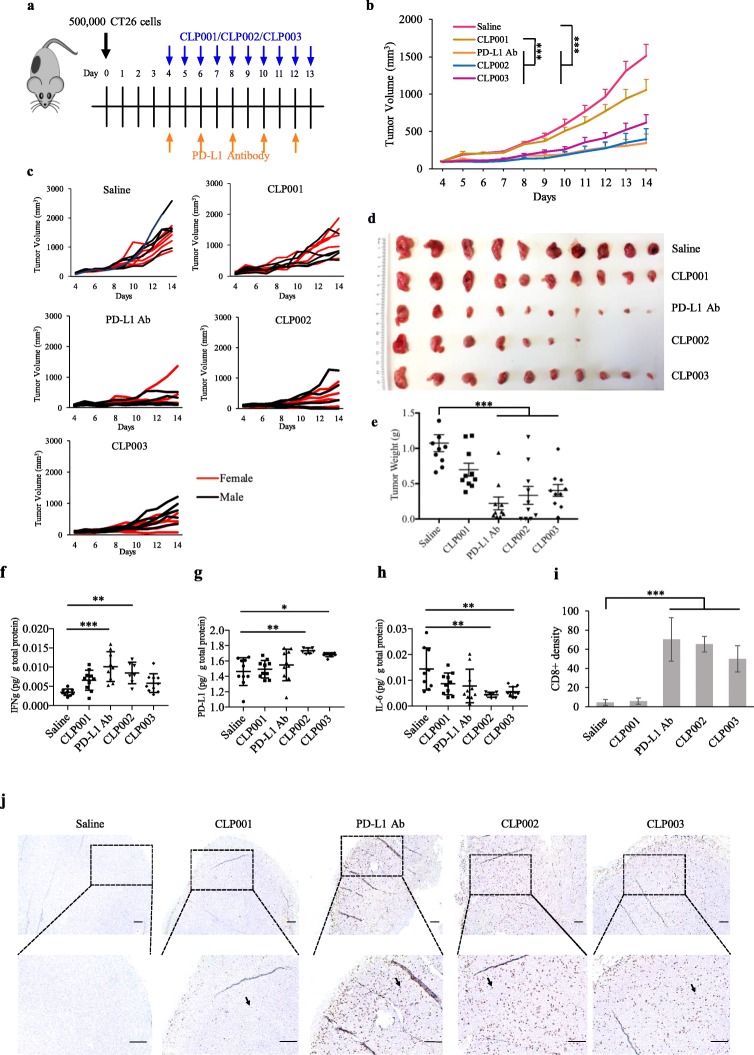
Methods: We developed a novel biopanning strategy to discover small peptide-based anti-PD-L1 inhibitors. The affinity and specificity of the peptides to PD-L1 were examined using various assays. Three-dimensional (3D) spheroid penetration study was performed to determine the tumor penetration capability of the peptides. Anti-tumor activity of the peptides was evaluated in mice bearing CT26 tumor cells.
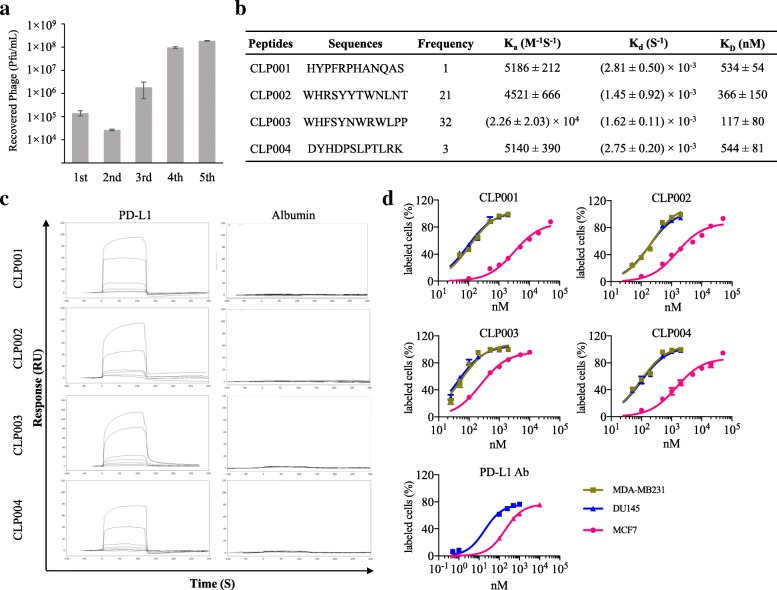
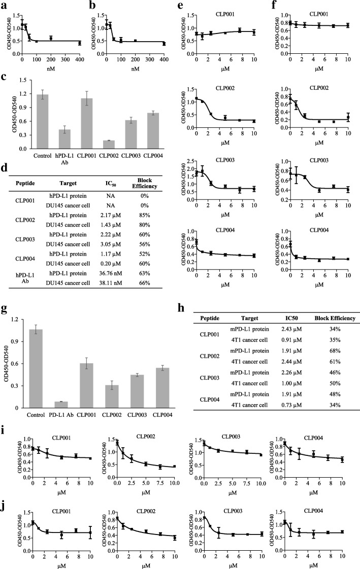
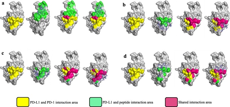
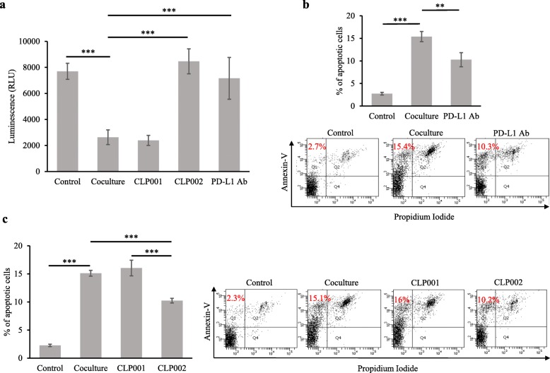
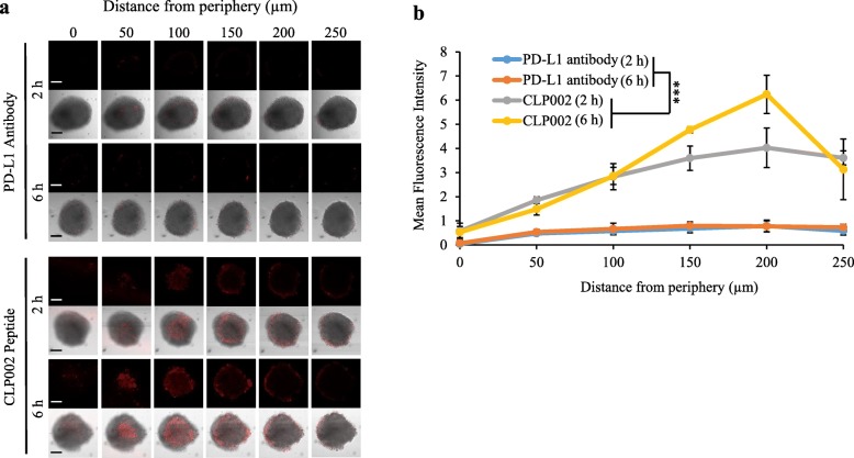
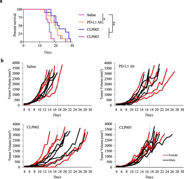
Results: We discover several anti-PD-L1 peptide inhibitors to block PD-1/PD-L1 interaction. The peptides exhibit high affinity and specificity to human PD-L1 protein as well as PD-L1-overexpressing human cancer cells MDA-MB-231 and DU-145. Molecular docking studies indicate that the peptide CLP002 specifically binds to PD-L1 at the residues where PD-L1 interacts with PD-1. The peptide also blocks the CD80/PD-L1 interaction, which may further enhance the immune response of tumor-infiltrating T cells. Compared to antibody, the peptide CLP002 exhibits better tumor penetration in a 3D tumor spheroid model. The peptide CLP002 restores proliferation and prevents apoptosis of T cells that are co-cultured with cancer cells. The peptide CLP002 also inhibits tumor growth and increases survival of CT26 tumor-bearing mice.
Conclusions: This study demonstrated the feasibility of using phage display to discover small peptide-based checkpoint inhibitors. Our results also suggested that the anti-PD-L1 peptide represents a promising low-molecular-weight checkpoint inhibitor for cancer immunotherapy.
Keywords: CT26; Checkpoint inhibitor; PD-1; PD-L1; Peptide; Phage display; Tumor penetration.
Conflict of interest statement
We are in the process of filing a patent for the anti-PD-L1 peptides discovered in this study.
Figures
Similar articles
-
Development of the Inhibitors that Target the PD-1/PD-L1 Interaction-A Brief Look at Progress on Small Molecules, Peptides and Macrocycles.Molecules. 2019 May 30;24(11):2071. doi: 10.3390/molecules24112071. Molecules. 2019. PMID: 31151293 Free PMC article. Review.
-
Novel anti-PD-L1 peptide selected from combinatorial phage library inhibits tumor cell growth and restores T-cell activity.J Drug Target. 2021 Aug;29(7):771-782. doi: 10.1080/1061186X.2021.1879087. Epub 2021 Jul 9. J Drug Target. 2021. PMID: 33478285
-
Design, synthesis and evaluation of PD-L1 peptide antagonists as new anticancer agents for immunotherapy.Bioorg Med Chem. 2021 Jan 15;30:115951. doi: 10.1016/j.bmc.2020.115951. Epub 2020 Dec 15. Bioorg Med Chem. 2021. PMID: 33360579
-
A Small Molecule Antagonist of PD-1/PD-L1 Interactions Acts as an Immune Checkpoint Inhibitor for NSCLC and Melanoma Immunotherapy.Front Immunol. 2021 May 14;12:654463. doi: 10.3389/fimmu.2021.654463. eCollection 2021. Front Immunol. 2021. PMID: 34054817 Free PMC article.
-
Structural Biology of the Immune Checkpoint Receptor PD-1 and Its Ligands PD-L1/PD-L2.Structure. 2017 Aug 1;25(8):1163-1174. doi: 10.1016/j.str.2017.06.011. Structure. 2017. PMID: 28768162 Review.
Cited by
-
Advanced detection of cervical cancer biomarkers using engineered filamentous phage nanofibers.Appl Microbiol Biotechnol. 2024 Feb 19;108(1):221. doi: 10.1007/s00253-024-13058-w. Appl Microbiol Biotechnol. 2024. PMID: 38372795 Free PMC article.
-
Computational Design of Miniproteins as SARS-CoV-2 Therapeutic Inhibitors.Int J Mol Sci. 2022 Jan 13;23(2):838. doi: 10.3390/ijms23020838. Int J Mol Sci. 2022. PMID: 35055023 Free PMC article.
-
PEGylated Cisplatin Nanoparticles for Treating Colorectal Cancer in a pH-Responsive Manner.J Immunol Res. 2022 Aug 5;2022:8023915. doi: 10.1155/2022/8023915. eCollection 2022. J Immunol Res. 2022. PMID: 36033392 Free PMC article.
-
Peptide ARHGEF9 Inhibits Glioma Progression via PI3K/AKT/mTOR Pathway.Dis Markers. 2023 Feb 18;2023:7146589. doi: 10.1155/2023/7146589. eCollection 2023. Dis Markers. 2023. Retraction in: Dis Markers. 2023 Aug 9;2023:9849123. doi: 10.1155/2023/9849123. PMID: 36852158 Free PMC article. Retracted.
-
Design of a novel chimeric peptide via dual blockade of CD47/SIRPα and PD-1/PD-L1 for cancer immunotherapy.Sci China Life Sci. 2023 Oct;66(10):2310-2328. doi: 10.1007/s11427-022-2285-6. Epub 2023 Apr 21. Sci China Life Sci. 2023. PMID: 37115491
References
Publication types
MeSH terms
Substances
Grants and funding
LinkOut - more resources
Full Text Sources
Other Literature Sources
Research Materials
Miscellaneous