. 2020 Mar;101(5):1118-1134.
doi: 10.1111/tpj.14579. Epub 2019 Nov 29.

The formation of perinucleolar bodies is important for normal leaf development and requires the zinc-finger DNA-binding motif in Arabidopsis ASYMMETRIC LEAVES2

Affiliations

The formation of perinucleolar bodies is important for normal leaf development and requires the zinc-finger DNA-binding motif in Arabidopsis ASYMMETRIC LEAVES2

Lilan Luo et al. Plant J. 2020 Mar.

Abstract

In Arabidopsis, the ASYMMETRIC LEAVES2 (AS2) protein plays a key role in the formation of flat symmetric leaves via direct repression of the abaxial gene ETT/ARF3. AS2 encodes a plant-specific nuclear protein that contains the AS2/LOB domain, which includes a zinc-finger (ZF) motif that is conserved in the AS2/LOB family. We have shown that AS2 binds to the coding DNA of ETT/ARF3, which requires the ZF motif. AS2 is co-localized with AS1 in perinucleolar bodies (AS2 bodies). To identify the amino acid signals in AS2 required for formation of AS2 bodies and function(s) in leaf formation, we constructed recombinant DNAs that encoded mutant AS2 proteins fused to yellow fluorescent protein. We examined the subcellular localization of these proteins in cells of cotyledons and leaf primordia of transgenic plants and cultured cells. The amino acid signals essential for formation of AS2 bodies were located within and adjacent to the ZF motif. Mutant AS2 that failed to form AS2 bodies also failed to rescue the as2-1 mutation. Our results suggest the importance of the formation of AS2 bodies and the nature of interactions of AS2 with its target DNA and nucleolar factors including NUCLEOLIN1. The partial overlap of AS2 bodies with perinucleolar chromocenters with condensed ribosomal RNA genes implies a correlation between AS2 bodies and the chromatin state. Patterns of AS2 bodies in cells during interphase and mitosis in leaf primordia were distinct from those in cultured cells, suggesting that the formation and distribution of AS2 bodies are developmentally modulated in plants.

Keywords: 45S ribosomal RNA genes; ETTIN/AUXIN RESEPONSE FACTOR3; chromocenter; epigenetic factor AS2; nucleolus; perinucleolar body; zinc-finger motif.


Conflict of interest statement

The authors declare no conflict of financial interest.

Figures

Figure 1
Figure 1
Observations of AS2‐YFP at the adaxial surfaces of cotyledons and leaf primordia. Expression of AS2‐YFP was induced by incubating 7‐day‐old transgenic Arabidopsis plants (7 days after sowing) with 0.05 µm 17β‐estradiol for 16 h. Aboveground parts of seedlings were fixed in 3.7% paraformaldehyde and chromosomes and nuclei were stained with DAPI. Cells at mitosis, with condensed chromosomes, are indicated by white arrows. (a) Fluorescence due to DAPI (cyan) and YFP (yellow) in cells at the adaxial surface of a cotyledon was monitored at 0.5‐µm intervals by confocal fluorescence microscopy. Merged images are also shown. Optical sections of a given cotyledon at the indicated depths are shown as typical examples. Bars, 100 µm. (b) Signals due to DAPI (cyan) and YFP (yellow) in cells at the adaxial surface of a leaf primordium are shown in one typical optical section. Bar, 100 µm. Magnified views of boxed regions in panels (a) and (b) are shown in (c) and (d), respectively. Bars, 10 µm.
Figure 2
Figure 2
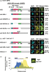
The zinc‐finger motif is required for localization of AS2 to AS2 bodies and the ICG and LZL regions are required for nuclear localization of AS2. (a. left) Schematic representation of AS2 and variant proteins with deletions in the AS2/LOB domain. DNA constructs encoding mutant proteins were fused to the sequence encoding to the N‐terminus of YFP and these fused constructs were linked to the estrogen‐inducible promoter. See text for details. (a. right) Images showing the signals from YFP fusion proteins in Arabidopsis plants (Col‐0) transformed with these DNA constructs. Three to five independent transgenic lines were established for analysis of AS2‐YFP and each as2‐variant‐YFP: four lines for AS2‐YFP; five lines for as2‐∆N2‐7‐YFP; and three lines each for as2‐∆N∆ZF2–24‐YFP, as2‐∆ICG25–80‐YFP, as2‐∆LZL81–109‐YFP and as2‐∆LZL‐NLS‐YFP. Expression of these genes was induced as described in the legend to Figure 1. Four to 27 cells that were YFP‐positive were observed in the adaxial epidermis of cotyledons of each transgenic line, as described in the legend to Figure 1. Signals due to DAPI (cyan), those due to YFP (yellow) and merged images in representative cells are shown. Numbers on the right side of the images show the ratios of total numbers of AS2 body‐positive cells to total numbers of YFP‐positive cells, which were obtained by adding the numbers of cells from the analysis of each transgenic line. Arrows in the sixth row (as2‐∆LZL‐NLS) show DAPI‐positive granules overlapping YFP‐negative areas. Bars, 10 µm. (b, left) Schematic representation of AS2 and as2‐∆N2–7. (b, right) Images showing the signals from YFP fusion proteins in as2‐1 and as1‐1 plants. Three transgenic lines were selected for analysis of fluorescence, which was visualized as described above. Signals due to DAPI (cyan) and YFP (yellow) and merged images are shown. Numbers on the right show the ratios of total AS2 body‐positive cells to total YFP‐positive cells in each transgenic line, counted as described above. Chromocenters and AS2 bodies that partially overlapped one another are indicated by white arrowheads. In (a) and (b), bars, 10 µm. (c) Partial overlap of the DAPI signal from the chromocenter with the YFP signal from an AS2 body formed by AS2‐YFP. The inset is a magnified view of the boxed region in panel a (top right, labeled c). Bar, 3 µm. Intensities of fluorescence signals due to YFP and DAPI were measured along the white arrow with ImageJ software (https://imagej.nih.gov/ij/).
Figure 3
Figure 3
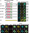
Amino acid residues in the zinc‐finger motif are critical for the formation of AS2 bodies. (a) Schematic representation of AS2 and variant proteins with amino acid substitutions in the ZF motif. Numbers below the wild‐type representation indicate positions of amino acid residues in AS2. Amino acid sequences in and around the ZF motif of the wild‐type are shown and mutated residues are depicted in red. The DNA constructs were linked to an estrogen‐inducible promoter. Details of the DNA constructs that encoded AS2‐YFP and as2‐variant‐YFP proteins can be found in the text. YFP is shown by the yellow bar at the bottom. Arabidopsis plants (Col‐0) were transformed with indicated constructs. (b) Images showing the signals from YFP fusion proteins in Arabidopsis plants (Col‐0) that had been transformed with the respective DNA constructs. Three independent transgenic lines were established for analysis of AS2‐YFP and each as2‐variant‐YFP protein. Expression of these genes was induced as described in the legend to Figure 1. Eight to 75 cells that were YFP‐positive were observed in the adaxial epidermis of cotyledons of each transgenic line, as described in the legend to Figure 2. Signals due to DAPI (cyan), those due to YFP (yellow) and merged images are shown. Numbers on the right side of the fluorescence images show the ratios of total numbers of AS2 body‐positive cells to total numbers of YFP‐positive cells, obtained by adding numbers of cells from the analysis of each transgenic line. Chromocenters and AS2 bodies that partially overlapped one another are indicated by white arrowheads. Bars, 10 µm. (c) Observation of 45S rDNA and 5S rDNA loci in cells of the aerial part of the AS2‐YFP‐expressing transgenic plant, as described above. DNA‐FISH was performed using 45S (top row) and 5S rRNA gene‐specific probes (bottom row). Signals due to DAPI (cyan), anti‐GFP (AS2‐YFP; yellow), FISH (red), and the merged images are shown. Chromocenters and AS2 bodies that partially overlapped one another are indicated by white arrowheads. FISH signals against 45S rDNA loci that partially overlapped with AS2 bodies are indicated by white arrows. Bars, 10 μm.
Figure 4
Figure 4
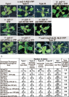
Variant as2 proteins that failed to form AS2 bodies did not rescue the as2‐1 mutation. Transgenic as2‐1 seeds harboring AS2‐YFP, as2‐variant‐YFPs and YFP, as indicated above the images, were grown on MS plates supplemented with 0.5 µm 17β‐estradiol under white light for 16 h and in darkness for 8 h daily, at 22ºC, as described in Experimental Procedures. The gross morphology of 14‐day‐old transgenic plants was analyzed and representative morphology is shown for each transgenic line in panels (a–h). On the basis of phenotypes of transgenic as2‐1 plants that harbored AS2‐YFP plants were classified into four types (Types I, II, III and non‐rescued as2‐like plants). Panel (i) shows numbers of plants belonging to five types (Types I to IV and non‐rescued as2‐like plants) and corresponding percentages for YFP signal‐positive plants of individual transgenic lines, as denoted by a number with # in parenthesis in the left‐hand column. Arrowheads and arrows in panel (a) indicate partially upwardly curled leaves and upwardly curled leaves, respectively. Transgenic as2‐1 plants harboring as2‐∆LZL‐NLS‐YFP [panels (h)] exhibited an unexpected phenotype (Type IV) in addition to the non‐complemented as2‐like phenotype. Bars, 1 cm.
Figure 5
Figure 5
Subcellular localization of AS2‐YFP in interphase and M phase cells in leaf primordia and cultured cell line MM2d. Expression of AS2‐YFP was induced by incubating 7‐day‐old seedlings of transgenic Arabidopsis plants with 0.05 µm 17β‐estradiol for 16 h (see Experimental Procedures). Seedlings were fixed in 3.7% paraformaldehyde, and chromosomes and nuclei were stained with DAPI. Fluorescence due to DAPI (cyan) and that due to YFP (yellow) was visualized by confocal fluorescence microscopy. Merged images of fluorescence due to DAPI and YFP are also shown. Stages of interphase (or G0, the differentiated phase) (a) and mitotic phases [panels (b–e)] were defined in terms of patterns of staining with DAPI. Panel (a) (top row) shows many interphase (or G0) cells and one anaphase cell (indicated by the white arrows) and magnified views of the boxed nuclei are shown in the lower row. Chromocenters and AS2 bodies that partially overlapped one another are indicated by white arrowheads. Images in (b–e) are seven‐image stacks from an individual sample or cell. The white arrow and the asterisk in (e) indicate a bridge‐like and a tail‐like structures, respectively (see text). The subcellular localization of AS2‐YFP in cells of the MM2d cultured cell line was analyzed similarly, as shown in (f–h). Cells were incubated for 16 h in the presence of 0.05 µm 17β‐estradiol and fixed as described above. Phases of the cell cycle were determined by fluorescence immunostaining of microtubules with antibodies raised in mouse against α‐tubulin from chicken and staining of chromosomal DNA with DAPI. Fluorescence from tubulins, DAPI and YFP was visualized by fluorescence microscopy as described in Experimental Procedures. Merged images (tubulin, red; DAPI, cyan; YFP, yellow) are shown on the right. Phragmoplast microtubules were observed between separating chromosomes (h). Numbers on the right represent ratios of AS2‐body‐containing cells to total YFP‐positive cells at each mitotic phase. Bars, 5 μm.
Figure 6
Figure 6
Summary of the association of mutations in the AS2/LOB domain with the ability to form AS2 bodies and the results of complementation tests with the AS2 variants used in the present study. Amino acid residues indicated in red were tested for their importance in the formation of AS2 bodies and development of normal leaf morphology, such as establishment of adaxial–abaxial polarity and development of proximal–distal polarity. Underlined residues are conserved in many ASL members of the AS2/LOB family (see Table S1) and were required for the formation of AS2 bodies and the functions of AS2 in leaf formation.

Similar articles

Cited by

References

    1. Banno, H. , Hirano, K. , Nakamura, T. , Irie, K. , Nomoto, S. , Matsumoto, K. and Machida, Y. (1993) NPK1, a tobacco gene that encodes a protein with a domain homologous to yeast BCK1, STE11, and Byr2 protein kinases. Mol. Cell Biol. 13, 4745–4752. - PMC - PubMed
    1. Barton, M.K. (2001) Leaving the meristem behind: regulation of KNOX genes. Genome Biol. 2, 1002.1–1002.3. - PMC - PubMed
    1. Boudonck, K. , Dolan, L. and Shaw, P.J. (1999) The movement of coiled Bodies visualized in living plant cells by the green fluorescent protein. Mol. Biol. Cell, 10, 2297–2307. - PMC - PubMed
    1. Bowman, J.L. and Floyd, S.K. (2008) Patterning and polarity in seed plant shoots. Annu. Rev. Plant Biol. 59, 67–88. - PubMed
    1. Byrne, M.E. , Barley, R. , Curtis, M. , Arroyo, J.M. , Dunham, M. , Hudson, A. and Martienssen, R.A. (2000) Asymmetric leaves1 mediates leaf patterning and stem cell function in Arabidopsis . Nature, 408, 967–971. - PubMed

Publication types