Intra-Regional Glu-GABA vs Inter-Regional Glu-Glu Imbalance: A 1H-MRS Study of the Neurochemistry of Auditory Verbal Hallucinations in Schizophrenia
- PMID: 31626702
- PMCID: PMC7147588
- DOI: 10.1093/schbul/sbz099
Intra-Regional Glu-GABA vs Inter-Regional Glu-Glu Imbalance: A 1H-MRS Study of the Neurochemistry of Auditory Verbal Hallucinations in Schizophrenia
Abstract
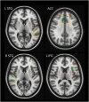
Glutamate (Glu), gamma amino-butyric acid (GABA), and excitatory/inhibitory (E/I) imbalance have inconsistently been implicated in the etiology of schizophrenia. Elevated Glu levels in language regions have been suggested to mediate auditory verbal hallucinations (AVH), the same regions previously associated with neuronal hyperactivity during AVHs. It is, however, not known whether alterations in Glu levels are accompanied by corresponding GABA alterations, nor is it known if Glu levels are affected in brain regions with known neuronal hypo-activity. Using magnetic resonance spectroscopy (MRS), we measured Glx (Glu+glutamine) and GABA+ levels in the anterior cingulate cortex (ACC), left and right superior temporal gyrus (STG), and left inferior frontal gyrus (IFG), in a sample of 77 schizophrenia patients and 77 healthy controls. Two MRS-protocols were used. Results showed a marginally significant positive correlation in the left STG between Glx and AVHs, whereas a significant negative correlation was found in the ACC. In addition, high-hallucinating patients as a group showed decreased ACC and increased left STG Glx levels compared to low-hallucinating patients, with the healthy controls in between the 2 hallucinating groups. No significant differences were found for GABA+ levels. It is discussed that reduced ACC Glx levels reflect an inability of AVH patients to cognitively inhibit their "voices" through neuronal hypo-activity, which in turn originates from increased left STG Glu levels and neuronal hyperactivity. A revised E/I-imbalance model is proposed where Glu-Glu imbalance between brain regions is emphasized rather than Glu-GABA imbalance within regions, for the understanding of the underlying neurochemistry of AVHs.
Keywords: GABA; Glx; MR spectroscopy (MRS); Magnetic resonance imaging (MRI); auditory verbal hallucinations; excitatory; glutamate; hallucinations; inhibitory (E/I) imbalance model; schizophrenia.
© The Author(s) 2019. Published by Oxford University Press on behalf of the Maryland Psychiatric Research Center.
Figures


Similar articles
-
Glutamate in dorsolateral prefrontal cortex and auditory verbal hallucinations in patients with schizophrenia: A 1H MRS study.Prog Neuropsychopharmacol Biol Psychiatry. 2017 Aug 1;78:132-139. doi: 10.1016/j.pnpbp.2017.05.020. Epub 2017 May 22. Prog Neuropsychopharmacol Biol Psychiatry. 2017. PMID: 28546056
-
Negative valence of hallucinatory voices as predictor of cortical glutamatergic metabolite levels in schizophrenia patients.Brain Behav. 2022 Jan;12(1):e2446. doi: 10.1002/brb3.2446. Epub 2021 Dec 7. Brain Behav. 2022. PMID: 34874613 Free PMC article.
-
Anterior Cingulate Glutamate and GABA Associations on Functional Connectivity in Schizophrenia.Schizophr Bull. 2019 Apr 25;45(3):647-658. doi: 10.1093/schbul/sby075. Schizophr Bull. 2019. PMID: 29912445 Free PMC article.
-
Glutamatergic and GABAergic metabolite levels in schizophrenia-spectrum disorders: a meta-analysis of 1H-magnetic resonance spectroscopy studies.Mol Psychiatry. 2022 Jan;27(1):744-757. doi: 10.1038/s41380-021-01297-6. Epub 2021 Sep 28. Mol Psychiatry. 2022. PMID: 34584230 Review.
-
Hearing voices in the head: Two meta-analyses on structural correlates of auditory hallucinations in schizophrenia.Neuroimage Clin. 2022;36:103241. doi: 10.1016/j.nicl.2022.103241. Epub 2022 Oct 19. Neuroimage Clin. 2022. PMID: 36279752 Free PMC article. Review.
Cited by
-
Language abnormalities in schizophrenia: binding core symptoms through contemporary empirical evidence.Schizophrenia (Heidelb). 2022 Nov 12;8(1):95. doi: 10.1038/s41537-022-00308-x. Schizophrenia (Heidelb). 2022. PMID: 36371445 Free PMC article. Review.
-
Medial Frontal Cortex GABA Concentrations in Psychosis Spectrum and Mood Disorders: A Meta-analysis of Proton Magnetic Resonance Spectroscopy Studies.Biol Psychiatry. 2023 Jan 15;93(2):125-136. doi: 10.1016/j.biopsych.2022.08.004. Epub 2022 Aug 17. Biol Psychiatry. 2023. PMID: 36335069 Free PMC article.
-
Glutamate- and GABA-Modulated Connectivity in Auditory Hallucinations-A Combined Resting State fMRI and MR Spectroscopy Study.Front Psychiatry. 2021 Feb 17;12:643564. doi: 10.3389/fpsyt.2021.643564. eCollection 2021. Front Psychiatry. 2021. PMID: 33679491 Free PMC article.
-
Meta-analysis and Open-source Database for In Vivo Brain Magnetic Resonance Spectroscopy in Health and Disease.bioRxiv [Preprint]. 2023 Jun 15:2023.02.10.528046. doi: 10.1101/2023.02.10.528046. bioRxiv. 2023. Update in: Anal Biochem. 2023 Sep 1;676:115227. doi: 10.1016/j.ab.2023.115227. PMID: 37205343 Free PMC article. Updated. Preprint.
-
Pilot-RCT Finds No Evidence for Modulation of Neuronal Networks of Auditory Hallucinations by Transcranial Direct Current Stimulation.Brain Sci. 2022 Oct 12;12(10):1382. doi: 10.3390/brainsci12101382. Brain Sci. 2022. PMID: 36291316 Free PMC article.
References
-
- Jardri R, Pouchet A, Pins D, Thomas P. Cortical activations during auditory verbal hallucinations in schizophrenia: a coordinate-based meta-analysis. Am J Psychiatry. 2011;168(1):73–81. - PubMed
-
- Larøi F, Aleman A.. Hallucinations - A Guide to Treatment and Management. Oxford, UK: Oxford university press; 2010.
-
- Kompus K, Westerhausen R, Hugdahl K. The “paradoxical” engagement of the primary auditory cortex in patients with auditory verbal hallucinations: a meta-analysis of functional neuroimaging studies. Neuropsychologia. 2011;49(12):3361–3369. - PubMed
Publication types
MeSH terms
Substances
LinkOut - more resources
Full Text Sources
Medical

