. 2019 Sep 19;47(16):8874-8887.
doi: 10.1093/nar/gkz642.

Cooperative DNA binding by proteins through DNA shape complementarity

Affiliations

Cooperative DNA binding by proteins through DNA shape complementarity

Stephen P Hancock et al. Nucleic Acids Res. .

Abstract

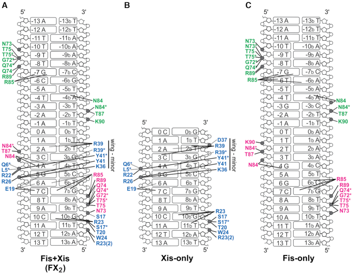
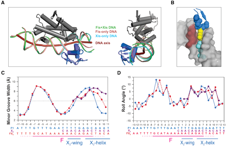
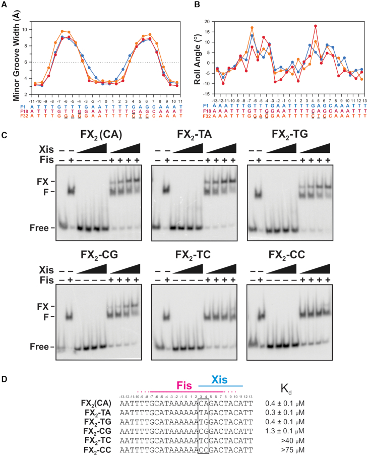
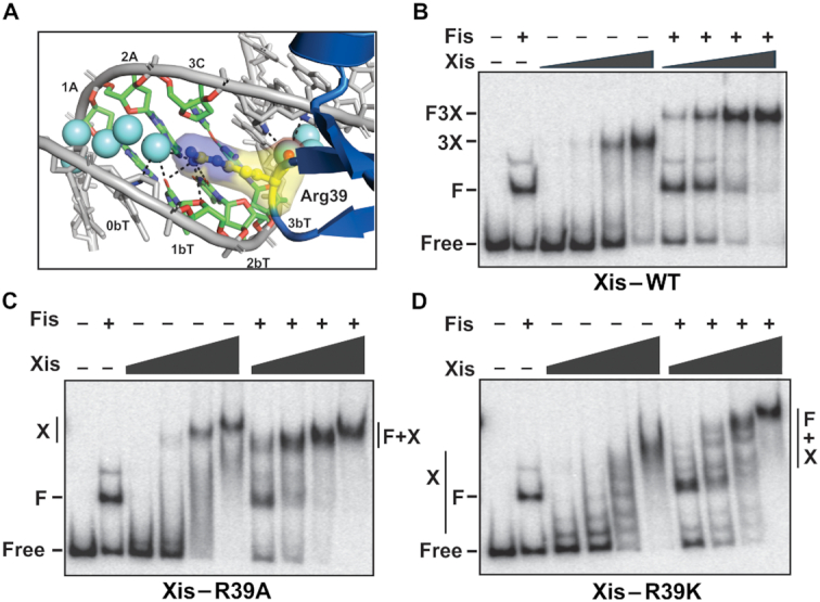
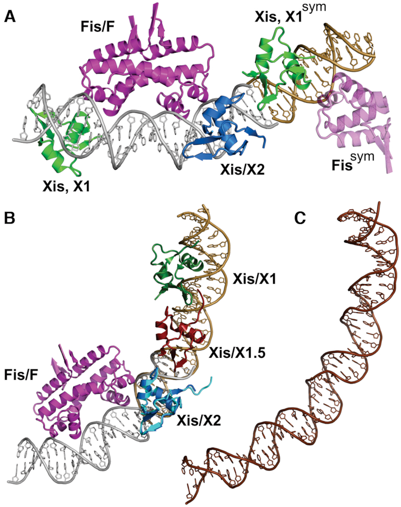
Localized arrays of proteins cooperatively assemble onto chromosomes to control DNA activity in many contexts. Binding cooperativity is often mediated by specific protein-protein interactions, but cooperativity through DNA structure is becoming increasingly recognized as an additional mechanism. During the site-specific DNA recombination reaction that excises phage λ from the chromosome, the bacterial DNA architectural protein Fis recruits multiple λ-encoded Xis proteins to the attR recombination site. Here, we report X-ray crystal structures of DNA complexes containing Fis + Xis, which show little, if any, contacts between the two proteins. Comparisons with structures of DNA complexes containing only Fis or Xis, together with mutant protein and DNA binding studies, support a mechanism for cooperative protein binding solely by DNA allostery. Fis binding both molds the minor groove to potentiate insertion of the Xis β-hairpin wing motif and bends the DNA to facilitate Xis-DNA contacts within the major groove. The Fis-structured minor groove shape that is optimized for Xis binding requires a precisely positioned pyrimidine-purine base-pair step, whose location has been shown to modulate minor groove widths in Fis-bound complexes to different DNA targets.


Figures

Figure 1.
Figure 1.
The phage λ excision reaction and Fis–Xis–DNA structures. (A) λ excision and integration reactions. The recombination reaction excising the phage genome (thick line) from the E. coli chromosome (dash line) occurs between specific recombination sites attL and attR and requires phage-encoded integrase (Int) and Xis plus bacterial nucleoid proteins IHF and Fis. The reverse reaction, integrative recombination, requires Int and IHF and is also stimulated by Fis but inhibited by Xis (3). (B) Organization of the attR recombination site with proteins and binding sites denoted. In the attR x attL recombination complex the bivalent Int recombinase simultaneously binds to the P2 and the bacterial DNA side of the crossover region (0) (12). The combined DNA bending activities of Xis, Fis and IHF stereo-specifically loop the DNA to enable formation of this bridge by the Int N- and C-terminal domains. (C–E) X-ray structures of three Fis–Xis–DNA ternary complexes: FX2, FX1-2-1Xis and FX1-2-2Xis, respectively. The DNA sequences of the oligonucleotides used for crystallography are given below with the boundaries of the Fis and Xis binding sites denoted; the Fis core sequence is in bold letters. Fis dimers (magenta and green subunits) bind DNA (grey) in a similar manner to that observed in Fis-DNA binary complexes. The Xis1–55 bound to the X2 (blue) site inserts its helix B into the major groove and its β-turn-β wing motif into the minor groove towards the center of the Fis binding interface. Xis1–55 bound within the X1 region (orange) in the FX1-2-2Xis structure inserts its wing motif into the minor groove at the outer edge of the Fis binding site and thus is in the reverse orientation. (F) FX1-2 DNA substituted with 5-bromodeoxyuridine (5-BrdU) at the sequence positions denoted with asterisks in panel D was used to verify the site of Xis1–55 binding in the FX1-2-1Xis structure. Difference density (blue mesh) of the 5-BrdU is rendered at 5σ. The right panels highlight nts 8 (top) and 12 (bottom) (numbered from the central T) that are substituted with 5-BrdU.
Figure 2.
Figure 2.
Molecular interactions between Fis, Xis, and DNA. (A) The FX2 structure over the X2 binding site. The side chains of Fis (orange) and Xis (yellow) residues that contact DNA along with DNA phosphate oxygen atoms (red) are highlighted. Only helices B–D of subunit A of the Fis dimer is displayed. (B) The DNA major groove interface with Fis and Xis. The recognition helices of Fis and Xis along with Fis Arg85, Xis Glu19 and Arg22, and two interconnected water molecules (cyan spheres) are displayed. Dashed lines represent hydrogen bonding. Fis Arg85 and Xis Glu19 most closely approach each other within the major groove and are directly hydrogen bonded to the same C7/G7b base pair. (C) Fo – Fc difference omit map showing a subset of interfacial water molecules between Fis and Xis within the major groove, along with the Fis Arg85 and Xis Glu19 side chains (omit density contoured at 5.0σ). Omit maps for the solvent molecules were individually generated and contoured at 4.5–5.0σ (blue) or 3.25σ (green). The C7/G7b base pair is colored. (D) View of the Fis–Xis binding region highlighting the layer of solvent molecules (cyan spheres) separating Fis and Xis residues in the major groove. Xis backbone atoms within the wing (Gly38–Glu40, blue) along with Arg39 side chain atoms (yellow) that insert into the minor groove are rendered as spheres; ordered waters within the minor groove also displayed. (E) Gel mobility shift assays evaluating DNA binding by Xis-wt and Glu19Ala on an attR DNA fragment with and without Fis. Xis concentrations increased from 50 to 1350 nM in 3-fold increments and Fis was added at 4 nM as designated. Xis-wt cooperatively binds to attR to forms a complex containing three Xis protomers (3X). Xis-wt binding is stimulated in the presence of Fis to assemble a Fis dimer + 3Xis – attR complex (F3X). DNA complexes by Xis-Glu19Ala alone are not detectable, but F3X complexes are formed at high mutant Xis concentrations demonstrating cooperative binding with Fis without the Glu19 side chain.
Figure 3.
Figure 3.
Contact diagrams of DNA complexes: (A) Fis + Xis (FX2), (B) Xis-X2 (derived from PDB codes 1RH6 (1.7 Å resolution) and 2OG0 (1.9 Å) with both rotamers of Arg23 depicted as found in 2IEF, 2.6 Å), and (C) Fis-F1 (PDB code 3IV5, 2.9 Å). Phosphates that are contacted directly by protein moieties are grey. Contacts made by Fis chain A (magenta) and chain B (green) and Xis (blue) residues are shown. Asterisks represent contacts made by protein backbone atoms. Direct hydrogen bonds are denoted by solid lines and water-bridged contacts by dashed lines.
Figure 4.
Figure 4.
DNA conformational changes in the Fis+Xis complex. (A) DNA backbones and curvatures in Fis, Xis and Fis + Xis complexes. The Fis-F1 DNA structure (PDB code 3IV5) was aligned with the FX2 structure over the Fis dimers (rmsd = 0.28 Å between Fis dimer backbone atoms). Side and end-on views with the DNA axis of FX2 (thick red line, calculated by CURVES (27) are shown. The Fis dimer of FX2 is displayed and colored grey, the DNA backbone from FX2 is colored green with the spheres denoting the most distal phosphate contacts by Fis Asn73; Xis is colored blue. The DNA backbone of the Fis-F1 complex is colored salmon. Xis from the Xis-X2 structure (PDB code 1RH6, colored slate) was aligned with the Xis of FX2 (rmsd = 0.24 Å over backbone atoms); the Xis-only DNA backbone is colored cyan. The FX2 and Fis–DNA backbones closely align up to T9b, the most distal Fis-DNA contact. The FX2 DNA axis then bends towards Xis in a manner similar to the DNA in the Xis-only structure as evidenced by the close alignment of the green and cyan DNA strands proximal to Xis. (B) A view of the Xis wing inserted into the minor groove in the FX2 complex to highlight the close fit. Rendering is similar to Figure 2D with the CA dinucleotide at position 3-4 on the DNA top strand (chain C) colored dark red. (C) Plot of DNA minor groove widths (minus van der Waals radii) over the FX2 (red), Fis-F1 (blue), Xis-X2 (purple) complexes. Dashed line indicates the average minor groove width for B DNA. See Supplementary Figure S2 for minor groove plots of the FX1-2-1Xis and FX1-2-2Xis complexes. (D) Plots of DNA roll angles along the length of the DNA are shown for the FX2 (red), Fis-F1 (blue), Xis-X2 (purple) complexes.
Figure 5.
Figure 5.
DNA base substitutions that are predicted to alter the conformation of Fis-bound DNA abrogate Xis recruitment by Fis. (A) Minor groove width plots for structures of Fis complexes with DNA containing sequence differences affecting the flexible pyrimidine-purine step within the Fis major groove interface. Blue plot is the F1 complex with a TG/CA dinucleotide step at ±(3–4) (PDB code 3IV5, (16)), red plot is the F18 complex with TG/CA at ±(4–5) (PDB code 3JRG, (16)) and orange plot is the F32 complex with no pyrimidine-purine step (pdb code 5E3O, (15)). (B) Roll angle plots of the same complexes in (A). (C) Gel mobility shift assays showing binding of Xis with and without Fis (4 nM) to 34 bp DNA duplexes containing the F-X2 binding sites with different dinucleotide steps at the 3–4 position. Free or Fis-bound DNA was incubated with increasing amounts of Xis (50, 150, 450, 1350 nM) for 20 min and subjected to PAGE. Xis does not bind without Fis to the short DNA fragment representing the native sequence (FX2 (CA)) under these conditions (8). (D) Sequences and apparent Kd values for Xis binding in the presence of Fis for each of the DNA variants at the 3–4 position tested. Means and standard deviations for the Kd values represent data from at least three replicate experiments.
Figure 6.
Figure 6.
Role of Arginine 39 in Xis–DNA binding with and without Fis. (A) Arg39–DNA interactions within the minor groove in the FX2 structure. Dashes represent hydrogen bonds between Arg39, water (cyan spheres), and DNA. (B–D) Gel mobility shift assays of (B) Xis-wt and mutants (C) Arg39Ala and (D) Arg39Lys binding to attR with and without Fis. For Xis-wt, 25, 75, 225, and 675 nM were added; F denotes the Fis-bound DNA complex, 3X denotes 3 Xis protomers bound to X1–X1.5–X2 and F3X denotes the Fis + 3Xis complex. For Xis mutants, 1.5, 3.0, 6.0 and 12 μM were added; X denotes DNA complexes with multiple Xis protomers and F + X denotes complexes with Fis and multiple Xis protomers.
Figure 7.
Figure 7.
Model of the Fis–Xis region of λ attR. (A) The FX1-2-2Xis structure is shown together with a portion of its symmetry mate on the X2 side. The DNA of the symmetry mate is colored brown. Xis protomers bound over the X1 sequence are green with the wing of Xis, X1sym oriented towards Xis bound at X2. Only half of the Fis dimer (transparent magenta) bound to the symmetry mate is displayed. A pseudo-continuous DNA helix extends from both DNA ends in the crystal lattice. (B) Model of the Fis–Xis complex over the F–X2–X1.5–X1 region. The Xis microfilament structure containing Xis bound to the X2–X1.5–X1 region (PDB code 2IEF, (6)) was aligned with the FX2 structure over the Xis protomers bound at X2 (Xis/X2 from FX2 colored blue and Xis/X2 from 2IEF colored cyan; rmsd over Xis backbone atoms = 0.42 Å). The DNA of 2IEF is brown. (C) The same DNA segment as shown in panel B (attR sequences 53–100) from the 11 Å cryoEM structure of the attR/attL intasome (PDB code 5J0N, (12)). The DNA is shown in the same orientation after alignment with the Fis–Xis model DNA in panel B. The cryoEM intasome structure did not include Fis.

Similar articles

Cited by

References

    1. Panne D., Maniatis T., Harrison S.C.. An atomic model of the interferon-beta enhanceosome. Cell. 2007; 129:1111–1123. - PMC - PubMed
    1. Kim S., Brostromer E., Xing D., Jin J., Chong S., Ge H., Wang S., Gu C., Yang L., Gao Y.Q. et al. .. Probing allostery through DNA. Science. 2013; 339:816–819. - PMC - PubMed
    1. Landy A. The lambda integrase site-specific recombination pathway. Microb. Spectr. 2015; 3:doi:10.1128/microbiolspec.MDNA3-0051-2014. - PMC - PubMed
    1. Ball C.A., Johnson R.C.. Efficient excision of phage lambda from the Escherichia coli chromosome requires the Fis protein. J. Bacteriol. 1991; 173:4027–4031. - PMC - PubMed
    1. Thompson J.F., Landy A.. Berg D, Howe M. Mobile DNA. 1989; Washington: ASM Press; 1–22.

Publication types

MeSH terms