

Fermentation of Blackberry with L. plantarum JBMI F5 Enhance the Protection Effect on UVB-Mediated Photoaging in Human Foreskin Fibroblast and Hairless Mice through Regulation of MAPK/NF-κB Signaling
- PMID: 31614689
- PMCID: PMC6835613
- DOI: 10.3390/nu11102429
Fermentation of Blackberry with L. plantarum JBMI F5 Enhance the Protection Effect on UVB-Mediated Photoaging in Human Foreskin Fibroblast and Hairless Mice through Regulation of MAPK/NF-κB Signaling
Abstract
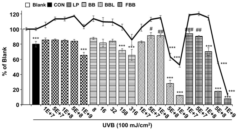
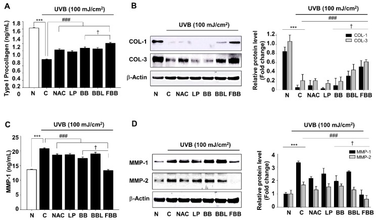
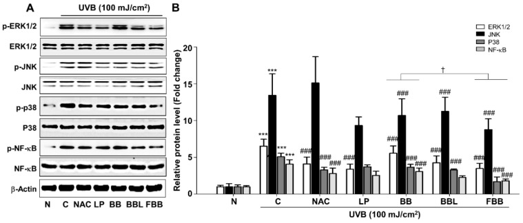
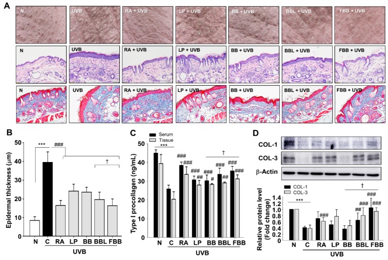
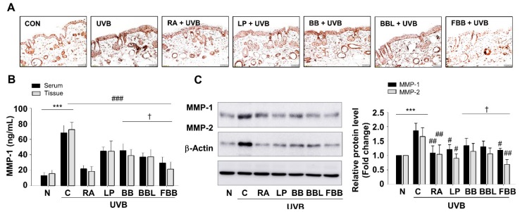
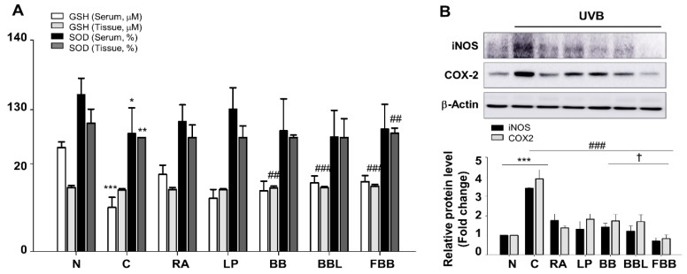
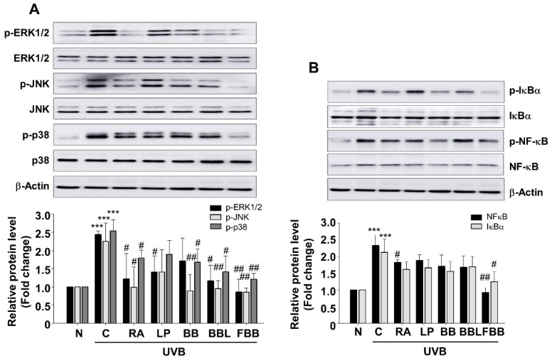
Chronic and extensive exposure of ultraviolet (UV)-irradiation causes human skin sunburn, inflammation, or photoaging, which is associated with downregulated collagen synthesis. This study investigated the effects of fermented blackberry (Rubus fruticosus B., FBB) by Lactobacillus plantarum JBMI F5 (LP) on UVB-induced photoaging in human foreskin fibroblast (Hs68) as well as in SKH-1 hairless mice. FBB pretreatment inhibited UVB-mediated type-1 procollagen degradation, matrix metalloproteinase (MMP)-1 and MMP-2 protein expression, and suppressed nuclear factor-κB (NF-κB) activation as well as mitogen-activated protein kinase (MAPK) phosphorylation in Hs68. In addition, FBB administration diminished the wrinkle formation in dorsal skin and epidermal thickening in UVB-irradiated hairless mice. Moreover, UVB-induced Type-1 procollagen reduction and antioxidant enzyme inactivation were reversed by FBB administration. These results suggest that FBB may have antiphotoaging effects on UVB-induced wrinkle formation by maintaining the extracellular matrix density in the dermis, which occurs via regulation of reactive oxygen species and related MAPK and NF-κB signaling. Therefore, FBB can be a potential candidate for protecting skin aging against UV irradiation.
Keywords: Lactobacillus plantarum; MMPs; fermented blackberry; photoaging; skin aging; type I procollagen.
Conflict of interest statement
The authors declare no potential conflicts of interests.
Figures
Similar articles
-
Hydrangea serrata (Thunb.) Ser. Extract Attenuate UVB-Induced Photoaging through MAPK/AP-1 Inactivation in Human Skin Fibroblasts and Hairless Mice.Nutrients. 2019 Mar 1;11(3):533. doi: 10.3390/nu11030533. Nutrients. 2019. PMID: 30823635 Free PMC article.
-
Hawthorn Polyphenol Extract Inhibits UVB-Induced Skin Photoaging by Regulating MMP Expression and Type I Procollagen Production in Mice.J Agric Food Chem. 2018 Aug 15;66(32):8537-8546. doi: 10.1021/acs.jafc.8b02785. Epub 2018 Jul 31. J Agric Food Chem. 2018. PMID: 30032605
-
Oral administration of Lactobacillus plantarum HY7714 protects hairless mouse against ultraviolet B-induced photoaging.J Microbiol Biotechnol. 2014 Nov 28;24(11):1583-91. doi: 10.4014/jmb.1406.06038. J Microbiol Biotechnol. 2014. PMID: 25112318
-
Epithelial-mesenchymal interaction mechanisms leading to the over-expression of neprilysin are involved in the UVB-induced formation of wrinkles in the skin.Exp Dermatol. 2016 Aug;25 Suppl 3:2-13. doi: 10.1111/exd.13083. Exp Dermatol. 2016. PMID: 27539896 Review.
-
Anti-photoaging and photoprotective compounds derived from marine organisms.Mar Drugs. 2010 Apr 8;8(4):1189-202. doi: 10.3390/md8041189. Mar Drugs. 2010. PMID: 20479974 Free PMC article. Review.
Cited by
-
The Role of Probiotics in Skin Care: Advances, Challenges, and Future Needs.Probiotics Antimicrob Proteins. 2024 Dec;16(6):2132-2149. doi: 10.1007/s12602-024-10319-y. Epub 2024 Jul 5. Probiotics Antimicrob Proteins. 2024. PMID: 38965196 Review.
-
METTL14 affects UVB-induced human dermal fibroblasts photoaging via miR-100-3p biogenesis in an m6A-dependent manner.Aging Cell. 2024 May;23(5):e14123. doi: 10.1111/acel.14123. Epub 2024 Feb 21. Aging Cell. 2024. PMID: 38380598 Free PMC article.
-
Lactobacillus plantarum improves LPS-induced Caco2 cell line intestinal barrier damage via cyclic AMP-PKA signaling.PLoS One. 2022 May 31;17(5):e0267831. doi: 10.1371/journal.pone.0267831. eCollection 2022. PLoS One. 2022. PMID: 35639684 Free PMC article.
-
A Randomized Double-Blind, Placebo-Controlled Study to Evaluate the Anti-Skin-Aging Effect of LactoSporin - The Extracellular Metabolite from Bacillus coagulans (Weizmannia coagulans) MTCC 5856 in Healthy Female Volunteers.Clin Cosmet Investig Dermatol. 2023 Mar 29;16:769-782. doi: 10.2147/CCID.S403418. eCollection 2023. Clin Cosmet Investig Dermatol. 2023. PMID: 37016604 Free PMC article. Clinical Trial.
-
Nicotinamide Mononucleotide Combined With Lactobacillus fermentum TKSN041 Reduces the Photoaging Damage in Murine Skin by Activating AMPK Signaling Pathway.Front Pharmacol. 2021 Mar 25;12:643089. doi: 10.3389/fphar.2021.643089. eCollection 2021. Front Pharmacol. 2021. PMID: 33841160 Free PMC article.
References
-
- Rittié L. UV-light-induced signal cascades and skin aging. Ageing Res. Rev. 2002;1:705–720. - PubMed
MeSH terms
Substances
LinkOut - more resources
Full Text Sources
Medical
Miscellaneous
