

Interleukin-2 induces extracellular matrix synthesis and TGF-β2 expression in retinal pigment epithelial cells
- PMID: 31608440
- PMCID: PMC6899885
- DOI: 10.1111/dgd.12630
Interleukin-2 induces extracellular matrix synthesis and TGF-β2 expression in retinal pigment epithelial cells
Abstract
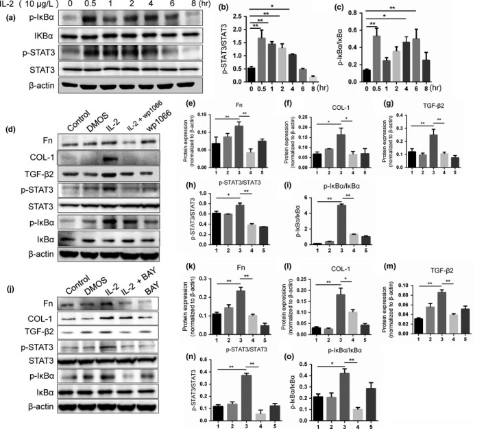
Macular fibrosis is a vital obstacle of vision acuity improvement of age-related macular degeneration patients. This study was to investigate the effects of interleukin 2 (IL-2) on epithelial-mesenchymal transition (EMT), extracellular matrix (ECM) synthesis and transforming growth factor β2 (TGF-β2) expression in retinal pigment epithelial (RPE) cells. 10 μg/L IL-2 was used to induce fibrosis in RPE cells for various times. Western blot was used to detect the EMT marker α-smooth muscle actin (α-SMA), ECM markers fibronectin (Fn) and type 1 collagen (COL-1), TGF-β2, and the activation of the JAK/STAT3 and NF-κB signaling pathway. Furthermore, JAK/STAT3 and NF-κB signaling pathways were specifically blocked by WP1066 or BAY11-7082, respectively, and the expression of α-SMA, COL-1, Fn and TGF-β2 protein were detected. Wound healing and Transwell assays were used to measure cell migration ability of IL-2 with or without WP1066 or BAY11-7082. After induction of IL-2, the expressions of Fn, COL-1, TGF-β2 protein were significantly increased, and this effect was correlated with IL-2 treatment duration, while α-SMA protein expression did not change significantly. Both WP1066 and BAY11-7082 could effectively downregulate the expression of Fn, COL-1 and TGF-β2 induced by IL-2. What's more, both NF-κB and JAK/STAT3 inhibitors could suppress the activation of the other signaling pathway. Additionally, JAK/STAT3 inhibitor WP1066 and NF-κB inhibitor BAY 11-7082 could obviously decrease RPE cells migration capability induced by IL-2. IL-2 promotes cell migration, ECM synthesis and TGF-β2 expression in RPE cells via JAK/STAT3 and NF-κB signaling pathways, which may play an important role in proliferative vitreoretinopathy.
Keywords: Interleukin 2; age-related macular degeneration; epithelial-mesenchymal transition; extracellular matrix synthesis; retinal pigment epithelial cells.
© 2019 The Authors. Development, Growth & Differentiation published by John Wiley & Sons Australia, Ltd on behalf of Japanese Society of Developmental Biologists.
Conflict of interest statement
All of the authors declare that there is no interest.
Figures
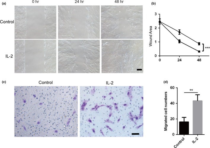
 , Control;
  , IL‐2). (c) Transwell assay between control group and 10 μg/L
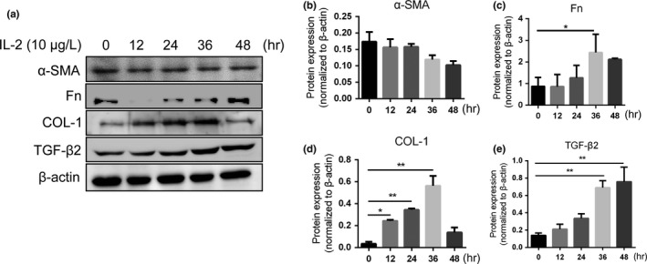
 , Control;
  , DMSO;
  , IL‐2 10 μg/L;
  , IL‐2 10 μg/L+WP1066;
  , WP1066). (j)
  , Control;
  , DMSO;
  , IL‐2 10 μg/L;
  , IL‐2 10 μg/L+BAY;
  , BAY). *p < .05, **p < .01
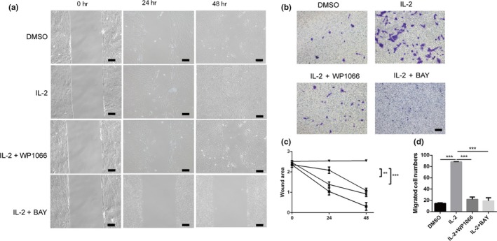
 , DMSO;
  , IL‐2;
  , IL‐2+WP1066;
  , IL‐2+BAY). (d) Migrated
 Similar articles
-
Effects of Interleukin-6 on posterior capsular opacification.Exp Eye Res. 2018 Jul;172:94-103. doi: 10.1016/j.exer.2018.03.013. Epub 2018 Apr 1. Exp Eye Res. 2018. PMID: 29617629
-
Interleukin-6 promotes migration and extracellular matrix synthesis in retinal pigment epithelial cells.Histochem Cell Biol. 2020 Dec;154(6):629-638. doi: 10.1007/s00418-020-01923-4. Epub 2020 Sep 30. Histochem Cell Biol. 2020. PMID: 32997263
-
TGF-β2 induces transdifferentiation and fibrosis in human lens epithelial cells via regulating gremlin and CTGF.Biochem Biophys Res Commun. 2014 May 16;447(4):689-95. doi: 10.1016/j.bbrc.2014.04.068. Epub 2014 Apr 19. Biochem Biophys Res Commun. 2014. PMID: 24755068
-
Blockade of Jagged/Notch pathway abrogates transforming growth factor β2-induced epithelial-mesenchymal transition in human retinal pigment epithelium cells.Curr Mol Med. 2014 May;14(4):523-34. doi: 10.2174/1566524014666140331230411. Curr Mol Med. 2014. PMID: 24694299 Review.
-
Role of JAK/STAT3 Signaling in the Regulation of Metastasis, the Transition of Cancer Stem Cells, and Chemoresistance of Cancer by Epithelial-Mesenchymal Transition.Cells. 2020 Jan 15;9(1):217. doi: 10.3390/cells9010217. Cells. 2020. PMID: 31952344 Free PMC article. Review.
Cited by
-
Causal relationships between inflammatory cytokines and myopia: an analysis of genetic and observational studies.Ann Med Surg (Lond). 2024 Jul 2;86(9):5179-5190. doi: 10.1097/MS9.0000000000002325. eCollection 2024 Sep. Ann Med Surg (Lond). 2024. PMID: 39239046 Free PMC article.
-
The effect of systemic levels of TNF-alpha and complement pathway activity on outcomes of VEGF inhibition in neovascular AMD.Eye (Lond). 2022 Nov;36(11):2192-2199. doi: 10.1038/s41433-021-01824-3. Epub 2021 Nov 8. Eye (Lond). 2022. PMID: 34750590 Free PMC article.
-
Genipin protects against mitochondrial damage of the retinal pigment epithelium under hyperglycemia through the AKT pathway mediated by the miR-4429/JAK2 signaling axis.Ann Transl Med. 2022 May;10(10):587. doi: 10.21037/atm-22-2219. Ann Transl Med. 2022. PMID: 35722358 Free PMC article.
-
Role of reactive oxygen species in epithelial-mesenchymal transition and apoptosis of human lens epithelial cells.Int J Ophthalmol. 2023 Dec 18;16(12):1935-1941. doi: 10.18240/ijo.2023.12.04. eCollection 2023. Int J Ophthalmol. 2023. PMID: 38111943 Free PMC article.
-
The Role of Inflammation in Age-Related Macular Degeneration.Int J Biol Sci. 2020 Sep 23;16(15):2989-3001. doi: 10.7150/ijbs.49890. eCollection 2020. Int J Biol Sci. 2020. PMID: 33061811 Free PMC article. Review.
References
-
- Anderluh, M. , Kocic, G. , Tomovic, K. , Kocic, H. , & Smelcerovic, A. (2019). DPP‐4 inhibition: Capital A, Cyrillic novel therapeutic approach to the treatment of pulmonary hypertension? Pharmacology & Therapeutics, 201, 1–7. - PubMed
-
- Battaglia, A. , Buzzonetti, A. , Baranello, C. , Fanelli, M. , Fossati, M. , Catzola, V. , … Fattorossi, A. (2013). Interleukin‐21 (IL‐21) synergizes with IL‐2 to enhance T‐cell receptor‐induced human T‐cell proliferation and counteracts IL‐2/transforming growth factor‐beta‐induced regulatory T‐cell development. Immunology, 139, 109–120. - PMC - PubMed
-
- Chambers, E. S. , Suwannasaen, D. , Mann, E. H. , Urry, Z. , Richards, D. F. , Lertmemongkolchai, G. , & Hawrylowicz, C. M. (2014). 1alpha,25‐dihydroxyvitamin D3 in combination with transforming growth factor‐beta increases the frequency of Foxp3(+) regulatory T cells through preferential expansion and usage of interleukin‐2. Immunology, 143, 52–60. - PMC - PubMed
MeSH terms
Substances
Grants and funding
- xjj2018099/Fundamental Research Funds for the Central Universities
- 2018M633528/Postdoctoral Natural Science Foundation
- 2018JQ8021/Natural Science Foundation of Shaanxi Province
- 81800812/National Natural Science Foundation of China
- 2016QN-04/The First Affiliated Hospital of Xi'an Jiaotong University Foundation
LinkOut - more resources
Full Text Sources
Miscellaneous
