

Scribble co-operatively binds multiple α1D-adrenergic receptor C-terminal PDZ ligands
- PMID: 31575922
- PMCID: PMC6773690
- DOI: 10.1038/s41598-019-50671-6
Scribble co-operatively binds multiple α1D-adrenergic receptor C-terminal PDZ ligands
Abstract
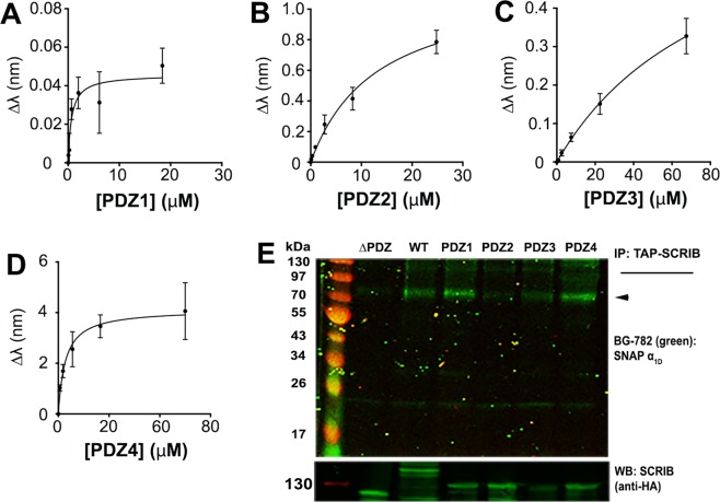
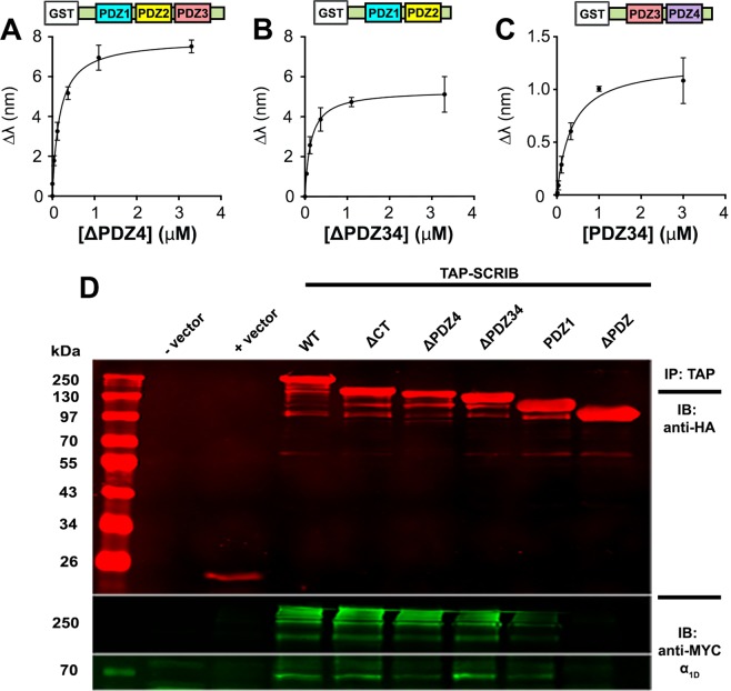
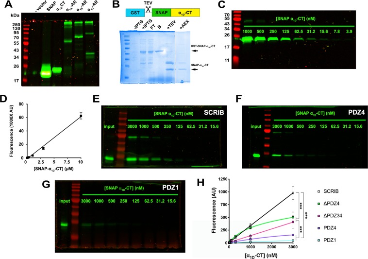
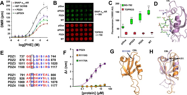
Many G protein-coupled receptors (GPCRs) are organized as dynamic macromolecular complexes in human cells. Unraveling the structural determinants of unique GPCR complexes may identify unique protein:protein interfaces to be exploited for drug development. We previously reported α1D-adrenergic receptors (α1D-ARs) - key regulators of cardiovascular and central nervous system function - form homodimeric, modular PDZ protein complexes with cell-type specificity. Towards mapping α1D-AR complex architecture, biolayer interferometry (BLI) revealed the α1D-AR C-terminal PDZ ligand selectively binds the PDZ protein scribble (SCRIB) with >8x higher affinity than known interactors syntrophin, CASK and DLG1. Complementary in situ and in vitro assays revealed SCRIB PDZ domains 1 and 4 to be high affinity α1D-AR PDZ ligand interaction sites. SNAP-GST pull-down assays demonstrate SCRIB binds multiple α1D-AR PDZ ligands via a co-operative mechanism. Structure-function analyses pinpoint R1110PDZ4 as a unique, critical residue dictating SCRIB:α1D-AR binding specificity. The crystal structure of SCRIB PDZ4 R1110G predicts spatial shifts in the SCRIB PDZ4 carboxylate binding loop dictate α1D-AR binding specificity. Thus, the findings herein identify SCRIB PDZ domains 1 and 4 as high affinity α1D-AR interaction sites, and potential drug targets to treat diseases associated with aberrant α1D-AR signaling.
Conflict of interest statement
The authors declare no competing interests.
Figures


Similar articles
-
Crystal structure of the human Scribble PDZ1 domain bound to the PDZ-binding motif of APC.FEBS Lett. 2019 Mar;593(5):533-542. doi: 10.1002/1873-3468.13329. Epub 2019 Feb 2. FEBS Lett. 2019. PMID: 30659601
-
Syntrophins regulate alpha1D-adrenergic receptors through a PDZ domain-mediated interaction.J Biol Chem. 2006 May 5;281(18):12414-20. doi: 10.1074/jbc.M508651200. Epub 2006 Mar 13. J Biol Chem. 2006. PMID: 16533813
-
Individual protomers of a G protein-coupled receptor dimer integrate distinct functional modules.Cell Discov. 2015;1:15011-. doi: 10.1038/celldisc.2015.11. Epub 2015 Jun 16. Cell Discov. 2015. PMID: 26617989 Free PMC article.
-
Endogenous N-terminal Domain Cleavage Modulates α1D-Adrenergic Receptor Pharmacodynamics.J Biol Chem. 2016 Aug 26;291(35):18210-21. doi: 10.1074/jbc.M116.729517. Epub 2016 Jul 5. J Biol Chem. 2016. PMID: 27382054 Free PMC article.
-
Extensions of PDZ domains as important structural and functional elements.Protein Cell. 2010 Aug;1(8):737-51. doi: 10.1007/s13238-010-0099-6. Epub 2010 Aug 28. Protein Cell. 2010. PMID: 21203915 Free PMC article. Review.
Cited by
-
N-glycosylation of α1D-adrenergic receptor N-terminal domain is required for correct trafficking, function, and biogenesis.Sci Rep. 2020 Apr 29;10(1):7209. doi: 10.1038/s41598-020-64102-4. Sci Rep. 2020. PMID: 32350295 Free PMC article.
-
Bidirectional protein-protein interactions control liquid-liquid phase separation of PSD-95 and its interaction partners.iScience. 2022 Jan 25;25(2):103808. doi: 10.1016/j.isci.2022.103808. eCollection 2022 Feb 18. iScience. 2022. PMID: 35198873 Free PMC article.
-
Introduction: A Short History of Adrenoceptor Research.Handb Exp Pharmacol. 2024;285:1-12. doi: 10.1007/164_2024_718. Handb Exp Pharmacol. 2024. PMID: 38797750
-
Unraveling the Functional Significance of Unstructured Regions in G Protein-Coupled Receptors.Biomolecules. 2023 Sep 22;13(10):1431. doi: 10.3390/biom13101431. Biomolecules. 2023. PMID: 37892113 Free PMC article. Review.
-
Membrane recruitment of the polarity protein Scribble by the cell adhesion receptor TMIGD1.Commun Biol. 2023 Jul 10;6(1):702. doi: 10.1038/s42003-023-05088-3. Commun Biol. 2023. PMID: 37430142 Free PMC article.
References
Publication types
MeSH terms
Substances
Grants and funding
LinkOut - more resources
Full Text Sources
Molecular Biology Databases
Research Materials
