. 2019 Sep 17:14:7609-7624.
doi: 10.2147/IJN.S208810. eCollection 2019.

Tuning the surface coating of IONs toward efficient sonochemical tethering and sustained liberation of topoisomerase II poisons

Affiliations

Tuning the surface coating of IONs toward efficient sonochemical tethering and sustained liberation of topoisomerase II poisons

Hana Michalkova et al. Int J Nanomedicine. .

Erratum in

Abstract

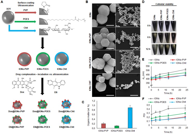
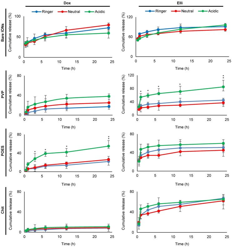
Background: Iron oxide nanoparticles (IONs) have been increasingly utilized in a wide spectrum of biomedical applications. Surface coatings of IONs can bestow a number of exceptional properties, including enhanced stability of IONs, increased loading of drugs or their controlled release.

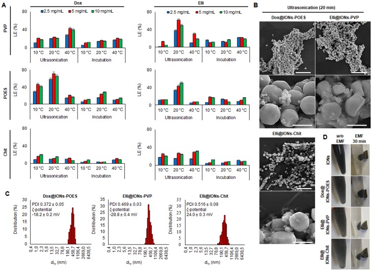
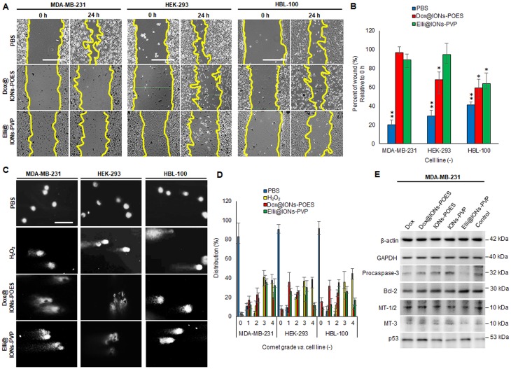
Methods: Using two-step sonochemical protocol, IONs were surface-coated with polyoxyethylene stearate, polyvinylpyrrolidone or chitosan for a loading of two distinct topo II poisons (doxorubicin and ellipticine). The cytotoxic behavior was tested in vitro against breast cancer (MDA-MB-231) and healthy epithelial cells (HEK-293 and HBL-100). In addition, biocompatibility studies (hemotoxicity, protein corona formation, binding of third complement component) were performed.

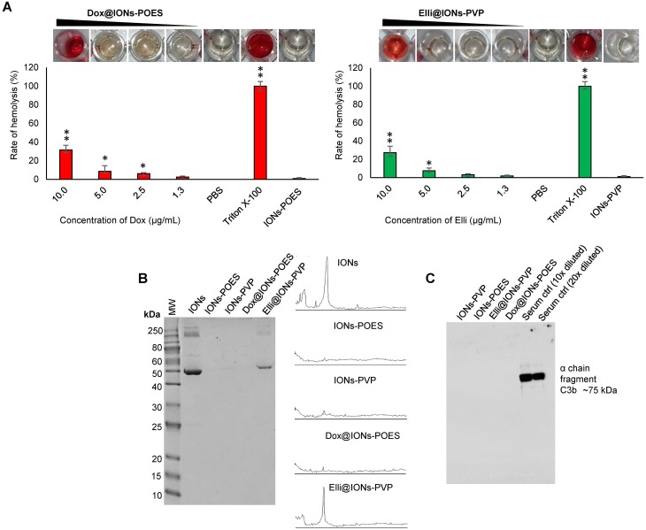
Results: Notably, despite surface-coated IONs exhibited only negligible cytotoxicity, upon tethering with topo II poisons, synergistic or additional enhancement of cytotoxicity was found in MDA-MB-231 cells. Pronounced anti-migratory activity, DNA fragmentation, decrease in expression of procaspase-3 and enhancement of p53 expression were further identified upon exposure to surface-coated IONs with tethered doxorubicin and ellipticine. Moreover, surface-coated IONs nanoformulations of topo II poisons exhibited exceptional stability in human plasma with no protein corona and complement 3 binding, and only a mild induction of hemolysis in human red blood cells.

Conclusion: The results imply a high potential of an efficient ultrasound-mediated surface functionalization of IONs as delivery vehicles to improve therapeutic efficiency of topo II poisons.

Keywords: doxorubicin; ellipticine; iron oxide; nanoparticles; release kinetics.


Conflict of interest statement

The authors report no conflicts of interest in this work.

Figures

Figure 1
Figure 1
Surface coating of IONs with biocompatible surfactant (POES) or polymers (PVP and Chit). (A) Schematic representation of surface coating of bare IONs with PVP, POES and Chit with a consequent tethering of cytotoxic substances Dox and Elli using incubation or ultrasonication, respectively. (B) SEM micrographs of morphology of bare IONs and their morphology after surface coatings. The scale bars, 400 nm (left) or 5 µm (right). (C) Content of organic matter in surface-coated formulations analyzed using CHNS/O analyzer. The values are expressed as the mean of three independent replicates (n=3). Vertical bars indicate standard error. (D) Photodocumentation of a colloidal stability of bare and surface-coated IONs. Time-evolution of (E) dhy and (F) PDI, both analyzed in Ringer's solution. The values are expressed as the mean of six independent replicates (n=6). The vertical bars + and − errors. Abbreviations: IONs, iron oxide nanoparticles; POES, polyoxyethylene stearate; PVP, polyvinylpyrrolidone; Chit, chitosan; SEM, scanning electron microscopy; dhy, hydrodynamic diameter; PDI, polydispersity index.
Figure 2
Figure 2
Optimization of loading of Dox and Elli onto surface-coated IONs. (A) Different amounts of surface-coating agents were tested for their LE with constant amounts for Dox and Elli (2 mg/mL). (B) SEM micrographs showing selected surface-coated IONs after 20 mins ultrasonication-mediated tethering of Dox or Elli. Scale bars, 5 µm (top), 400 nm (bottom). (C) Distribution of dhy of Dox/Elli-loaded surface-modified IONs with the highest LEs. Inserted are PDI and ζ-potential values of IONs dispersed in Ringer´s solution. (D) Photographs of bare IONs and selected topo II poisons-tethered surface-coated IONs after the application of an EMF (Nd-Fe-B permanent magnet, 30 mins). Abbreviations: IONs, iron oxide nanoparticles; LE, loading efficiency; Dox, doxorubicin; Elli, ellipticine; SEM, scanning electron microscopy; PDI, polydispersity index; EMF, external magnetic field.
Figure 3
Figure 3
In vitro cumulative release kinetic profiles of Dox and Elli from bare and surface-coated IONs determined in various physiological pH conditions (intracellular, pH 6.9, endosomal, pH 5.0 and plasma, pH 7.4). The values are expressed as the mean of six independent replicates (n=6). Vertical bars indicate + and −errors. The p-values were calculated for each time-point and denoted if found to be significantly different, *P<0.05, **P<0.01. Abbreviations: Dox, doxorubicin; Elli, ellipticine; IONs, iron oxide nanoparticles.
Figure 4
Figure 4
Evaluation of potential synergic effects of IONs to Dox and Elli cytotoxicity. (A) Isobolograms demonstrating synergistic/antagonistic effects of selected surface-coated IONs and Dox/Elli within all tested cell lines. (B) Total iron accumulation in intracellular region of treated cells (6 hrs) analyzed by AAS. The values are expressed as the mean of three independent replicates (n=3). The vertical bars indicate standard error. (C) Internalization kinetics of Dox@IONs-POES and Elli@IONs-PVP analyzed using CLSM in all tested cell lines during the first 6 hrs of treatment. Scale bar, 15 µm. Abbreviations: Dox, doxorubicin; Elli, ellipticine; IONs, iron oxide nanoparticles; AAS, atomic absorption spectroscopy; CLSM, confocal laser scanning microscopy; CI, combination index.
Figure 5
Figure 5
(A) Representative micrographs of wound-healing assay showing the marked effect of Dox/Elli-tethered surface-modified IONs on a migration of tested cell lines. Representative pictures demonstrate the artificial gaps at the experimental start-point (0 hrs) and migration of cells after 24 hrs cultivation. Yellow lines indicate the approximate borders of the initial gap. Scale bar, 400 µm. (B) Quantitation of relative free areas from wound-healing assay. The values are expressed as the mean of three independent replicates (n=3). Vertical bars indicate + and −errors. *P<0.05, **P<0.01 related to the initial gap area. (C) SCGE of cells following exposure to Dox@IONs-POES and Elli@IONs-PVP. PBS (pH 7.4) and 60 µM H2O2 were employed as negative and positive controls. Scale bar, 100 µm. (D) Quantitation of index of damage upon 12 hrs exposure. The values are expressed as the mean of three independent replicates (n=3). Vertical bars indicate + and −errors. (E) Representative immunoblots of whole-cell lysate of MDA-MB-231 cells. β-Actin and GAPDH served as loading controls. Abbreviations: Dox, doxorubicin; Elli, ellipticine; IONs, iron oxide nanoparticles; SCGE, single-cell gel electrophoresis; PBS, phosphate-buffered saline; GAPDH, glyceraldehyde-3-phosphate dehydrogenase.
Figure 6
Figure 6
Examination of in vitro biocompatibility of Dox@IONs-POES and Elli@IONs-PVP. (A) Hemolysis of Dox@IONs-POES and Elli@IONs-PVP assayed on human RBCs. PBS (pH 7.4) and 0.1% Triton X-100 were utilized as negative and positive controls, respectively. Amount of tested IONs-POES and IONs-PVP without tethered Dox and Elli is adequate to the highest amount of IONs in Dox@IONs-POES and Elli@IONs-PVP treatments. Upper images depict representative photographs of tested samples. The values are expressed as the mean of three independent replicates (n=3). Vertical bars indicate + and −errors. *P<0.05, **P<0.01 related to the IONs-POES and IONs-PVP without tethered topo II poisons. (B) Protein corona patterns obtained after 30 mins incubation of annotated formulations with human plasma followed by extensive washing, elution, and loading onto 12% SDS-PAGE. As a control, human plasma (1,000× diluted) was loaded to the first lane. Figures on the right side show protein coronas quantified by densitometric analysis. (C) Immunoblot of C3b binding from human serum from male AB clotted whole blood. Abbreviations: Dox, doxorubicin; Elli, ellipticine; IONs, iron oxide nanoparticles; RBCs, red blood cells.

Similar articles

Cited by

References

    1. Bobo D, Robinson KJ, Islam J, Thurecht KJ, Corrie SR. Nanoparticle-based medicines: a review of FDA-approved materials and clinical trials to date. Pharm Res. 2016;33(10):2373–2387. doi:10.1007/s11095-016-1958-5 - DOI - PubMed
    1. Skorjanc T, Benyettou F, Olsen JC, Trabolsi A. Design of organic macrocycle-modified iron oxide nanoparticles for drug delivery. Chem Eur J. 2017;23(35):8333–8347. doi:10.1002/chem.201605246 - DOI - PubMed
    1. Chertok B, Moffat BA, David AE, et al. Iron oxide nanoparticles as a drug delivery vehicle for MRI monitored magnetic targeting of brain tumors. Biomaterials. 2008;29(4):487–496. doi:10.1016/j.biomaterials.2007.08.050 - DOI - PMC - PubMed
    1. Zhao Z, Zhou Z, Bao J, et al. Octapod iron oxide nanoparticles as high-performance T(2) contrast agents for magnetic resonance imaging. Nat Commun. 2013;4:2266. doi:10.1038/ncomms3266 - DOI - PubMed
    1. Eguchi H, Umemura M, Kurotani R, et al. A magnetic anti-cancer compound for magnet-guided delivery and magnetic resonance imaging. Sci Rep. 2015;5:14. doi:10.1038/srep09194 - DOI - PMC - PubMed