BCL6 BTB-specific inhibition via FX1 treatment reduces Tfh cells and reverses lymphoid follicle hyperplasia in Indian rhesus macaque (Macaca mulatta)
- PMID: 31571234
- PMCID: PMC7236024
- DOI: 10.1111/jmp.12438
BCL6 BTB-specific inhibition via FX1 treatment reduces Tfh cells and reverses lymphoid follicle hyperplasia in Indian rhesus macaque (Macaca mulatta)
Abstract
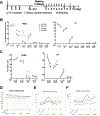
Background: The BTB domain of B-cell lymphoma 6 (BCL6) protein was identified as a therapeutic target for B-cell lymphoma. This study compared the pharmacokinetics (PK) of the BCL6 BTB inhibitor (FX1) between mice and macaques, as well as evaluating its lymphoid suppressive effect in uninfected macaques with lymphoid hyperplasia.
Materials and methods: Eight uninfected adult Indian rhesus macaques (Macaca mulatta) were used in the study, four animals carrying lymphoid tissue hyperplasia. Plasma FX1 levels were measured by HPLC-MS/MS. Lymph node biopsies were used for H&E and immunohistochemistry staining, as well as mononuclear cell isolation for flow cytometry analysis.
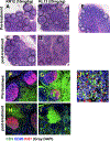
Results: Inhibition of the BCL6 BTB domain with FX1 led to a reduction in the frequency of GC, Tfh CD4+ , and Tfh precursor cells, as well as resolving lymphoid hyperplasia, in rhesus macaques.
Conclusions: B-cell lymphoma 6 inhibition may represent a novel strategy to reduce hyperplastic lymphoid B-cell follicles and decrease Tfh cells.
Keywords: B-cell lymphoma 6; T follicular helper; germinal center reaction; lymphoid hyperplasia.
© 2019 John Wiley & Sons A/S. Published by John Wiley & Sons Ltd.
Conflict of interest statement
Figures


Similar articles
-
BCL6 BTB-specific inhibitor reversely represses T-cell activation, Tfh cells differentiation, and germinal center reaction in vivo.Eur J Immunol. 2021 Oct;51(10):2441-2451. doi: 10.1002/eji.202049150. Epub 2021 Sep 16. Eur J Immunol. 2021. PMID: 34287839 Free PMC article.
-
BCL6 Inhibitor-Mediated Downregulation of Phosphorylated SAMHD1 and T Cell Activation Are Associated with Decreased HIV Infection and Reactivation.J Virol. 2019 Jan 4;93(2):e01073-18. doi: 10.1128/JVI.01073-18. Print 2019 Jan 15. J Virol. 2019. PMID: 30355686 Free PMC article.
-
BCL6 inhibitor FX1 attenuates inflammatory responses in murine sepsis through strengthening BCL6 binding affinity to downstream target gene promoters.Int Immunopharmacol. 2019 Oct;75:105789. doi: 10.1016/j.intimp.2019.105789. Epub 2019 Aug 8. Int Immunopharmacol. 2019. PMID: 31401377
-
The life and death of the germinal center.Ann Diagn Pathol. 2020 Feb;44:151421. doi: 10.1016/j.anndiagpath.2019.151421. Epub 2019 Nov 13. Ann Diagn Pathol. 2020. PMID: 31751845 Review.
-
T Cells That Help B Cells in Chronically Inflamed Tissues.Front Immunol. 2018 Aug 23;9:1924. doi: 10.3389/fimmu.2018.01924. eCollection 2018. Front Immunol. 2018. PMID: 30190721 Free PMC article. Review.
Cited by
-
Anti-PD-1 therapy triggers Tfh cell-dependent IL-4 release to boost CD8 T cell responses in tumor-draining lymph nodes.J Exp Med. 2024 Apr 1;221(4):e20232104. doi: 10.1084/jem.20232104. Epub 2024 Feb 28. J Exp Med. 2024. PMID: 38417020 Free PMC article.
-
Small-molecule BCL6 inhibitor protects chronic cardiac transplant rejection and inhibits T follicular helper cell expansion and humoral response.Front Pharmacol. 2023 Mar 17;14:1140703. doi: 10.3389/fphar.2023.1140703. eCollection 2023. Front Pharmacol. 2023. PMID: 37007047 Free PMC article.
-
Targeting Bcl6 in the TREX1 D18N murine model ameliorates autoimmunity by modulating T-follicular helper cells and germinal center B cells.Eur J Immunol. 2022 May;52(5):825-834. doi: 10.1002/eji.202149324. Epub 2022 Feb 15. Eur J Immunol. 2022. PMID: 35112355 Free PMC article.
-
BCL6 BTB-specific inhibitor reversely represses T-cell activation, Tfh cells differentiation, and germinal center reaction in vivo.Eur J Immunol. 2021 Oct;51(10):2441-2451. doi: 10.1002/eji.202049150. Epub 2021 Sep 16. Eur J Immunol. 2021. PMID: 34287839 Free PMC article.
References
-
- Cardenas MG, Yu W, Beguelin W, Teater MR, Geng H, Goldstein RL, Oswald E, Hatzi K, Yang SN, Cohen J, Shaknovich R, Vanommeslaeghe K, Cheng H, Liang D, Cho HJ, Abbott J, Tam W, Du W, Leonard JP, Elemento O, Cerchietti L, Cierpicki T, Xue F, MacKerell AD Jr., Melnick AM. 2016. Rationally designed BCL6 inhibitors target activated B cell diffuse large B cell lymphoma. J Clin Invest 126:3351–3362. - PMC - PubMed
-
- Ueda C, Akasaka T, Ohno H. 2002. Non-immunoglobulin/BCL6 gene fusion in diffuse large B-cell lymphoma: prognostic implications. Leuk Lymphoma 43:1375–1381. - PubMed
Publication types
MeSH terms
Substances
Grants and funding
LinkOut - more resources
Full Text Sources
Research Materials
Miscellaneous

