

The Anti-Apoptotic Effect of ASC-Exosomes in an In Vitro ALS Model and Their Proteomic Analysis
- PMID: 31540100
- PMCID: PMC6770878
- DOI: 10.3390/cells8091087
The Anti-Apoptotic Effect of ASC-Exosomes in an In Vitro ALS Model and Their Proteomic Analysis
Abstract
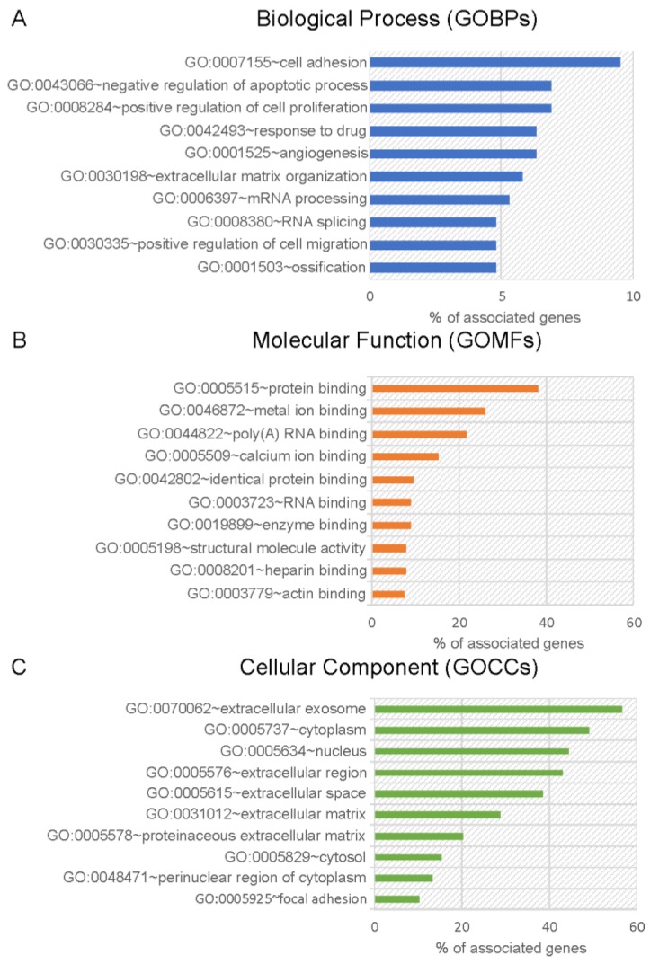
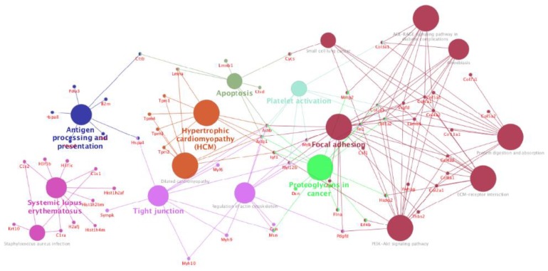
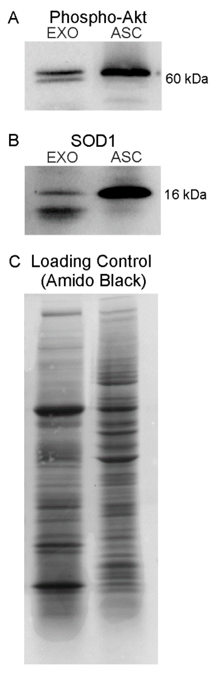
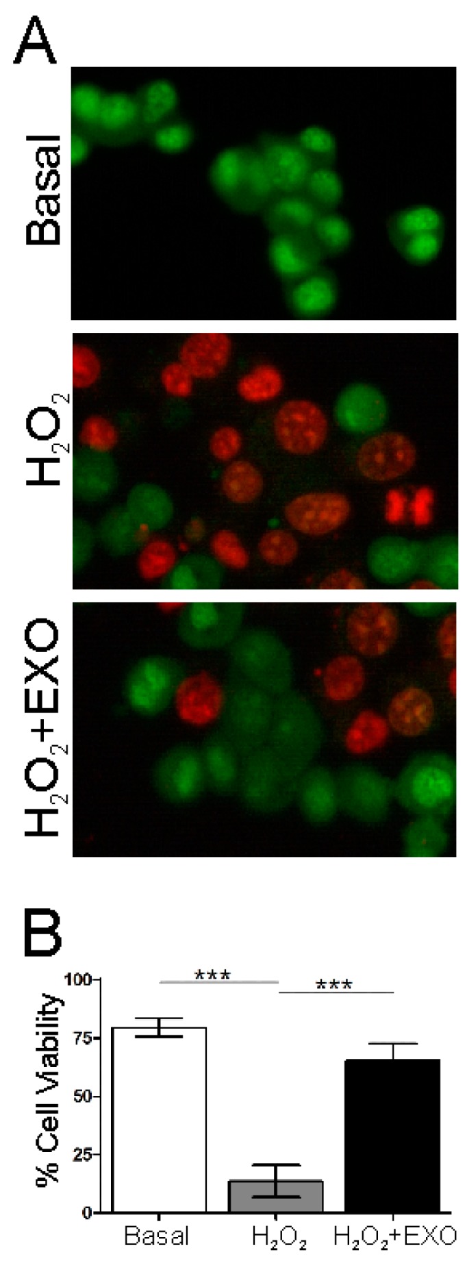
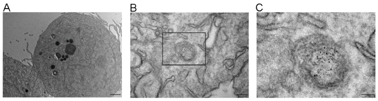
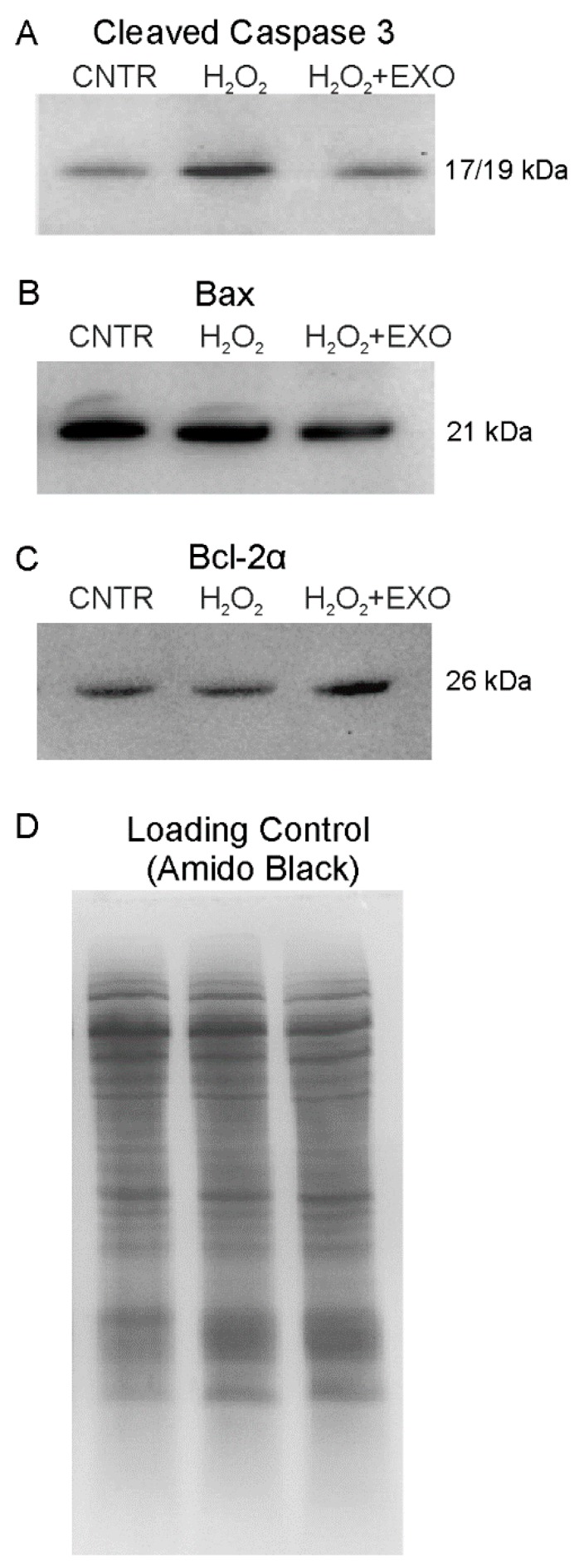
Stem cell therapy represents a promising approach in the treatment of several neurodegenerative disorders, including amyotrophic lateral sclerosis (ALS). The beneficial effect of stem cells is exerted by paracrine mediators, as exosomes, suggesting a possible potential use of these extracellular vesicles as non-cell based therapy. We demonstrated that exosomes isolated from adipose stem cells (ASC) display a neuroprotective role in an in vitro model of ALS. Moreover, the internalization of ASC-exosomes by the cells was shown and the molecules and the mechanisms by which exosomes could exert their beneficial effect were addressed. We performed for the first time a comprehensive proteomic analysis of exosomes derived from murine ASC. We identified a total of 189 proteins and the shotgun proteomics analysis revealed that the exosomal proteins are mainly involved in cell adhesion and negative regulation of the apoptotic process. We correlated the protein content to the anti-apoptotic effect of exosomes observing a downregulation of pro-apoptotic proteins Bax and cleaved caspase-3 and upregulation of anti-apoptotic protein Bcl-2 α, in an in vitro model of ALS after cell treatment with exosomes. Overall, this study shows the neuroprotective effect of ASC-exosomes after their internalization and their global protein profile, that could be useful to understand how exosomes act, demonstrating that they can be employed as therapy in neurodegenerative diseases.
Keywords: amyotrophic lateral sclerosis; apoptosis; extracellular vesicles; mesenchymal stem cells; proteomic profiling; therapy.
Conflict of interest statement
The authors declare that no conflicts of interest.
Figures


Similar articles
-
Exosome derived from murine adipose-derived stromal cells: Neuroprotective effect on in vitro model of amyotrophic lateral sclerosis.Exp Cell Res. 2016 Jan 1;340(1):150-8. doi: 10.1016/j.yexcr.2015.12.009. Epub 2015 Dec 18. Exp Cell Res. 2016. PMID: 26708289
-
Adipose-derived stem cell exosomes alleviate pathology of amyotrophic lateral sclerosis in vitro.Biochem Biophys Res Commun. 2016 Oct 21;479(3):434-439. doi: 10.1016/j.bbrc.2016.09.069. Epub 2016 Sep 15. Biochem Biophys Res Commun. 2016. PMID: 27641665
-
Extracellular Vesicles From Adipose Stem Cells Prevent Muscle Damage and Inflammation in a Mouse Model of Hind Limb Ischemia: Role of Neuregulin-1.Arterioscler Thromb Vasc Biol. 2020 Jan;40(1):239-254. doi: 10.1161/ATVBAHA.119.313506. Epub 2019 Oct 31. Arterioscler Thromb Vasc Biol. 2020. PMID: 31665908
-
Adipose-Derived Stem Cells Secretome and Its Potential Application in "Stem Cell-Free Therapy".Biomolecules. 2021 Jun 13;11(6):878. doi: 10.3390/biom11060878. Biomolecules. 2021. PMID: 34199330 Free PMC article. Review.
-
Exosomes as a potential novel therapeutic tools against neurodegenerative diseases.Pharmacol Res. 2016 Nov;113(Pt B):816-822. doi: 10.1016/j.phrs.2016.02.002. Epub 2016 Feb 6. Pharmacol Res. 2016. PMID: 26855317 Review.
Cited by
-
Adipose stem cells-released extracellular vesicles as a next-generation cargo delivery vehicles: a survey of minimal information implementation, mass production and functional modification.Stem Cell Res Ther. 2022 May 3;13(1):182. doi: 10.1186/s13287-022-02849-5. Stem Cell Res Ther. 2022. PMID: 35505389 Free PMC article.
-
Recent Advances on Extracellular Vesicles in Central Nervous System Diseases.Clin Interv Aging. 2021 Feb 10;16:257-274. doi: 10.2147/CIA.S288415. eCollection 2021. Clin Interv Aging. 2021. PMID: 33603351 Free PMC article. Review.
-
Mesenchymal stem cell-derived exosomes enriched with miR-218 reduce the epithelial-mesenchymal transition and angiogenesis in triple-negative breast cancer cells.Eur J Med Res. 2023 Nov 15;28(1):516. doi: 10.1186/s40001-023-01463-2. Eur J Med Res. 2023. PMID: 37968694 Free PMC article.
-
Combinational treatments of RNA interference and extracellular vesicles in the spinocerebellar ataxia.Front Mol Neurosci. 2022 Oct 13;15:1043947. doi: 10.3389/fnmol.2022.1043947. eCollection 2022. Front Mol Neurosci. 2022. PMID: 36311034 Free PMC article. Review.
-
The Emerging Role of Neural Cell-Derived Exosomes in Intercellular Communication in Health and Neurodegenerative Diseases.Front Neurosci. 2021 Aug 31;15:738442. doi: 10.3389/fnins.2021.738442. eCollection 2021. Front Neurosci. 2021. PMID: 34531720 Free PMC article. Review.
References
-
- Marconi S., Bonaconsa M., Scambi I., Squintani G.M., Rui W., Turano E., Ungaro D., D’Agostino S., Barbieri F., Angiari S., et al. Systemic treatment with Adipose-Derived Mesenchymal Stem Cells ameliorates clinical and pathological features in the Amyotrophic Lateral Sclerosis Murine Model. Neuroscience. 2013;248:333–343. doi: 10.1016/j.neuroscience.2013.05.034. - DOI - PubMed
Publication types
MeSH terms
Substances
LinkOut - more resources
Full Text Sources
Medical
Research Materials
Miscellaneous
