. 2020 Apr;27(4):1286-1299.
doi: 10.1038/s41418-019-0415-5. Epub 2019 Sep 17.

Trim32 suppresses cerebellar development and tumorigenesis by degrading Gli1/sonic hedgehog signaling

Affiliations

Trim32 suppresses cerebellar development and tumorigenesis by degrading Gli1/sonic hedgehog signaling

Minglei Wang et al. Cell Death Differ. 2020 Apr.

Abstract

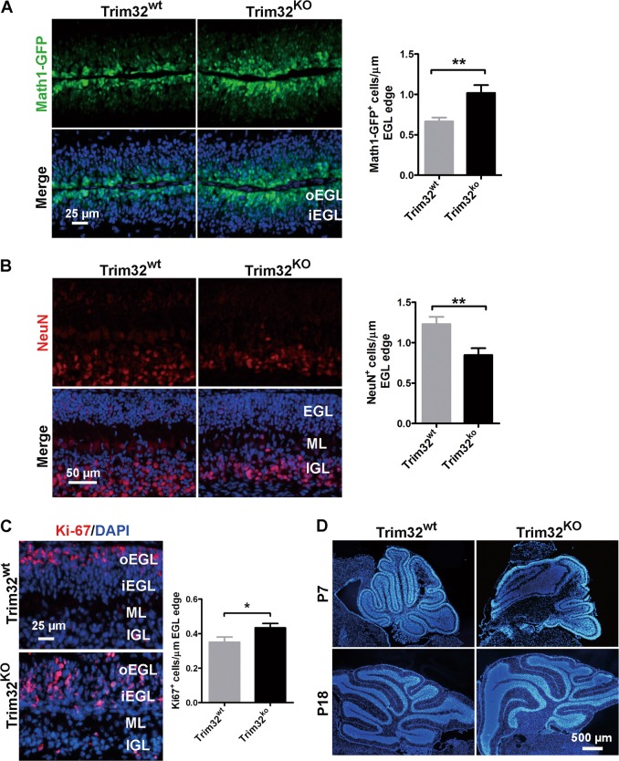
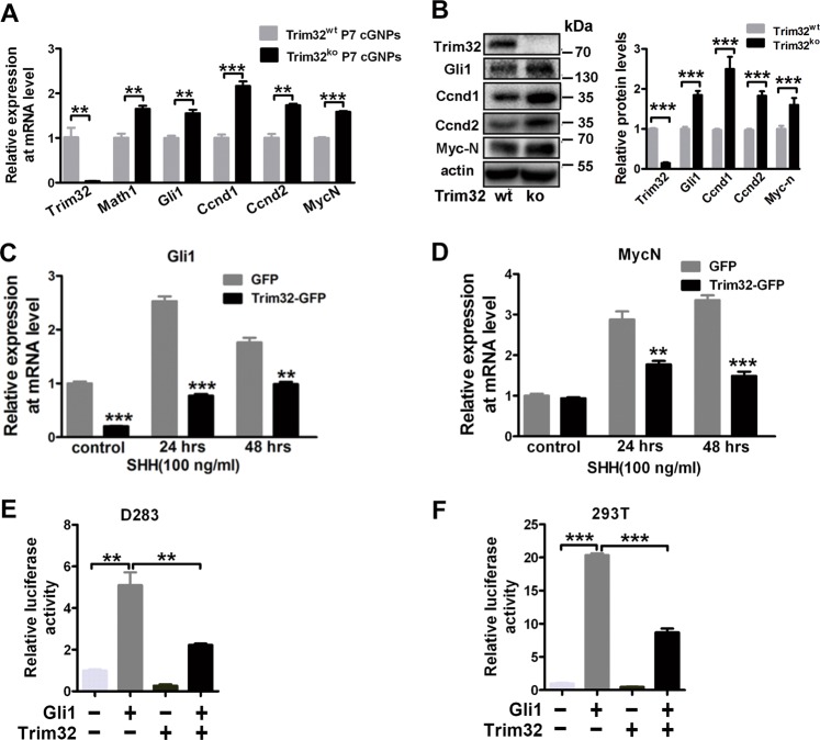
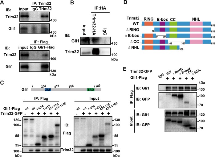
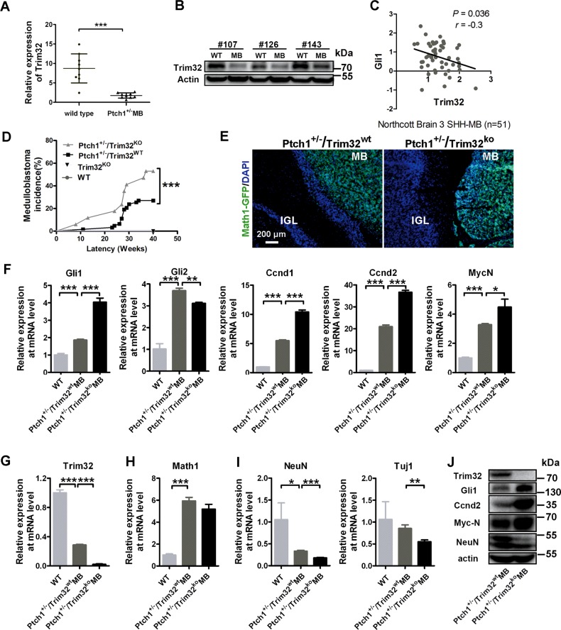
Sonic hedgehog (SHH) signaling is crucial for the maintenance of the physiological self-renewal of granule neuron progenitor cells (GNPs) during cerebellar development, and its dysregulation leads to oncogenesis. However, how SHH signaling is controlled during cerebellar development is poorly understood. Here, we show that Trim32, a cell fate determinant, is distributed asymmetrically in the cytoplasm of mitotic GNPs, and that genetic knockout of Trim32 keeps GNPs at a proliferating and undifferentiated state. In addition, Trim32 knockout enhances the incidence of medulloblastoma (MB) formation in the Ptch1 mutant mice. Mechanistically, Trim32 binds to Gli1, an effector of SHH signaling, via its NHL domain and degrades the latter through its RING domain to antagonize the SHH pathway. These findings provide a novel mechanism that Trim32 may be a vital cell fate regulator by antagonizing the SHH signaling to promote GNPs differentiation and a tumor suppressor in MB formation.


Conflict of interest statement

The authors declare that they have no competing interest.

Figures

Fig. 1
Fig. 1
Trim32 is selectively expressed in inner EGL and displays an uneven cytoplasmic distribution during GNP differentiation. a RT-qPCR analysis of Trim32 in P0, P7, P14, and adult mouse cerebella. Data are expressed as means ± SD (n = 3). An asterisk indicates P< 0.05 and triple asterisks indicate P< 0.001. b Immunoblotting analysis of Trim32, Gli1, and MycN in P0, P7, P14, and adult mouse cerebellums. c Immunofluorescence staining of Trim32 (red) in the EGL of P7 Math1-GFP transgenic mouse cerebellum. Nuclei were counterstained with DAPI (blue). EGL external granule layer, ML molecular layer, PCL Purkinje cells layer, and IGL internal granule layer. The scale bars represent 100 µm in the first panel and 25 µm in the second panel. d Confocal analysis (left) and quantification of immunofluorescence intensities (right) of distribution of Trim32 (green) in the dividing GNPs in different phases of the cell cycle. The dashed line highlights the cells that are in the indicated phase of cell cycle. The cell-cycle phases were identified by PH3 staining (red) for DNA. Nuclei were counterstained with DAPI (blue). The scale bars represent 10 µm. Data are expressed as means ± SD (n = 3). ns indicates P > 0.05 and triple asterisks indicate P< 0.001
Fig. 2
Fig. 2
Trim32 knockout enhances GNP proliferation in the postnatal developing cerebellum. a Expression of the Math1-GFP protein (GFP, green) in P7 mouse cerebellar sections from the Math1-GFP/Trim32wt mice and Math1-GFP/Trim32KO mice. Nuclei were counterstained with DAPI (blue). Graph in a representing Math1 positive cells normalized to the length of the EGL edge. The scale bar represents 25 µm. Immunofluorescence staining of NeuN (red, b) and Ki67 (red, c) in P7 mouse cerebellar sections from the Math1-GFP/Trim32wt mice and Math1-GFP/Trim32KO mice. Nuclei were counterstained with DAPI (blue). Graph in b and c respectively representing NeuN or Ki67-positive cells normalized to the length of the EGL edge. The scale bar in b represents 50 µm. The scale bar in c represents 25 µm. oEGL outer external granule layer, iEGL inner external granule layer, ML molecular layer, and IGL internal granule layer. d P7 and P18 cerebellar midsagittal sections were stained for DAPI to show the overall morphology of cerebellum in Trim32wt mice and Trim32ko mice. The scale bar represents 500 µm
Fig. 3
Fig. 3
Trim32 antagonizes the SHH signaling activity. a RT-qPCR analysis of Trim32, the granule neuronal progenitor marker Maht1, and SHH target genes in P7 cerebellar granule neuronal progenitors (cGNPs) from Trim32wt mice and Trim32KO mice. Data are expressed as means ± SD (n = 3). Double asterisks indicate P< 0.01 and triple asterisks indicate P< 0.001. b Immunoblotting analysis of Trim32, Gli1, Ccnd1, Ccnd2, and MycN in P7 mouse cerebellum from Trim32wt mice and Trim32KO mice. c, d RT-PCR mRNA expression of the SHH target genes Gli1 and MycN in HEK293T cells overexpressing Trim32-GFP or GFP control vector. The cells were cultured with or without SHH for 24 and 48 h. Data are expressed as means ± SD (n = 3). Double asterisks indicate P< 0.01 and triple asterisks indicate P< 0.001. Gli-RE-luciferase activity in human medulloblastoma cell line D283 (e) and HEK293T cells (f), following transfected the indicated vectors. The luciferase activity was evaluated relative to Renilla activity. The means ± SD from three experiments are shown
Fig. 4
Fig. 4
Trim32 interacts with Gli1 though the NHL domain. a In vivo assay for binding between Trim32 and Gli1. Expression vectors encoding TRIM32-GFP and Flag-tagged Gli1 were transfected into HEK293T cells. Whole cell lysates were immunoprecipitated with anti-Trim32 or anti-FLAG antibody and immunoblotted with anti-Trim32 and anti-Gli1 antibodies. b Co-immunoprecipitation of exogenously expressed Trim32-HA with the endogenous Gli1 in HEK293T cells. c Co-immunoprecipitation of exogenously expressed Trim32-GFP with the Gli1 truncated mutants. d Schematic drawing of full-length Trim32 (Trim32-wt) and deletion mutants. Trim32 contains a RING finger (R), two B-boxes (B), a coiled-coil region (Coil), and an NHL domain (NHL). e Co-immunoprecipitation of exogenously expressed Flag-tagged Gli1 with the Trim32 truncated mutants shown as in d
Fig. 5
Fig. 5
Trim32 promotes Gli1 ubiquitination and degradation. a HEK293T cells transfected with Gli1 and increasing doses of Trim32 were collected for immunoblotting with the indicated antibodies. b Quantification of Gli1 and Trim32 protein levels (normalized to actin) from a. The means ± SEM from three experiments are shown. c Cells stably expressing scramble RNA or shRNA targeting Trim32 (#1, #2) were harvested for immunoblotting with the indicated antibodies. d HEK293T cells were transfected with plasmids encoding Flag-tagged Gli1 and control GFP vector or Trim32 or Trim32-ΔR, treated with 20 µg/ml cycloheximide (CHX) and harvested at the indicated times points. The levels of Gli1 and Trim32 in the lysates were investigated by immunoblotting. e Quantification of Gli1 remaining protein levels (normalized to actin) from d. The means ± SEM from three experiments are shown. f Cells were transfected with scramble RNA or shRNA targeting Trim32 (#1), treated with 20 µg/ml cycloheximide (CHX), collected at the indicated time points, and then immunoblotted with the indicated antibodies. g Quantification of Gli1 protein levels (normalized to actin) from f. The means ± SEM from three experiments are shown. h Cells transfected with Gli1 or Trim32 were treated with or without 10 µM MG132 for 8 h, collected, and then immunoblotted with the indicated antibodies. Graph representing quantification of Gli1 protein levels (normalized to actin). i Anti-ubiquitin immunoblotting of immunoprecipitated exogenous Gli1 in HEK293T cells transfected with the indicated plasmids. j The effect of Trim32-wt or Trim32 deletion mutant (Trim32-ΔR) to the activation of Gli-RE-luciferase induced by Gli1 in HEK293T cells. The means ± SD from three experiments are shown. Double asterisks indicate P< 0.01
Fig. 6
Fig. 6
Trim32 knockout increases the incidence of MB in Ptch1+/− mice. Quantitative PCR mRNA (a; mean ± SD; n = 9) and protein levels (b; n = 3) of Trim32 in mouse medulloblastomas (MB) from Ptch1+/− mice and normal cerebella. c RNA expression level of Trim32 negatively correlated with Gli1 (n = 51) in human SHH MB samples. Data are analyzed from Oncomine database. d Kaplan–Meier analysis of MB incidence in 63 Ptch1+/−/Trim32wt mice (gray line) versus 17 Ptch1+/−/Trim32KO mice (black line). Ptch1+/−/Trim32wt mice and Ptch1+/−/Trim32KO mice, obtained by interbreeding for at least three generations the progeny of Ptch1 heterozygous and Trim32 knock-out mice, were then monitored for the onset of medulloblastoma; (triple asterisks indicate P< 0.001, Logrank test). e Expression of the Math1-GFP protein (GFP, green) in the Math1-GFP mouse medulloblastomas and adjacent cerebellar cortices sections from Ptch1+/−/Trim32wt mice and Ptch1+/−/Trim32KO mice. Nuclei were counterstained with DAPI (blue). The scale bar represents 200 µm. MB medulloblastoma, IGL internal granule layer. RT-qPCR analysis of SHH target genes (f), Trim32 (g), the granule neuronal progenitor marker Maht1 (h), and the differentiated granule cell markers including Tuj1 and NeuN (i) in adult mouse cerebellums and medulloblastomas from Ptch1+/−/Trim32wt mice and Ptch1+/−/Trim32KO mice. Data are expressed as means ± SD (n = 3). An asterisk indicates P< 0.05, double asterisks indicate P< 0.01, and triple asterisks indicate P < 0.001. j Immunoblotting analysis of Trim32, Gli1, Ccnd2, MycN, and NeuN in MB from Ptch1+/−/Trim32wt mice and Ptch1+/−/Trim32KO mice

Similar articles

Cited by

References

    1. Roussel MF, Hatten ME. Cerebellum development and medulloblastoma. Curr Top Dev Biol. 2011;94:235–82. - PMC - PubMed
    1. Hatten ME, Roussel MF. Development and cancer of the cerebellum. Trends Neurosci. 2011;34:134–42. - PMC - PubMed
    1. Espinosa JS, Luo L. Timing neurogenesis and differentiation: insights from quantitative clonal analyses of cerebellar granule cells. J Neurosci. 2008;28:2301–12. - PMC - PubMed
    1. Yang R, Wang M, Wang J, Huang X, Yang R, Gao WQ. Cell division mode change mediates the regulation of cerebellar granule neurogenesis controlled by the sonic hedgehog signaling. Stem Cell Rep. 2015;5:816–28. - PMC - PubMed
    1. Dahmane N, Ruiz i Altaba A. Sonic hedgehog regulates the growth and patterning of the cerebellum. Development. 1999;126:3089–100. - PubMed

Publication types

MeSH terms

LinkOut - more resources