. 2019 Sep 16;9(1):13383.
doi: 10.1038/s41598-019-50055-w.

ECM alterations in Fndc3a (Fibronectin Domain Containing Protein 3A) deficient zebrafish cause temporal fin development and regeneration defects

Affiliations

ECM alterations in Fndc3a (Fibronectin Domain Containing Protein 3A) deficient zebrafish cause temporal fin development and regeneration defects

Daniel Liedtke et al. Sci Rep. .

Abstract

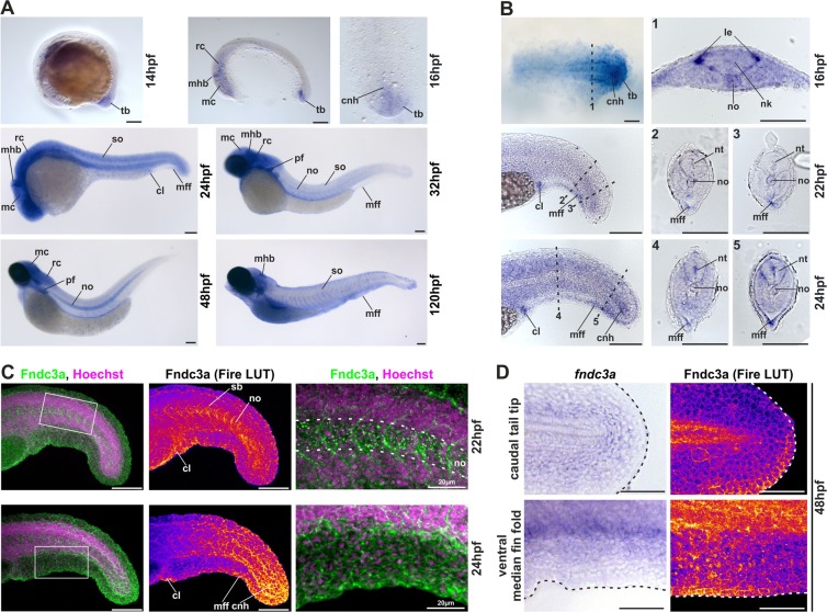
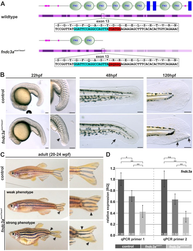
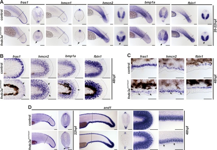
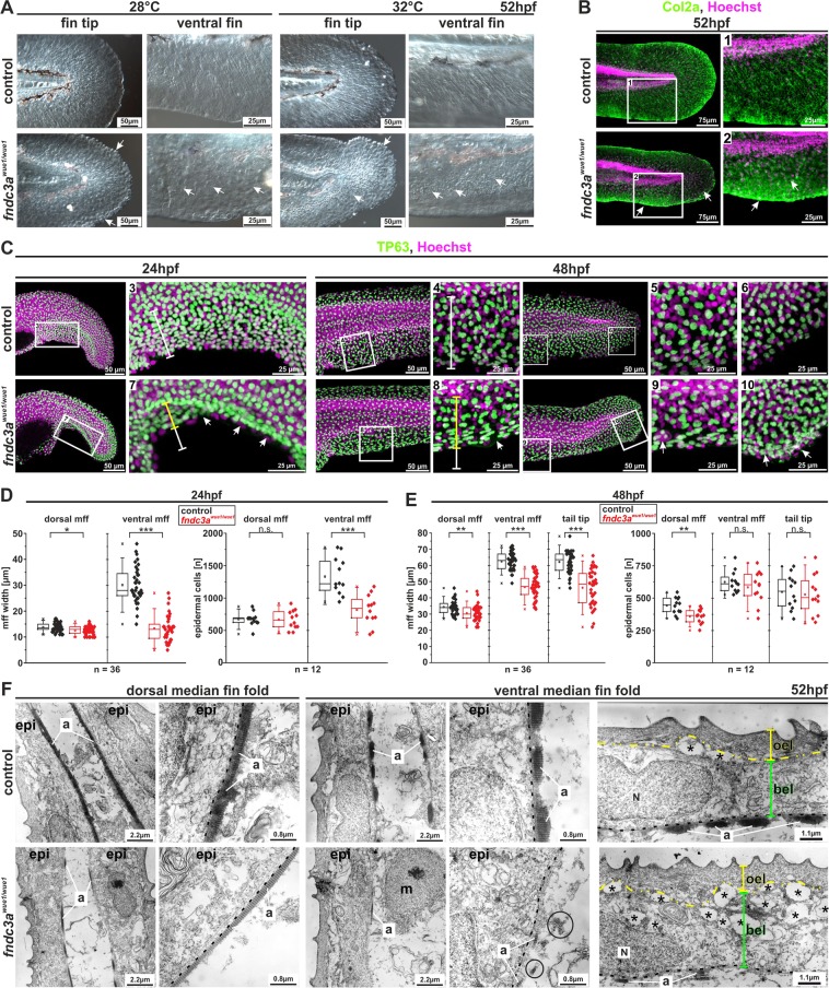
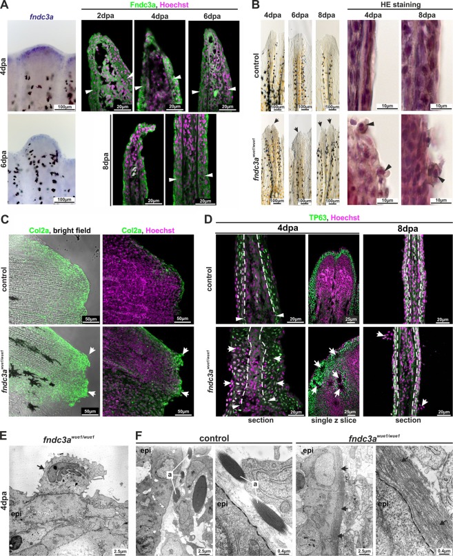
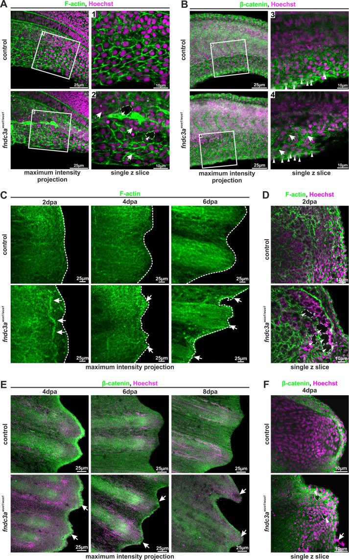
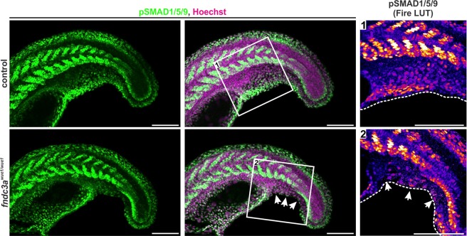
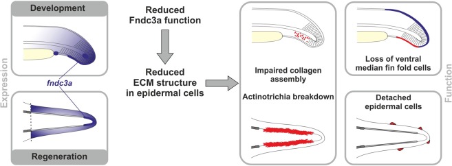
Fin development and regeneration are complex biological processes that are highly relevant in teleost fish. They share genetic factors, signaling pathways and cellular properties to coordinate formation of regularly shaped extremities. Especially correct tissue structure defined by extracellular matrix (ECM) formation is essential. Gene expression and protein localization studies demonstrated expression of fndc3a (fibronectin domain containing protein 3a) in both developing and regenerating caudal fins of zebrafish (Danio rerio). We established a hypomorphic fndc3a mutant line (fndc3awue1/wue1) via CRISPR/Cas9, exhibiting phenotypic malformations and changed gene expression patterns during early stages of median fin fold development. These developmental effects are mostly temporary, but result in a fraction of adults with permanent tail fin deformations. In addition, caudal fin regeneration in adult fndc3awue1/wue1 mutants is hampered by interference with actinotrichia formation and epidermal cell organization. Investigation of the ECM implies that loss of epidermal tissue structure is a common cause for both of the observed defects. Our results thereby provide a molecular link between these developmental processes and foreshadow Fndc3a as a novel temporal regulator of epidermal cell properties during extremity development and regeneration in zebrafish.


Conflict of interest statement

The authors declare no competing interests.

Figures

Figure 1
Figure 1
Localization of fndc3a RNA and protein during embryonic zebrafish development. (A,B) Expression of fndc3a mRNA was detected in the tail bud and the median fin fold from 14 hpf onwards. fndc3a is rather broadly expressed during embryogenesis, but was highly expressed in caudal and pectoral fins, somites, notochord cells and distinct brain regions. (C,D) Detection of Fndc3a protein via immunofluorescence indicated similar regional localization as fndc3a mRNA in 22–48 hpf embryos. Furthermore, it showed intracellular accumulation of Fndc3a at notochord cells, at somite boundaries and epidermal cells at this stage. In-situ stained embryos shown in (A,B) differ in proteinase K incubation and NBT/BCIP staining times to visualize weak fndc3a expression in different tissues and stages. Dashed lines in (B) indicate planes of the corresponding numbered sections 1–5, in (C) notochord boundary and in (D) fin fold border. Fire LUT in (C,D) shows pseudo-colored images of Fndc3a immunofluorescence and marks regions of high and low intensities (highest to lowest signal: yellow, red, blue, black). cnh: chordo neural hinge; cl: cloaca; le: lateral edge; mc: mesencephalon; mff: median fin fold; mhb: midbrain hindbrain boundary (marked with chevron); nk: neural keel; no: notochord; nt: neural tube; pf: pectoral fin; sb: somite boundary; so: somites; tb: tail bud; rc: rhombencephalon. Scale bars: 100 µm, except higher magnification in (C): 20 µm.
Figure 2
Figure 2
Generation and phenotype of fndc3awue1/wue1 zebrafish mutants. (A) The CRISPR/Cas9 system was used to target exon 13 in the zebrafish fndc3a gene coding for the third fibronectin type III domain (nucleotides marked in light blue indicate sgRNA target sequence and in red the region of mutated sequence). (B) fndc3awue1/wue1 mutants showed straightened tail buds (22 hpf; n = 19/40), kinked tails (48 hpf; n = 27/100), and caudal fin deformations (120 hpf; n = 9/41) during the first days of embryonic development. (C) A fraction of adult fndc3awue1/wue1 mutants displayed weak (n = 15/71) to strong (n = 6/71) caudal fin phenotypes and tail malformations. (D) qPCR quantification of relative fndc3a expression levels in genotypic different groups of embryos indicated reduction of fndc3a transcripts in fndc3awue1/+ and fndc3awue1/wue1 (ΔΔCt calculation; significance levels of a 2-sided paired student t-test are given). Investigation of protein domains shown in A has been performed via the SMART database (Simple Modular Architecture Research Tool; http://smart.embl-heidelberg.de). Black arrows indicate developmental malformations. Scale bars for whole embryos: 250 µm; scale bars for tail magnifications: 100 µm.
Figure 3
Figure 3
Normal development of the ventral median fin fold is altered in fndc3awue1/wue1 mutants. (A) Investigation of fras1, hmcn1, hmcn2, bmp1a, and fbln1 expression in the tail bud 20–22 hpf indicated loss of mesenchymal cells in the median fin fold in fndc3awue1/wue1 mutants (arrows; fras1: 9/32; hmcn1: 11/26; hmcn2: 24/43; bmp1a: 22/45; fbln1: 16/32). (B) Expression of mesenchymal markers in posterior fins of 48 hpf old embryos was slightly changed following loss of fndc3a (fras1: 10/24; hmcn2: 12/25; bmp1a: 8/21; fbln1: 9/24). (C) Ventral fin folds in 48 hpf old embryos did not show loss of mesenchymal markers after fndc3awue1/wue1 mutation. (D) Expression of and1, an essential marker for actinotrichia formation, in fndc3awue1/wue1 in the ventral fin fold was partly lost in 22 hpf embryos (12/25), but recovered in 48 hpf old embryos to a reduced expression level (control n = 16; weaker expression in fndc3awue1/wue1 embryos n = 10/18). Dashed lines in (A,D) indicate planes of shown sections. Dashed lines in (B,C) indicate fin boundaries. Scale bars: 50 µm
Figure 4
Figure 4
The fndc3awue1/wue1 mutation results in structural defects in epidermal cells during fin development. Visualization of actinotrichia by either differential interference contrast microscopy (A) or immunofluorescence staining of Col2a (B) showed loss of mature actinotrichia in fndc3awue1/wue1 mutants 52 hpf (control n = 0/17; fndc3awue1/wue1 n = 19/26; white arrows indicate lost actinotrichia and Col2a accumulation in apical cells of the median fin fold). (C) Investigation of fndc3awue1/wue1 mutants showed reduced median fin fold width and reduced TP63 positive epidermal cell number in the ventral median fin. (D,E) Quantification of median fin fold width and TP63 positive cells in 24 and 48 hpf embryos (Mann-Whitney U test; p < 0.05; two-tailed, significant U value changes are indicated with asterisks). (F) Ultrastructural analysis of dorsal and ventral fins of control and fndc3awue1/wue1 mutants revealed breakdown of actinotrichia fibers and cellular malformations in cells of the basal epidermal layer in the ventral fin folds of 52 hpf old embryos (circles indicate misplaced fibers; dashed lines indicate the basal membrane; yellow brackets indicate outer epidermal cells and green brackets indicate basal epidermal cells; black asterisks indicate cavities). a: actinotrichia; bel: basal epidermal layer; epi: epidermis; mff: median fin fold; m: mesenchymal cell; N: nucleus; oel: outer epidermal layer.
Figure 5
Figure 5
Interference with Fndc3a function during fin regeneration results in epidermal cells defects. (A) Expression of fndc3a and localization of Fndc3a in regenerates could be detected in epidermal cell layers. (B) Phenotypical and histological investigations of fin regeneration in fndc3awue1/wue1 mutants indicated abnormal regeneration by showing unorganized fin borders at the regenerative front and aberrant epidermal cells (arrows; abnormal regenerate phenotypes, control: 3/19; fndc3awue1/wue1: 13/19). (C) Col2a protein localization visualized via immunofluorescence on 8 dpa regenerates showed accumulation of collagen in abnormal cells of fndc3awue1/wue1 mutants (white arrows; n = 6 for each group). (D) TP63 staining in regenerates of fndc3awue1/wue1 mutants 4 and 8 dpa indicated disorganization of epidermal cells (arrows indicate epidermal thickening, cavities within the regenerate and abnormal cells; n = 4 for each group). (E) TEM analysis showed accumulation of electron dense material in the Golgi apparatus of abnormal cells attached to the epidermal layer (arrow). (F) TEM analyses of actinotrichia in fin regenerates 4 dpa revealed loss of these fibers located near the basal membrane of epidermal cells in fndc3a depleted fish (arrows). Arrowheads indicate amputation site. a: actinotrichia; epi: epidermis; dashed lines indicate basal membrane boundary.
Figure 6
Figure 6
Correct ECM structure in the median fin fold and regenerating caudal fins is hampered in fndc3awue1/wue1 mutants. (A,B) F-actin in the median fin fold was visualized by phalloidin staining and localization of β-catenin by immunofluorescence (n = 6 for each group, 22–24 hpf). Cellular organization of ventral median fin fold cells and ECM matrix was symmetrically structured in control embryos and showed nuclear localization of active Wnt signals in apical cells (white arrowheads in B). fndc3awue1/wue1 mutants depicted cellular alterations and unstructured ECM assembly by showing irregular cell shapes (arrows in A), cavities within the fin fold (dashed lines in A) and speckled accumulation of β-catenin between cells (arrows in B). Nuclear localization of β-catenin in apical cells was maintained (arrowheads in B). (C,D) Fin regenerates of fndc3awue1/wue1 mutants stained for F-actin showed regenerate abnormalities (arrows in C), irregular regenerate borders (dashed lines in C) and cellular cavities (dashed lines in D; n = 4 for each group). (E,F) Fin regenerates of fndc3awue1/wue1 mutants stained for β-catenin depicted divergent ECM assembly (arrows in E), appearance of abnormal cells loosely attached to the regenerate (arrows in F) and cavities (dashed lines in F; n = 3 for each group). Images either show maximum intensity projections (30 to 40 single z-slices; z-distance: 1.5 µm) or a representative higher resolution single z slice.
Figure 7
Figure 7
Investigation of TGF-beta/BMP signals in the median fin fold of fndc3awue1/wue1 mutants. Images show tail structures of representative control and fndc3awue1/wue1 mutant 22 hpf embryos stained for phosphorylated SMAD (pSMAD1/5/9) by immunofluorescence. Lack of pSMAD positive cells is observed in ventral median fin fold structures in fndc3awue1/wue1 mutants. All pictures display maximum intensity projections. White arrows indicate loss of pSMAD signals. Fire LUT shows pseudo-colored pSMAD1/5/9 signals. Dashed white lines indicate median fin fold borders. Scale bars: 50 µm.
Figure 8
Figure 8
Model of fndc3a function during zebrafish median fin fold development and caudal fin regeneration. Expression and localization of fndc3a was detected during early phases of median fin fold development and in caudal fin regenerates. Reduced Fndc3a function resulted in prominent changes of ECM structure in epidermal cells. During development this results in impaired collagen assembly of actinotrichia fibers and loss of ventral median fin fold cells. During regeneration altered ECM structure results in actinotrichia breakdown and detached epidermal cells.

Similar articles

Cited by

References

    1. Yano T, Tamura K. The making of differences between fins and limbs. J Anat. 2013;222:100–113. doi: 10.1111/j.1469-7580.2012.01491.x. - DOI - PMC - PubMed
    1. Amaral Danielson B., Schneider Igor. Fins into limbs: Recent insights from sarcopterygian fish. genesis. 2017;56(1):e23052. doi: 10.1002/dvg.23052. - DOI - PubMed
    1. Yano T, Abe G, Yokoyama H, Kawakami K, Tamura K. Mechanism of pectoral fin outgrowth in zebrafish development. Development. 2012;139:2916–2925. doi: 10.1242/dev.075572. - DOI - PubMed
    1. Masselink W, et al. A somitic contribution to the apical ectodermal ridge is essential for fin formation. Nature. 2016;535:542–546. doi: 10.1038/nature18953. - DOI - PubMed
    1. van den Boogaart JG, Muller M, Osse JW. Structure and function of the median finfold in larval teleosts. The Journal of experimental biology. 2012;215:2359–2368. doi: 10.1242/jeb.065615. - DOI - PubMed

Publication types

Substances

LinkOut - more resources