Review
. 2019 Jun 21;6(17):1900101.
doi: 10.1002/advs.201900101. eCollection 2019 Sep 4.

Immunomodulatory Nanosystems

Affiliations
Review

Immunomodulatory Nanosystems

Xiangru Feng et al. Adv Sci (Weinh). .

Abstract

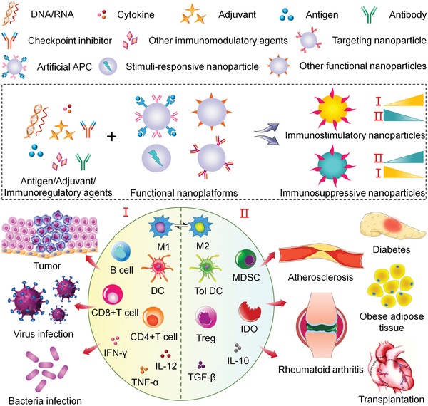
Immunotherapy has emerged as an effective strategy for the prevention and treatment of a variety of diseases, including cancer, infectious diseases, inflammatory diseases, and autoimmune diseases. Immunomodulatory nanosystems can readily improve the therapeutic effects and simultaneously overcome many obstacles facing the treatment method, such as inadequate immune stimulation, off-target side effects, and bioactivity loss of immune agents during circulation. In recent years, researchers have continuously developed nanomaterials with new structures, properties, and functions. This Review provides the most recent advances of nanotechnology for immunostimulation and immunosuppression. In cancer immunotherapy, nanosystems play an essential role in immune cell activation and tumor microenvironment modulation, as well as combination with other antitumor approaches. In infectious diseases, many encouraging outcomes from using nanomaterial vaccines against viral and bacterial infections have been reported. In addition, nanoparticles also potentiate the effects of immunosuppressive immune cells for the treatment of inflammatory and autoimmune diseases. Finally, the challenges and prospects of applying nanotechnology to modulate immunotherapy are discussed.

Keywords: disease treatment; immunostimulation; immunosuppression; immunotherapy; nanotechnology.


Conflict of interest statement

The authors declare no conflict of interest.

Figures

Scheme 1
Scheme 1
Engineering of immunostimulatory nanoparticles and immunosuppressive nanoparticles based on functional nanoplatforms, and their applications in treatment of various diseases by regulating immune‐related cells, cytokines, and enzymes.
Figure 1
Figure 1
Separated delivery of antigen and adjuvant via γPGA NPs for effective anticancer immunotherapy. A) Synthetic process of CNNPs. B) Near‐infrared (NIR) fluorescence image of IR800‐labeled CNNP‐OVA and IR800‐labeled soluble OVA at different time points after injection through footpad of mice. The axillary LN was circled. Mean fluorescence intensity (MFI) was calculated. C) Histological evaluation of LNs at 24 h after coadministration of CNNP‐OVA‐FITC and CNNP‐IC‐rodamine B. D) Tumor volume and E) survival curve of vaccinated mice after tumor challenge. Reproduced with permission.138 Copyright 2017, Elsevier.
Figure 2
Figure 2
PD‐1 blockade NVs for melanoma immunotherapy. A) Schematic illustration of preparation process of PD‐1 NVs. B) DsRed‐PD‐1 NVs and Cy5.5‐labeled NVs bound with B16F10 cell membrane after incubation for 2 h. WGA Alexa‐Fluor 488 dye highlighted cell membrane. C) Fluorescence of Cy5.5‐labeled free NVs and PD‐1 NVs after intravenous injection. D) Distribution of PD‐1 NVs and free NVs shown by in vivo imagine system (IVIS). E) B16F10 tumor growth curves and F) survival rates of the mice in different groups. Reproduced with permission.142 Copyright 2018, John Wiley & Sons.
Figure 3
Figure 3
R848‐loaded nanoparticles improved cancer immunotherapy by regulating the polarization of TAMs. A) Preparation of CDNPs by cross‐linking CD with lysine and loading of R848 through host–guest interaction. B) Fluorescence imaging of CDNP accumulation in the tumor and organs at 24 h after administration. C) Quantified distribution of CDNP. D) Uptake of CDNPs by TAMs in tumor bearing mice detected by confocal laser scanning microscopy (CLSM) at (a,b) 60 min or (c,d) 24 h postinjection. Scale bars: 10 µm (b, d, expanded). E) Quantification of IL‐12 in TAMs within tumors at 24 h after different administrations. F) Change of individual tumor area at day 8 after treatment with distinct formulations. Reproduced with permission.146 Copyright 2018, Springer Nature.
Figure 4
Figure 4
Prodrug nanoparticles containing OXA and NLG919 improved immunotherapy by dual modulation of tumor immune microenvironment. A) Self‐assembly of ASPN from stimuli‐responsive OXA prodrug and NLG919 prodrug. B) Schematic illustration of synergistic antitumor effect from activated CD8+ T cells and suppressed Tregs. C) H&E staining of lung metastasis in 4T1 tumor‐bearing mice at the end of study. D) Survival curve of 4T1 tumor‐bearing mice during therapy. Reproduced with permission.126 Copyright 2018, John Wiley and Sons.
Figure 5
Figure 5
Smart nanoreactor enhanced antitumor effect to both primary and distant tumors by combination of PDT with PD‐L1 blockade. A) Synthetic process of DPEG‐coating mitochondria‐targeting nanoreactor loaded with catalase and Ce6, and schematic depiction of using nanoreactor to improve PDT process. B) Transmission electron microscopy (TEM) image of synthetic nanoreactor. C) Changes of tumor volume and D) cytotoxic T cell infiltration in primary tumors after treatments. E) Tumor growth curve for nonirradiated tumors. Reproduced with permission.161 Copyright 2018, American Chemical Society.
Figure 6
Figure 6
ROS‐responsive nanoplatform for enhanced radiation therapy and abscopal effect. A) Mechanism of ACF‐MnO2 NPs eliminating primary and abscopal tumors by tumor oxygenation and HIF‐1 dysfunction. B) Immunofluorescence images of primary tumor slices stained with PD‐L1 Ab. C,D) Tumor volume changes of C) primary and D) distant tumors in CT26 mice model. E) Primary tumor growth in 4T1‐bearing mice after different treatments. Reproduced with permission.163 Copyright 2018, American Chemical Society.
Figure 7
Figure 7
Chemo‐photothermal therapy potentiated antitumor immunity against primary and metastatic tumors. A) Synthesis of SGNPs with PDA coating. B–E) Frequencies of antigen‐specific CD8+ T cells and NK cells in tumor‐infiltrating LN in B,C) primary tumors and D,E) contralateral tumors. F,G) Tumor volume curve of F) treated primary tumors and G) untreated contralateral tumors. Reproduced with permission.34 Copyright 2018, Springer Nature.
Figure 8
Figure 8
Double‐layered polypeptide nanoparticles induced potent protection against influenza. A) Schematic illustration of core nanoparticle fabrication and double‐layered nanoparticle formation. B) Evaluation of M2e IgG binding ability to p09M2e, VtnM2e, and SHM2e. C–E) Examination of splenocyte cytokine secretion, including C) IFN‐γ, D) IL‐4, and E) IL‐2. F) Survival rate of immunized mice challenged with H5N1 virus. Reproduced with permission.257 Copyright 2018, National Academy of Sciences.
Figure 9
Figure 9
Antivirulence vaccination against bacteria infection via in situ capture of bacterial toxins. A) Fabrication process of nanotoxoid carrying pathogen‐specific virulence factors and its protection against toxic effect. B) Fluorescence imaging of B220 (green), IgD (blue), and GL‐7 (red) in draining LN at different magnifications. C) Multivalent Ab responses in mice vaccinated with different formulations. Reproduced with permission.261 Copyright 2017, John Wiley and Sons.
Figure 10
Figure 10
Reversible magnetic manipulation of RGD nanocaging controlled macrophage polarization. A) Fabrication of RGD‐Au NP‐magnetic nanocage heterodimer nanostructure, and manipulation of macrophage polarization via a magnetic field. B) Immunofluorescence micrographs of host macrophages staining actin (green), Arg‐1 (red), and nuclei (blue) following 24 h of RGD caging or uncaging. RGD caging–uncaging group received RGD caging for the initial 12 h followed by uncaging in the next 12 h. C) Quantification of cell density, area, and aspect ratio or macrophages after 24 h. Reproduced with permission.300 Copyright 2018, American Chemical Society.
Figure 11
Figure 11
Targeted delivery of anti‐CD3 Ab to LN to suppress transplant rejection. A) Schematic illustration of IR800‐NP synthesis process and conjugation with MECA79 monoclonal Ab. B,C) Detection of B) MECA79‐IR800‐NPs and C) nontargeted IR800‐NPs trafficking in the draining LN by staining IR800 (red), PNAd (green), and nuclei (blue). D) TEM of IR800‐NPs. E) Survival of C57BL/6 recipients receiving different treatments after transplantation with BALB/C hearts. F) Proportion of Tregs in draining LN of recipient mice by flow cytometric analysis and representative flow plots. Reproduced from with permission.42 Copyright 2018, American Society for Clinical Investigation.

Similar articles

Cited by

References

    1. Dunn G. P., Bruce A. T., Ikeda H., Old L. J., Schreiber R. D., Nat. Immunol. 2002, 3, 991. - PubMed
    1. Finlay B. B., McFadden G., Cell 2006, 124, 767. - PubMed
    1. Quail D. F., Joyce J. A., Nat. Med. 2013, 19, 1423. - PMC - PubMed
    1. Pearson R. M., Casey L. M., Hughes K. R., Miller S. D., Shea L. D., Adv. Drug Delivery Rev. 2017, 114, 240. - PMC - PubMed
    1. Ilinskaya A., Dobrovolskaia M., Br. J. Pharmacol. 2014, 171, 3988. - PMC - PubMed

LinkOut - more resources