. 2019 Nov;189(11):2311-2322.
doi: 10.1016/j.ajpath.2019.07.016. Epub 2019 Sep 6.

Lactoferrin CpG Island Hypermethylation and Decoupling of mRNA and Protein Expression in the Early Stages of Prostate Carcinogenesis

Affiliations

Lactoferrin CpG Island Hypermethylation and Decoupling of mRNA and Protein Expression in the Early Stages of Prostate Carcinogenesis

Corey M Porter et al. Am J Pathol. 2019 Nov.

Abstract

Lactoferrin (LTF) is an iron-binding protein canonically known for its innate and adaptive immune functions. LTF may also act as a tumor suppressor with antiproliferative action. LTF is inactivated genetically or epigenetically in various cancers, and a CpG island spanning the transcriptional start site of LTF is hypermethylated in prostate cancer cell lines. We, therefore, hypothesized that LTF expression is silenced via CpG island hypermethylation in the early stages of prostate tumorigenesis carcinogenesis. Targeted methylation analysis was performed using a combination of methylated-DNA precipitation and methylation-sensitive restriction enzymes, and laser-capture microdissection followed by bisulfite sequencing on DNA isolated from prostate tissue samples, including both primary and metastatic disease. LTF mRNA in situ hybridization and LTF protein immunohistochemistry were also performed. We report that the LTF CpG island is frequently and densely methylated in high-grade prostatic intraepithelial neoplasia, primary prostate carcinoma, and metastases. We further report a decoupling of lactoferrin mRNA and protein expression, including in lesions where LTF mRNA has presumably been silenced via CpG island methylation. We conclude that LTF mRNA expression is silenced in prostate tumorigenesis via hypermethylation, supporting a role for LTF as a prostate cancer tumor suppressor gene. Likewise, the frequency at which the LTF CpG island is methylated across samples suggests it is an important and conserved step in prostate cancer initiation.


Figures

Figure 1
Figure 1
Combination of methylated-DNA precipitation and methylation-sensitive restriction enzymes (COMPARE-MS) demonstrates hypermethylation in primary and metastatic prostate cancer. A: Heat maps of COMPARE-MS results. White indicates no methylation, and dark red indicates 100% methylation. COMPARE-MS results demonstrate that organ donor and benign areas of prostates, obtained at radical prostatectomy, have low levels of methylation, whereas primary and metastatic prostate cancer has higher levels of methylation. Benign prostatic hyperplasia (BPH) has an intermediate level of methylation. B:In silico analysis of lactoferrin methylation versus mRNA expression using The Cancer Genome Atlas data from cBioPortal. LTF, lactoferrin; RSEM, RNA-seq by expectation maximization.
Figure 2
Figure 2
Lactoferrin CpG island is hypermethylated in prostatic intraepithelial neoplasia (PIN) and prostate cancer. Clones obtained from bisulfite sequencing were analyzed with a bisulfite read mapper tool, Methyl Map. Each circle indicates average methylation across sequences from a case for each CpG site. White indicates unmethylated in all sequences, and red indicates methylated in all sequences. Benign samples generally had low methylation; however, PIN both near and away from cancer and cancer showed higher levels of methylation across all CpG sites.
Figure 3
Figure 3
Lactoferrin (LTF) protein and mRNA do not correlate with each other or with methylation. A: Percentage positive pixels for immunohistochemistry (IHC) and RNA in situ hybridization (RISH) plotted against percentage methylation in sequences reveals no correlation between methylation and protein expression. B: LTF protein expression assessed via IHC shows abundant LTF protein. Adjacent section to image in C. C: LTF mRNA expression assessed via RISH shows little to no expression in areas with abundant protein in B. Original magnification, ×40 (B and C). PIN, prostatic intraepithelial neoplasia.
Figure 4
Figure 4
Lactoferrin (LTF) protein and mRNA expression do not correlate, as assessed with tissue microarrays. A: LTF protein is often present when there is no LTF mRNA expression. B and C: Log2 of total positive intensity shows that atrophy is the only cell type with protein at comparable levels (B) to mRNA (C). Atrophy was also the highest expressing lesion type for both protein and mRNA. C: For mRNA, normal prostate shows greater expression compared with primary or metastatic prostate cancer. *P < 0.05, **P < 0.005, ***P < 0.0005, and ****P < 0.0001. Original magnification, ×200 (A). IHC, immunohistochemistry; PIN, prostatic intraepithelial neoplasia; PPIB, peptidylprolyl isomerase B; RISH, RNA in situ hybridization.
Figure 5
Figure 5
Distant metastases have varying low levels of lactoferrin (LTF) mRNA and protein that do not correspond. A: LTF protein and mRNA vary by site and do not correspond. B and C: Log of total positive intensity shows a wide variety in protein (B) and mRNA (C) by metastatic site. *P < 0.05, **P < 0.005, ***P < 0.0005, and ****P < 0.0001. Original magnification, ×200 (A). IHC, immunohistochemistry; PPIB, peptidylprolyl isomerase B; RISH, RNA in situ hybridization.
Figure 6
Figure 6
Lactoferrin (LTF) receptor [intelectin-1/2 (ITLN-1/2)] expression does not explain discordant LTF mRNA and protein expression. Immunohistochemistry (IHC) for intelectin, the canonical LTF receptor. A: Small intestine (positive control). B: ITLN-1/2 is localized to basal epithelial cells in normal, noninflamed prostate. C: Top panels: ITLN-1/2 is localized to basal epithelial cells and LTF mRNA and protein is absent in normal, noninflamed prostate. Middle panels: ITLN-1/2 staining is observed in luminal cells concurrent with LTF mRNA and protein in some regions of prostatic atrophy. Bottom panels: ITLN-1/2 is not expressed in cases where there is LTF protein expression but no mRNA. Example shown is an LTF IHC-positive region in cancer. Original magnification, ×200 (AC). RISH, RNA in situ hybridization.
Supplemental Figure S1
Location of all assays in relation to the lactoferrin (LTF) gene structure. A: Targeted location of primers, RNA in situ hybridization probes, and antibody (Ab). B: Location of primers used for combination of methylated-DNA precipitation and methylation-sensitive restriction enzymes (COMPARE-MS) and bisulfite sequencing. bPCR, bisulfite PCR; UTR, untranslated region.
Supplemental Figure S2
Lactoferrin (LTF) antibody, immunohistochemistry (IHC) assay, and RNA in situ hybridization (RISH) assay validation with positive and negative controls. Top panel: The Abcam lactoferrin antibody produces one band at the correct size by Western blot analysis using prostate tissue lysates, whole blood cell (WBC) lysate (positive control), and recombinant LTF (positive control). The lactoferrin antibody does not cross-react with transferrin (negative control). Middle and bottom panels: Positive and negative controls for lactoferrin IHC assay using formalin-fixed, paraffin-embedded cell plugs of PC3 cells (negative for lactoferrin, negative control) transfected with both known forms of lactoferrin [soluble lactoferrin (sLTF) and ΔLTF; positive controls]. Original magnification, ×200.
Supplemental Figure S3
Intelectin-1/2 ITLN1/2) RNA in situ hybridization (RISH) assay validation with positive and negative controls. PC3 and LNCAP cells are negative for ITLN1/2 by RISH (negative controls). PC3 cells transfected with an ITLN1/2 expression vector (PC3 + ITLN1/2) are positive for ITLN1/2 by RISH (positive control). Original magnification, ×200.
Supplemental Figure S4
Example of laser-capture microdissection (LCM) for prostate epithelium. Example given is of prostate glands before LCM and where epithelium has been specifically targeted and removed by LCM (after LCM). Original magnification, ×200.
Supplemental Figure S5
Example of image analysis for lactoferrin (LTF) immunohistochemistry (IHC). Light blue lines indicate the annotated regions included in the image analysis. These were identical to the regions where laser-capture microdissection (LCM) was performed. Dark blue lines indicate regions excluded from image analysis (eg, glandular lumens). Orange, yellow, and blue pixels demonstrate a gland where image analysis has been performed and the positive regions (orange and yellow pixels) and negative regions (blue pixels) for lactoferrin have been determined. Original magnification, ×200.
Supplemental Figure S6
Immunohistochemistry (IHC) with two separate lactoferrin (LTF) antibodies (Abcam EPR4338 and Sigma L3262) demonstrates an identical staining pattern for lactoferrin protein in areas that were negative for LTF RNA by RNA in situ hybridization (RISH) [peptidylprolyl isomerase B (PPIB) included as a positive control for RNA integrity for RISH]. Original magnification, ×200.
Supplemental Figure S7
Positive staining, as assessed by immunohistochemistry (IHC), plotted against positive staining, as assessed by RNA in situ hybridization (RISH), in normal, atrophy, prostatic intraepithelial neoplasia, cancer, and distant metastases.
Supplemental Figure S8
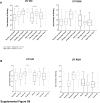
A: Assessment of lactoferrin (LTF) immunohistochemistry (IHC) and RNA in situ hybridization (RISH) in relation to tumor grade. B: Assessment of LTF IHC and RISH in relation to patient race, African American (AA) or European American (EA). *P < 0.05, **P < 0.005, and ****P < 0.0001. PIN, prostatic intraepithelial neoplasia.
Supplemental Figure S9
Example of lactoferrin (LTF) immunohistochemistry (IHC) in a liver metastasis. Brown staining cells appear to be infiltrating immune cells positive for lactoferrin, whereas the tumor cells are negative for lactoferrin, as indicated in the figure. Original magnification, ×200.

Similar articles

Cited by

References

    1. Sfanos K.S., De Marzo A.M. Prostate cancer and inflammation: the evidence. Histopathology. 2012;60:199–215. - PMC - PubMed
    1. De Marzo A.M., Platz E.A., Sutcliffe S., Xu J., Gronberg H., Drake C.G., Nakai Y., Isaacs W.B., Nelson W.G. Inflammation in prostate carcinogenesis. Nat Rev Cancer. 2007;7:256–269. - PMC - PubMed
    1. Nelson W.G., De Marzo A.M., DeWeese T.L., Isaacs W.B. The role of inflammation in the pathogenesis of prostate cancer. J Urol. 2004;172:S6–S12. - PubMed
    1. Sfanos K.S., Wilson B.A., De Marzo A.M., Isaacs W.B. Acute inflammatory proteins constitute the organic matrix of prostatic corpora amylacea and calculi in men with prostate cancer. Proc Natl Acad Sci. 2009;106:3443–3448. - PMC - PubMed
    1. Brock J.H. The physiology of lactoferrin. Biochem Cell Biol. 2002;80:1–6. - PubMed

Publication types

MeSH terms