. 2019 Oct 23;39(43):8457-8470.
doi: 10.1523/JNEUROSCI.0016-19.2019. Epub 2019 Sep 6.

Degeneration of Injured Axons and Dendrites Requires Restraint of a Protective JNK Signaling Pathway by the Transmembrane Protein Raw

Affiliations

Degeneration of Injured Axons and Dendrites Requires Restraint of a Protective JNK Signaling Pathway by the Transmembrane Protein Raw

Yan Hao et al. J Neurosci. .

Abstract

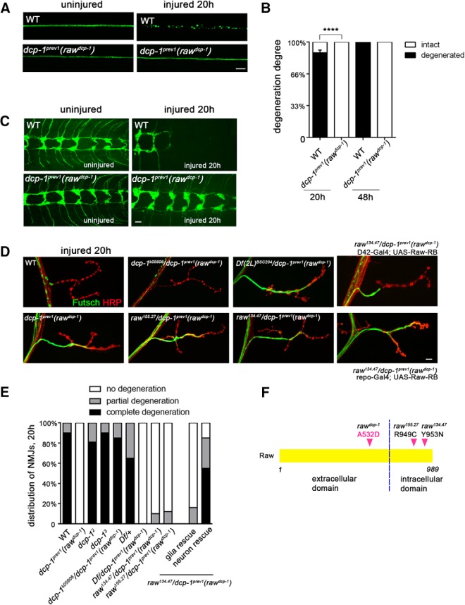
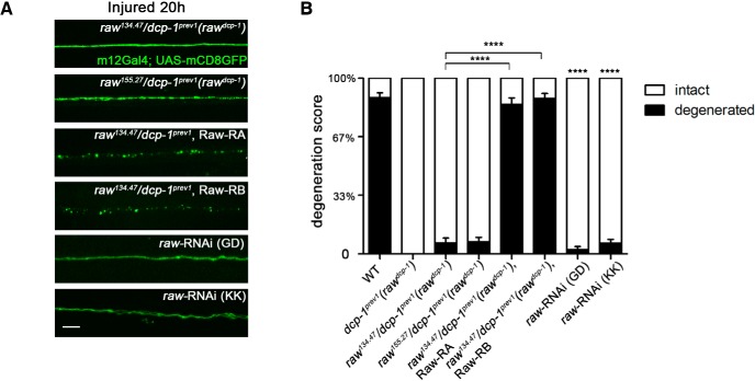
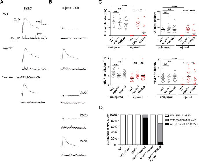
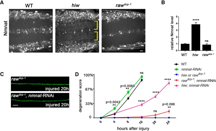
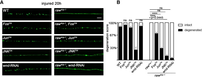
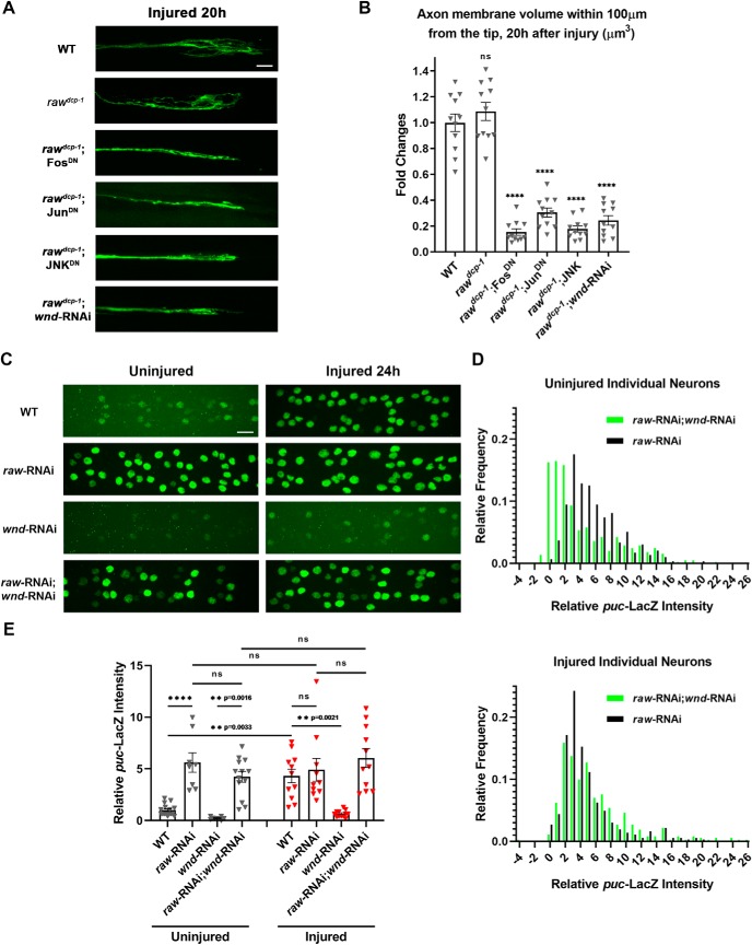
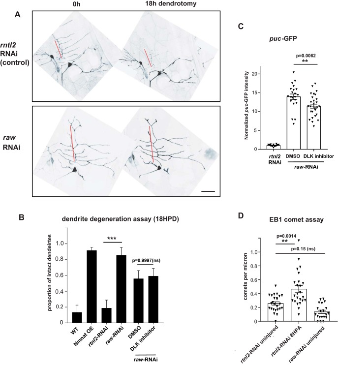
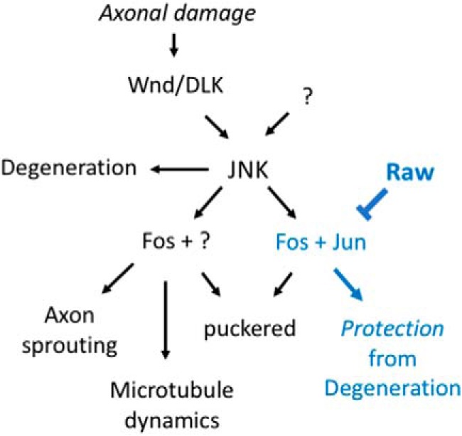
The degeneration of injured axons involves a self-destruction pathway whose components and mechanism are not fully understood. Here, we report a new regulator of axonal resilience. The transmembrane protein Raw is cell autonomously required for the degeneration of injured axons, dendrites, and synapses in Drosophila melanogaster In both male and female raw hypomorphic mutant or knock-down larvae, the degeneration of injured axons, dendrites, and synapses from motoneurons and sensory neurons is strongly inhibited. This protection is insensitive to reduction in the levels of the NAD+ synthesis enzyme Nmnat (nicotinamide mononucleotide adenylyl transferase), but requires the c-Jun N-terminal kinase (JNK) mitogen-activated protein (MAP) kinase and the transcription factors Fos and Jun (AP-1). Although these factors were previously known to function in axonal injury signaling and regeneration, Raw's function can be genetically separated from other axonal injury responses: Raw does not modulate JNK-dependent axonal injury signaling and regenerative responses, but instead restrains a protective pathway that inhibits the degeneration of axons, dendrites, and synapses. Although protection in raw mutants requires JNK, Fos, and Jun, JNK also promotes axonal degeneration. These findings suggest the existence of multiple independent pathways that share modulation by JNK, Fos, and Jun that influence how axons respond to stress and injury.SIGNIFICANCE STATEMENT Axonal degeneration is a major feature of neuropathies and nerve injuries and occurs via a cell autonomous self-destruction pathway whose mechanism is poorly understood. This study reports the identification of a new regulator of axonal degeneration: the transmembrane protein Raw. Raw regulates a cell autonomous nuclear signaling pathway whose yet unknown downstream effectors protect injured axons, dendrites, and synapses from degenerating. These findings imply that the susceptibility of axons to degeneration is strongly regulated in neurons. Future understanding of the cellular pathway regulated by Raw, which engages the c-Jun N-terminal kinase (JNK) mitogen-activated protein (MAP) kinase and Fos and Jun transcription factors, may suggest new strategies to increase the resiliency of axons in debilitating neuropathies.

Keywords: Drosophila; MAP kinase signaling; Wallerian degeneration; axon degeneration; axon injury; motoneuron.


Figures

Figure 1.
Figure 1.
A new mutation that strongly inhibits Wallerian degeneration of motoneuron and sensory neuron axons and synapses, identified in dcp-1prev1 animals, maps to raw. A, Single motoneuron axons in third instar larvae are labeled with a membrane-targeted reporter transgene, UAS-mCD8::GFP, driven by m12-Gal4. In control (WT) animals, axons distal to the injury sites are completely fragmented within 20 h after nerve crush injury. However, axons remain completely intact in dcp-1prev1 mutant background. B, Quantification of the degree of axonal degeneration in different genotypes and different time points is shown in black, whereas white bars indicate the percentage of axons that remain intact. C, The axons and nerve terminals of class IV sensory neurons in larval ventral nerve cord were labeled by membrane bound mCD8::GFP with the ppk-Gal4 driver. In WT animals, these terminals were completely fragmented and cleared within 20 h after injury (the remaining terminals are from uninjured segments) but remain intact in dcp-1prev1 animals. D, Representative images of the degeneration phenotype at muscle 4 NMJs 20 h after injury. In WT animals, the cytoskeleton marker Futsch (shown in green) disappeared from the NMJ, whereas the neuronal membrane (labeled by anti-HRP in red) became highly fragmented. In contrast, homozyogous dcp-1prev1 mutations failed to degenerate, however other homozygous mutations in dcp-1: dcp-12 and dcp-13, and trans-heterozygous mutants dcp-1k05606/ dcp-1prev1 showed normal degeneration. NMJs in the dcp-1prev1 animals show no sign of degeneration. Genotypes are described in further detail in Table 1. Trans-heterozygotes of dcp-1prev1 with a deficiency deleted for raw and with previously identified mutations in raw, raw155.27/ dcp-1prev1 and raw134.47/ dcp-1prev1, uncovered the degeneration phenotype. Expression of UAS-Raw in raw134.47/ dcp-1prev1 background using a motoneuron driver D42-Gal4 significantly rescues the protective phenotype, whereas expression of UAS-Raw using a pan-glia driver repo-Gal4 fails to rescue. E, Quantification of NMJ degeneration. Black bars represent the percentage of NMJs that were completely degenerated 20 h after injury, gray bars represent percentage of NMJs that were partially degenerated, and white bars represent the percentage of intact NMJs. F, Schematic of the protein structure of Raw, which has a single transmembrane domain indicated by the black dotted line. The point mutation in raw in the dcp-1prev1 (rawdcp-1) changes amino acid 532, which lies in the extracellular domain, from Alanine to Aspartic acid. Previously described missense mutations raw134.47 and raw155.27 lie with the intracellular domain. Scale bars, 20 μm, error bars represent SEM; ****p < 0.0001, one-way ANOVA test; n ≥ 25 axons or axon terminals from 5 animals per condition.
Figure 2.
Figure 2.
Raw functions cell autonomously in neurons to promote Wallerian degeneration. A, Representative images of the degeneration phenotype for single motoneuron axons labeled via the m12-Gal4 driver expressing UAS-mCD8::GFP 20 h after injury. Expression of either of two spliced isoforms of Raw cDNA (Raw-RA or Raw-RB) in m12-Gal4-expressing neurons rescued the protective phenotype in raw134.47/dcp-1prev1 background animals. Moreover, depletion of Raw in motoneurons using raw-RNAi lines in combination with UAS-Dcr2 also strongly delays axonal degeneration. B, Quantification of axonal degeneration of the animals with genotypes shown in A. Scale bars, 20 μm. Error bars indicate SEM. ****p < 0.0001, one-way ANOVA test; n ≥ 25 axons from 5 animals per condition.
Figure 3.
Figure 3.
Injured synapses retain functional neurotransmission properties in raw mutants. A, Example traces for EJP and mEJP potentials recorded from muscle 6 NMJs in control/WT (Canton S), rawdcp-1(dcp-1prev1) homozygous animals and animals expressing UAS-Raw-RA cDNA in motoneurons (via the D42-Gal4 driver) in a rawdcp-1/raw134.47 mutant background. Recordings were done at segment 3 or 4 from naive (uninjured) animals, and in B, in animals 20 h after a ventral nerve crush that injured the nerves innervating these segments. NMJs in rawdcp-1/raw134.47 mutants expressing Raw cDNA in motoneurons showed three phenotypic classes: 6/20recorded NMJs of this genotype retained both EJPs and mEJPs similarly to uninjured NMJs, in left example traces; 12/20 retained mEJPs but lost EJPs, in right traces, and 2/20 lacked both EJPs and mEJPs, similarly to WT injured, indicating partial rescue of the raw phenotype. C, Quantification of EJP amplitude, mEJP amplitude, quantal content (corrected for nonlinear summation), and mEJP frequency (Hz) for recordings from uninjured (black) and 20 h postinjury (red) larvae. D, Percentage of NMJs in the three different phenotypic classes depicted in A. n ≥ 15 NMJs per condition. ****p < 0.0001.
Figure 4.
Figure 4.
Protection in raw mutants is not sensitive to levels of Nmnat enzyme. A, Endogenous Nmnat levels in neuropil are increased in hiw mutants (noted with yellow brackets), however are not detectably changed in raw mutants (rawdcp-1). B, Quantification of the relative levels of Nmnat protein in the neuropil regions of WT, hiwΔN and rawdcp-1 animals. C, Example axons from m12-Ga4 expressing neurons, labeled by coexpression of UAS-mCD8::GFP, UAS-Dcr2 and UAS-nmnat-RNAi, shown 20 h after injury. D, Quantification of axonal degeneration in different time points for the noted genotypes. Comparing to WT animals (black line), knock-down of nmnat by RNAi (green line) modestly promotes axonal degeneration. Although knock-down of nmnat in the hiw mutant background significantly rescues the axonal protection phenotype in hiw mutant (compare pink line to blue line), knock-down of nmnat in rawdcp-1 mutant background only modestly changes the axonal protective effect of rawdcp-1 (compare red line to blue line). Scale bar, 20 μm. Error bars indicate SEM. ****p < 0.0001; ns (p = 0.5314), one-way ANOVA test; n = 5 animals per genotype in B, n ≥ 25 axons from 5 animals per condition in D.
Figure 5.
Figure 5.
Protection of injured axons from degeneration in raw mutants requires the transcription factors Fos and Jun and the JNK MAP kinase. A, Single motoneuron axons are labeled by membrane bound mCD8::GFP driven by the m12-Gal4 driver. As shown in Figure 1, axonal degeneration is strongly delayed in rawdcp-1 animals. Expression of dominant negative forms of Jun and Fos (JunDN and FosDN) partially rescue the protective effect in rawdcp-1 animals. B, Quantification of axon degeneration scores in indicated genotypes. Scale bar, 20 μm. Error bars indicate SEM. ****p < 0.0001, *0.01 < p < 0.05 (p = 0.0445). ns (p > 0.05), one-way ANOVA test; n ≥ 25 axons from 5 animals per condition.
Figure 6.
Figure 6.
Raw has no effect upon axonal injury signaling and axonal regeneration. A, Representative examples of proximal stump of sprouting m12-Gal4-expressing motoneuron axons 20 h after nerve crush. B, Quantification of new axonal growth based on measured volume of axonal membrane within 100 μm of the injured axon tip. C, Example images showing puc-lacZ reporter expression in motoneuron nuclei that express UAS-raw-RNAi (vdrc 101255), UAS-wnd-RNAi (BL 35369) or both, together with UAS-Dcr2, via the pan-neuronal driver BG380-Gal4. D, Distribution of puc-lacZ expression levels measured in individual motoneuron nuclei coexpressing raw-RNAi; wnd-RNAi (green) compared with raw-RNAi alone (black). E, Quantification of relative puc-lacZ intensities (normalized to uninjured control) in different genotypes. Individual triangles represent mean intensities per larva measured from 30 to 35 dorsal motoneurons in segments 3–7 of the larval nerve cord. Scale bars, 20 μm. Error bars indicate SEM. ****p < 0.0001, ns, one-way ANOVA test (p > 0.05), n ≥ 11 axons in ≥ 5 animals per genotype in B, n ≥ 8 animals in 3 independent trials in E.
Figure 7.
Figure 7.
Raw is required for dendrite degeneration. Individual dendrite branches of ddaE sensory neurons, labeled by expression of UAS-mCD8-GFP with the 221-Gal4 driver, were surgically transected using a pulsed dye laser in flies coexpressing expressing Dcr2 and either control RNAi (rtnl2 RNAi, VDRC 33320) or raw RNAi (VDRC 101255). A, Example images of ddaE neurons 0 and 18 h postdendrotomy are shown. Degeneration is characterized by membrane blebbing and loss of continuity in the primary comb (red line). Scale bar, 20 μm. B, Quantification of dendrites that remain intact 18 h postdendrotomy is shown in the graph. Reduction of raw expression via RNAi protects dendrites comparably to overexpression of the protective enzyme Nmnat, as in Chen et al. (2016). Rearing animals in the presence of GNE-3511, which inhibits DLK (Patel et al., 2015) and Wnd-mediated phenotypes in ddaE neurons (Feng et al., 2019), caused no significant change to the raw-RNAi phenotype compared with animals reared in the presence of the DMSO vehicle (B), but did cause a modest reduction in expression of the puc-GFP reporter (C). D, In contrast to a previously described mechanism for dendrite protection (Chen et al., 2012), raw mutants do not show increased microtubule dynamics. The number of growing microtubule plus ends labeled by EB1::GFP were measured in UAS-Dicer2;221Gal4, UAS-EB1::GFP larvae expressing UAS- rtnl2 (control) RNAi or UAS-raw-RNAi. Statistical significance was calculated using one-way ANOVA. ***p < 0.001, **0.001 < p < 0.01, ns represents p > 0.05 (p = 0.15). n ≥ 14 ddaE neurons per condition.
Figure 8.
Figure 8.
Working model: Raw regulates the ability of injured axons and dendrites to degenerate by restraining a JNK-dependent transcriptional program. Signaling downstream of the Wallenda/DLK kinase after axonal injury and in other conditions of stress (Asghari Adib et al., 2018) regulates multiple JNK-dependent responses. These include promotion of axonal degeneration, which may occur locally in injured axons independently of the nucleus (Miller et al., 2009; Yang et al., 2015), and downstream events that involve transcription factors including Fos and Jun: regenerative axon sprouting (Xiong et al., 2010; Stone et al., 2014), global upregulation of microtubule dynamics (Stone et al., 2010; Chen et al., 2012), and protection of neurites from degeneration (Chen et al., 2012, 2016; Xiong and Collins, 2012). Our data suggest that Raw regulates protective signaling without influencing sprouting or increasing microtubule dynamics. Because the nuclear localization of Jun(Jra) is altered (increased) in raw mutants (Jemc et al., 2012), we propose that this specificity might occur via the regulation of different transcription factor complexes. Raw may therefore provide a new inroad to functionally separate pathways that share dependence upon the JNK MAP kinase.

Similar articles

Cited by

References

    1. Abe N, Cavalli V (2008) Nerve injury signaling. Curr Opin Neurobiol 18:276–283. 10.1016/j.conb.2008.06.005 - DOI - PMC - PubMed
    1. Asghari Adib E, Smithson LJ, Collins CA (2018) An axonal stress response pathway: degenerative and regenerative signaling by DLK. Curr Opin Neurobiol 53:110–119. 10.1016/j.conb.2018.07.002 - DOI - PMC - PubMed
    1. Babetto E, Beirowski B, Russler EV, Milbrandt J, DiAntonio A (2013) The phr1 ubiquitin ligase promotes injury-induced axon self-destruction. Cell Rep 3:1422–1429. 10.1016/j.celrep.2013.04.013 - DOI - PMC - PubMed
    1. Bates KL, Higley M, Letsou A (2008) Raw mediates antagonism of AP-1 activity in Drosophila. Genetics 178:1989–2002. 10.1534/genetics.107.086298 - DOI - PMC - PubMed
    1. Bauer Huang SL, Saheki Y, VanHoven MK, Torayama I, Ishihara T, Katsura I, van der Linden A, Sengupta P, Bargmann CI (2007) Left-right olfactory asymmetry results from antagonistic functions of voltage-activated calcium channels and the raw repeat protein OLRN-1 in C. elegans. Neural Dev 2:24. 10.1186/1749-8104-2-24 - DOI - PMC - PubMed

Publication types

MeSH terms

Substances

LinkOut - more resources