. 2019 Aug 7:7:134.
doi: 10.3389/fcell.2019.00134. eCollection 2019.

Herpes Simplex Virus, Alzheimer's Disease and a Possible Role for Rab GTPases

Affiliations

Herpes Simplex Virus, Alzheimer's Disease and a Possible Role for Rab GTPases

Elaine L Bearer et al. Front Cell Dev Biol. .

Abstract

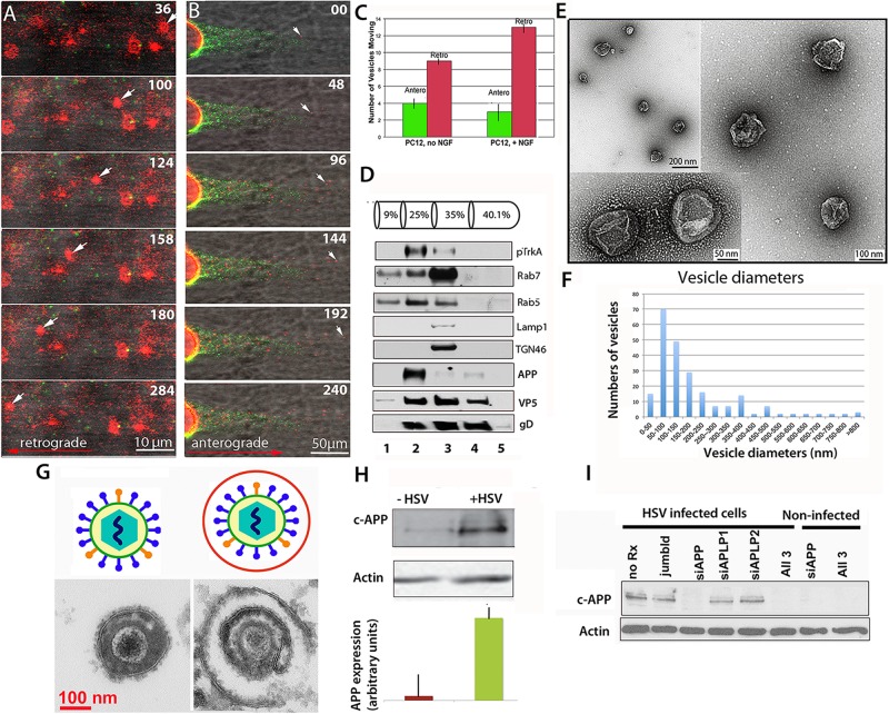
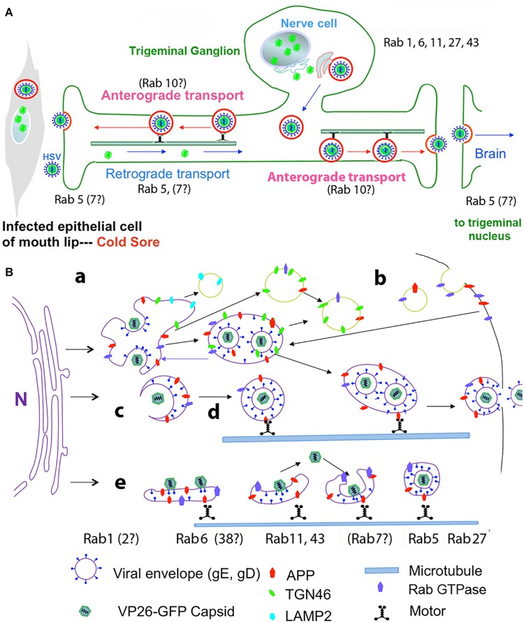
Herpes simplex virus (HSV) is a common pathogen, infecting 85% of adults in the United States. After reaching the nucleus of the long-lived neuron, HSV may enter latency to persist throughout the life span. Re-activation of latent herpesviruses is associated with progressive cognitive impairment and Alzheimer's disease (AD). As an enveloped DNA virus, HSV exploits cellular membrane systems for its life cycle, and thereby comes in contact with the Rab family of GTPases, master regulators of intracellular membrane dynamics. Knock-down and overexpression of specific Rabs reduce HSV production. Disheveled membrane compartments could lead to AD because membrane sorting and trafficking are crucial for synaptic vesicle formation, neuronal survival signaling and Abeta production. Amyloid precursor protein (APP), a transmembrane glycoprotein, is the parent of Abeta, the major component of senile plaques in AD. Up-regulation of APP expression due to HSV is significant since excess APP interferes with Rab5 endocytic trafficking in neurons. Here, we show that purified PC12-cell endosomes transport both anterograde and retrograde when injected into the squid giant axon at rates similar to isolated HSV. Intracellular HSV co-fractionates with these endosomes, contains APP, Rab5 and TrkA, and displays a second membrane. HSV infected PC12 cells up-regulate APP expression. Whether interference with Rabs has a specific effect on HSV or indirectly affects membrane compartment dynamics co-opted by virus needs further study. Ultimately Rabs, their effectors or their membrane-binding partners may serve as handles to reduce the impact of viral re-activation on cognitive function, or even as more general-purpose anti-microbial therapies.

Keywords: Alzheimer’s disease; HSV (herpes simplex virus); Rab GTPases; axonal transport; dynein; endosomes; kinesin; squid giant axon.


Figures

FIGURE 1
FIGURE 1
Axonal transport of endosomes isolated from PC12 cells in the squid giant axon and roles for APP and Rabs. (A) Membrane-bound vesicles purified from PC12 cells (Fraction 2) are transported retrograde after injection into a freshly dissected giant axon of the squid. Inert green fluorescent beads were co-injected. Most vesicles remain stationary, but one in this series (arrow) moves retrograde. Also see Supplementary Videos S1 and S2. (B) Vesicles (red), also move anterograde even when hCAPP peptide is co-injected., Peptide inhibits transport of beads (green) conjugated with hCAPP peptide, as previously described (Satpute-Krishnan et al., 2006). See Supplementary Video S3. (C) More vesicles move retrograde than anterograde and this difference increases after NGF treatment of the PC12 cells (n = 5, error bars = SEM). (D–F) Characterization of the vesicle preparation used in panels (A–C). (D) Western blotting of fractions from PC12 cell organelle preparation showing co-fractionation at 25% sucrose of pTrkA, the high affinity NGF receptor, and Rab5 in fraction 2, the fraction that was injected in panels (A,B). In contrast Rab7, LAMP1 and TGN46 concentrate in the slightly denser 35% fraction. Fraction numbers (1 ml) are indicated at the bottom. Fractionation of vesicles after radioactive NGF labeling showed that this Rab5B concentrates with I125-labeled NGF and phospho-TrkA in the 25% fraction (not shown). In parallel gradients of HSV-infected PC12 cells, APP also sediments in fraction 2 along with viral proteins, VP5 (capsid) and gD (envelope). (E) Examples of vesicles imaged by negative stain electron-microscopy demonstrate 50–300 nm diameter membrane-bound vesicles in fraction 2 from experiments shown in panels (A–C). (F) Distribution of vesicle sizes in fraction 2. Three different fractionation experiments were examined by negative-stain electron-microscopy and vesicle sizes measured (n = 216). Average diameter across all three experiments was 180 nm, median size was 136 nm, and range was 42–1,200 nm. 75% of the vesicles fell between 50 and 300 nm. For the fraction injected in panel (A) (n = 42), the average size was 186, the median size was 140 nm, and range 10–500 nm. 80% of the vesicles fell between 50 and 300 nm (graph not shown). (G) Intracellular viral particles are found within a second membrane. Diagram (top). Thin section electron-microscopy (bottom) of extracellular (left) and intracellular (right) virions detects a second membrane surrounding enveloped particles within the cytoplasm of cells that is not present around extracellular virions. (H) HSV infection increases expression of APP in vitro. Western blots of PC12 cells with and without HSV infection demonstrated a large increase (25-fold) of APP as detected by the Zymed anti-C-APP antibody (Cheng et al., 2011). Shown is the 110 kD band representing full-length APP. Comparison of blot intensity by Image J (lower panel) quantifies the average change in arbitrary units. (Bars indicate range of triplicates.) (I) Suppression of APP by siRNA. APP expression in infected cells can be completely and specifically suppressed with siRNA in PC12 cells. In Vero cells, this suppression led to a dramatic decrease in production of infective virions (Cheng et al., 2011). Methods: PC12 cells were grown in a 15 cm dish plated at 2.3 × 10⁷ PC12 cells per plate (Wu et al., 2001; Cui et al., 2007). For labeling, cells were grown on coverslips and treated for 10 min with 5 μl of Vibrant DiI cell labeling solution (Molecular Probes/Thermo Fisher D3911) in 100 μl culture media. Cells were harvested and collected at 3k × g, resuspended in 0.5 ml 250 mM sucrose (28.6%), 10 mM Hepes, 1 mM imidazole, (pH 7.2), and debris pelleted at 800 × g. Supernatant was adjusted to 40% sucrose, put into a Beckman centrifuge tube and a sucrose gradient layered above (as shown in panel D) and spun at 100 × g in a TiSw501 Beckman rotor (Wu et al., 2001; Delcroix et al., 2003). Fractions were suctioned off, and sent frozen to Marine Biological Laboratory (MBL). Fraction 2 was co-injected with 1/100 dilution of deactivated beads (panel A) or C-APP conjugated beads (panel B). Green fluorescent beads (BioDesign) were prepared as described with or without peptide and de-activated in glycine (Satpute-Krishnan et al., 2006; Seamster et al., 2012). (Images captured with a 40× water immersion long-working distance lens on a 510 Zeiss scanning confocal microscope at 4s intervals for repeated sessions of 100 frames. PC12 cells were synchronously infected with HSV-GFP26 as described (Cheng et al., 2011) and vesicles (including intracellular viral particles) subjected to the same sucrose density gradient in parallel. For Western blots, fractions were TCA-precipitated, pelleted, resuspended in Laemmli buffer and loaded onto 10% SDS-PAGE. Blotting was performed as previously described (Wu et al., 2001; Delcroix et al., 2003; Wu et al., 2007; Xu et al., 2016). HSV blots were as described (Satpute-Krishnan et al., 2003). For electron-microscopy, 2 μl of vesicle fraction 2 was mounted onto formvar-coated, carbon shadowed de-ionized copper grids and negatively stained with 2 μl of filtered 1% aqueous uranyl acetate (EMSciences) as described for HSV (Bearer et al., 2000). Electron microscopy was performed on a Siemens CX200 electron microscope at MBL.)
FIGURE 2
FIGURE 2
Diagrams of HSV intracellular dynamics. (A) Trigeminal ganglion neurons extend two processes – one outward to the mucous membranes of the lip, and the other inward to synapse in the trigeminal nucleus in the brain. Virus produced by the mucosal epithelial cells secondarily enters the sensory termini and travels retrograde to the neuronal nucleus where it may enter latency or be immediately replicated. DNA replication and capsid formation occur in the nucleus, secondary envelopment occurs in membrane systems within the cytoplasm, and motile virus travels out either process, to the lip or into the brain. Rab GTPases have been implicated in all these steps. (B) Cartoon showing viral interactions with intracellular membrane systems during egress in the cell body. This cartoon is based on observations in epithelial cells but a similar process is believed to occur in the neuronal cell body. Note that Rabs are also involved in the trafficking of APP. Colors and symbols representing various molecular and anatomical features are indicated at the bottom. Processes involving Rabs are indicated by letters: (a) nucleocapsids exit the nucleus; (b) recycling of viral glycoproteins to and from the plasma membrane may be involved in secondary envelopment; (c) nucleocapsids bud into Golgi-derived cellular transport vesicles; (d) enveloped virus inside a transport vesicle moves anterograde on microtubules; (e) capsids move in and out of cellular membranes during transit from Golgi to cell surface, or within axons to and from termini. Various Rabs are required for maintenance and dynamics of these cellular systems, but not all have been implicated in HSV egress (modified from Cheng et al., 2011).

Similar articles

Cited by

References

    1. Agola J. O., Sivalingam D., Cimino D. F., Simons P. C., Buranda T., Sklar L. A., et al. (2015). Quantitative bead-based flow cytometry for assaying Rab7 GTPase interaction with the Rab-interacting lysosomal protein (RILP) effector protein. Methods Mol. Biol. 1298 331–354. 10.1007/978-1-4939-2569-8_28 - DOI - PMC - PubMed
    1. Ahmad L., Mostowy S., Sancho-Shimizu V. (2018). Autophagy-virus interplay: from cell biology to human disease. Front. Cell Dev. Biol. 6:155. 10.3389/fcell.2018.00155 - DOI - PMC - PubMed
    1. Bearer E. L. (2004). Perspectives on herpes-APP interactions. Aging Cell 3 81–84. 10.1111/j.1474-9728.2004.00089.x - DOI - PMC - PubMed
    1. Bearer E. L. (2012). HSV, axonal transport and Alzheimer’s disease: in vitro and in vivo evidence for causal relationships. Future Virol. 7 885–899. 10.2217/fvl.12.81 - DOI - PMC - PubMed
    1. Bearer E. L., Breakefield X. O., Schuback D., Reese T. S., LaVail J. H. (2000). Retrograde axonal transport of herpes simplex virus: evidence for a single mechanism and a role for tegument. Proc. Natl. Acad. Sci. U.S.A. 97 8146–8150. 10.1073/pnas.97.14.8146 - DOI - PMC - PubMed