Review
. 2020 Jan 1;100(1):211-269.
doi: 10.1152/physrev.00038.2018. Epub 2019 Aug 22.

Extracellular Nucleotides and P2 Receptors in Renal Function

Affiliations
Review

Extracellular Nucleotides and P2 Receptors in Renal Function

Volker Vallon et al. Physiol Rev. .

Abstract

The understanding of the nucleotide/P2 receptor system in the regulation of renal hemodynamics and transport function has grown exponentially over the last 20 yr. This review attempts to integrate the available data while also identifying areas of missing information. First, the determinants of nucleotide concentrations in the interstitial and tubular fluids of the kidney are described, including mechanisms of cellular release of nucleotides and their extracellular breakdown. Then the renal cell membrane expression of P2X and P2Y receptors is discussed in the context of their effects on renal vascular and tubular functions. Attention is paid to effects on the cortical vasculature and intraglomerular structures, autoregulation of renal blood flow, tubuloglomerular feedback, and the control of medullary blood flow. The role of the nucleotide/P2 receptor system in the autocrine/paracrine regulation of sodium and fluid transport in the tubular and collecting duct system is outlined together with its role in integrative sodium and fluid homeostasis and blood pressure control. The final section summarizes the rapidly growing evidence indicating a prominent role of the extracellular nucleotide/P2 receptor system in the pathophysiology of the kidney and aims to identify potential therapeutic opportunities, including hypertension, lithium-induced nephropathy, polycystic kidney disease, and kidney inflammation. We are only beginning to unravel the distinct physiological and pathophysiological influences of the extracellular nucleotide/P2 receptor system and the associated therapeutic perspectives.

Keywords: P2 receptors; kidney; nucleotides; renal hemodynamics; transport function.


Conflict of interest statement

Over the past 36 mo, V. Vallon has served as a consultant and received honoraria from Astra-Zeneca, Bayer, Boehringer Ingelheim, Janssen Pharmaceutical, Eli Lilly, and Merck and has received grant support for investigator-initiated research from Astra-Zeneca, Bayer, Boehringer Ingelheim, Fresenius, and Janssen. R. Unwin is an employee of AstraZeneca IMED ECD CVRM (Gothenburg, Sweden). B. K. Kishore received research grant support and is collaborating with AstraZeneca and has United States patents issued for the use of P2Y2 and P2Y12 antagonists for the treatment of nephrogenic diabetes insipidus. E. W. Inscho and J. Leipziger have no conflicts of interest, financial or otherwise.

Figures

None
Graphical abstract
FIGURE 1.
FIGURE 1.
The integrated extracellular nucleotides/P2 receptor system. A variety of stimuli including volume or flow-mediated stretch, damage, or agonist binding can trigger ATP release into the extracellular space via exocytosis or through specific channels. Ectonucleotidases present on the plasma membrane of the same or adjacent cells rapidly catalyze the sequential hydrolysis of ATP to ADP, AMP, and ultimately adenosine. Adenosine acts on P1 receptors, whereas ATP acts on P2X and some P2Y receptors, while ADP acts on P2Y receptors. Activation of P2X receptors leads to increased ion permeability and an increase in cytosolic calcium; P2X7 and P2X4 receptors are also potentially pore-forming and can increase permeability to larger molecules. P2Y receptor activation couples through G proteins to phospholipase C or adenylate cyclase activation, also leading to increases in intracellular calcium, as well as in cAMP for downstream signaling and effects on renal cell functions. The figure indicates the primary agonists in humans; see TABLE 1 for more details. IP3, inositol trisphosphate; DAG, diacylglycerol. [Modified from Shirley et al. (474), with permission from Elsevier.]
FIGURE 2.
FIGURE 2.
P2 receptor expression in the renal epithelium. Most P2X and P2Y receptor subtypes can be found throughout the nephron, although their relative expression has been difficult to define, as well as their exact function in each nephron segment. There is clearly some overlap in function and even between P2X and P2Y receptors. Message (mRNA in red) detection has been easier than protein (in black) by immunohistochemistry. Also shown are the corresponding renal tubular segment expression patterns for Na+-K+-2Cl cotransporter (NKCC2), Na+-Cl cotransporter (NCC), and epithelial sodium channel (ENaC), which have been proposed to be P2 receptor regulated. [Modified from Shirley et al. (474), with permission from Elsevier.]
FIGURE 3.
FIGURE 3.
Some examples of P2 receptor-mediated effects of ATP on the vascular segments (A) and the nephron and collecting duct system (B). A: P2 receptor activation induces distinct effects on the renal vasculature, including constriction of the afferent arteriole and vasa recta, with the former contributing to renal autoregulation of glomerular filtration rate and renal blood flow. B: P2 receptor activation affects multiple transport processes along the nephron and collecting duct system, including the inhibition of Na+ reabsorption in multiple segments and of water reabsorption in the connectin tubule (CNT)/cortical collecting duct (CCD). TAL, thick ascending limb; JGA, juxtaglomerular apparatus; DCT, distal convoluted tubule; TGF, tubuloglomerular feedback; ENaC, epithelial sodium channel; PC, principal cell; AVP, arginine vasopressin. See text for details.
FIGURE 4.
FIGURE 4.
Renal microvascular responses to ATP or P2 receptor activation. A: changes in intrarenal microvessel diameter in response to exogenous ATP in blood-perfused rat juxtamedullary nephrons. Represented are arcuate arteries, interlobular arteries, and efferent and afferent arterioles. Exogenous ATP was applied from the adventitial surface via superfusion. Following a 5-min control period (CON), increasing concentrations of ATP from 0.1 to 100 μM were applied at 5-min intervals as indicated by the dashed lines and shaded bars. Each protocol ended with a 5-min recovery period (REC). [Modified from Inscho et al. (226).] B: sustained afferent arteriole responses to ATP and selected P2X and P2Y receptor agonists. Data are expressed as percentage of control diameter before agonist exposure. Top panel illustrates responses to P2X agonists, α,β-methylene ATP, and β,γ-methylene ATP compared with ATP. Bottom panel illustrates responses to P2Y agonists 2-methylthio-ATP (2-MeSATP), UDP, UTP, and ATP-γ-S compared with ATP. [Modified from Inscho et al. (223).] C: in situ effects of P2 receptor agonists on vasa recta diameter. P2 receptor agonists ATP, UTP, BzATP, and 2-MeSATP evoked a greater constriction of vasa recta at pericyte sites (dark gray bars) compared with non‐pericyte sites (light gray bars). *P < 0.01 vs. non-pericyte. Line graph shows concentration‐dependent effect of agonist‐evoked change in vasa recta diameter at pericyte sites for all P2 receptor agonists shown above. [Modified from Crawford et al. (110), with permission from John Wiley and Sons.]
FIGURE 5.
FIGURE 5.
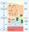
Nucleotide/P2 receptor system and tubuloglomerular feedback. Numbers in circles refer to the following sequence of events: 1) increase in concentration-dependent uptake of Na+, K+, and Cl via the furosemide-sensitive Na+-K+-2Cl cotransporter (NKCC2) in macula densa cells; 2) basolateral release of ATP; 3 and 4) transport-dependent, intra- and/or extracellular generation of adenosine (ADO); 5 and 6) the extracellular generation involves local ecto-NTPDase1 (CD39) and ecto-5′-nucleotidase (CD73); 7) extracellular ADO activates adenosine A1 receptors triggering an increase in cytosolic Ca2+ on extraglomerular mesangium cells (MC; not shown) and smooth muscle cells of the afferent arteriole (VSMC) resulting in afferent arteriolar vasoconstriction; 8) extracellular ATP activates P2 receptors on extraglomerular MC and increases cytosolic Ca2+; the intensive coupling between extraglomerular MC, intraglomerular MC (not shown), and VSMC of the afferent arteriole by gap junctions contributes to the propagation of the Ca2+ signal; 9 and 10) ATP release and P2 receptor activation also contribute to the cell to cell propagation of the Ca2+ signal, including to VSMC where activation of P2X1 receptors causes afferent arteriolar vasoconstriction; 11) extracellular ATP may also activate P2Y2 receptors on the basolateral membrane of macula densa cells with unknown function. See text for further explanations. [Modified from Vallon et al. (525).]
FIGURE 6.
FIGURE 6.
Regulation of renal transport processes by flow-induced and cilium-mediated nucleotide release. Model of ciliary-activated release of ATP, which in turn activates P2X and P2Y receptors to increase intracellular calcium from endoplasmatic reticulum (ER) stores and stimulation of inositol trisphosphate (IP3) receptor-mediated enhancement of, in this example, apical endocytosis in the proximal tubule. 1) Exposure to flow-induced shear stress (FSS) bends the apical cilium, which causes entry of extracellular Ca2+ via a ciliary-localized cation channel [possibly polycystin-2 (PC2)] thereby increasing intracellular Ca2+ ([Ca2+]i). 2) Bending of the primary cilium causes release of ATP into the tubular lumen (via nucleotide transporters or other mechanisms) which in turn activates P2Y or P2X receptors in the apical membrane and increases [Ca2+]i. 3) The latter enhances the endocytic capacity at the base of microvilli, possibly by modulating actin dynamics to increase the size and volume of individual apical clathrin-coated pits. Addition of ATP bypasses the need for FSS in enhancing endocytic capacity. [Modified from Raghavan et al. (420) and Raghavan and Weisz (421), with permission from Wolters Kluwer Health, Inc.]
FIGURE 7.
FIGURE 7.
P2Y2 receptor activation determines the inhibitory effect of ATP as well as of dietary salt on epithelial sodium channel (ENaC) open probability (Po) in connecting tubule (CNT)/cortical collecting duct (CCD). A: representative continuous current traces from cell-attached patches on principal cells (containing ≥2 ENaCs) before and after application of ATP (100 µM) in CNTs/CCDs harvested from wild-type (WT) and P2Y2−/− mice fed a regular 0.32% Na+ diet. Patches clamped to voltage potential of −60 mV and inward Li+ currents through ENaC are downward. Dashed lines indicate the respective current levels with c denoting the closed state. The summary graph on the right shows that ATP reduced ENaC Po, which was blunted in the absence of P2Y2 receptors. *P < 0.05 vs. wild type (WT). B: an increase in dietary NaCl intake for 1 wk reduced ENaC Po in WT. This effect was absent in P2Y2−/− mice. *P < 0.05 vs. WT. C: inhibition of local ATP signaling [hexokinase (+glucose) to degrade local ATP plus suramin to prevent P2 receptor activation] prevented the regulation of ENaC Po by dietary NaCl intake in cell-attached patches from WT mice. *P < 0.05 vs. WT control. [Modified from Pochynyuk et al. (405), with permission from FASEB; permission conveyed through Copyright Clearance Center, Inc.]
FIGURE 8.
FIGURE 8.
The inhibition of epithelial sodium channel (ENaC) open probability (Po) by dietary salt is mediated by the ATP/UTP/Cx30/P2Y2 receptor system in the aldosterone-sensitive distal nephron. Low salt intake enhances aldosterone concentrations and suppresses apical release of ATP and UTP. This reduces activation of apical P2Y2 receptors on principal cells (PC), and the inner leaflet of the lipid bilayer maintains a high concentration of negatively charged phosphatidylinositol 4,5-bisphosphate (PIP2), which binds to positively charged domains of the NH2 terminus of the β-subunit of ENaC, thereby keeping the ENaC channel open to reabsorb Na+. High salt intake enhances the apical release of ATP and UTP, at least in part via connexin30 (Cx30) in intercalated cells (IC). The nucleotides activate P2Y2 receptors and, thereby, phospholipase C (PLC), which hydrolyzes and lowers the concentration of negatively charged PIP2. This releases the NH2 terminus of β-ENaC and induces a conformation change that lowers ENaC Po and activity, thereby increasing Na+ excretion. See text for details. IP3, inositol trisphosphate. [Modified from Vallon and Rieg (527).]
FIGURE 9.
FIGURE 9.
A proposed model for cell volume-dependent ATP release and water transport inhibition by P2Y2 receptor activation in inner medullary collecting duct (IMCD) cells. Arginine vasopressin (AVP) activates vasopressin V2 receptor (V2R) to stimulate aquaporin-2-mediated water entry which increases cell volume. The latter increases basolateral and apical release of ATP (and potentially UTP?) by unknown mechanisms. P2Y2 receptor activation inhibits water reabsorption via multiple signaling pathways (see text for details) and stimulates volume regulatory potassium channels as shown in other cell types. In response to water loading, the ATP/UTP/P2Y2 receptor system maintains cell volume, which tends to increase by the relative hypotonic extracellular fluid, and accelerates the excretion of free water before circulating AVP levels drop. AC, adenylyl cyclase; AQP2, -3, -4, aquaporin-2, -3, -4; COX1, cyclooxygenase 1; CREB, cAMP responsive element binding protein; Cx37, connexin 37; DAG, diacylglycerol; EP3, PGE2 E3 receptor; ET-1, endothelin-1; ETB, endothelin B receptor; Gi, inhibitory G protein; Gq, Gs, stimulatory G protein; IP3, inositol trisphosphate; P256, P261, phosphorylation of AQP2; Panx1, pannexin 1; PGE2, prostaglandin E2; PLC, phospholipase C; PKA, protein kinase A; PKC, protein kinase C; Ub, ubiquitin; UT-A1, urea transporter A1. [Modified from Vallon and Rieg (527).]
FIGURE 10.
FIGURE 10.
A model of P2X7 receptor participation in cell proliferation, inflammation, and cell death in an archetypal cell, typically, though not exclusively, an immune cell, e.g., a macrophage. Release of mature interleukin (IL)-1β from macrophages requires a cellular priming signal delivered via Toll-like receptors in the case of microbial infection or inflammatory cytokine receptors (e.g., tumor necrosis factor receptor) in sterile inflammation, which leads to inflammatory gene transcription and accumulation of pro-IL-1β (‟Signal 1ˮ). P2X7 receptor activation requires relatively high local concentrations of ATP (in the high micromolar to millimolar range) usually released following cell injury, although localized cell surface concentrations may be sufficiently high in other settings. P2X7 receptor stimulation leads to an initial inorganic cation permeability when two ATP binding sites are occupied, progressing to pore formation when all three binding sites are occupied. An associated decrease in intracellular potassium is required for assembly of the NLRP3 inflammasome, the central caspase-1 activating platform. The P2X7 receptor then triggers opening of large-diameter membrane pores, including the pannexin-1 hemichannel. This event leads through still undefined mechanisms to activation of caspase-1, rapid maturation and release of IL-1β, and the related cytokine IL-18, and cell death (“Signal 2”). Elevations of cytosolic calcium in the absence of large membrane pore opening are thought to underlie the seemingly paradoxical mitogenic effect to P2X7 receptor activation seen in some settings (e.g., tumor growth). This P2X7 receptor-activated processes are believed to be important in a number of inflammatory diseases, including nephropathies in which inflammation, proliferation, and cell death may play a part, for example, glomerulonephritis, diabetic nephropathy, and polycystic kidney disease. See text for details. LPS, lipopolysaccharide; TNF, tumor necrosis factor; TLR, Toll-like receptor; TNF-R, tumor necrosis factor receptor; NMDG, N-methyl-d-glucamine. [Modified from Booth et al. (42), with permission from Dustri-Verlag.]

Similar articles

Cited by

References

    1. Abbracchio MP, Boeynaems JM, Barnard EA, Boyer JL, Kennedy C, Miras-Portugal MT, King BF, Gachet C, Jacobson KA, Weisman GA, Burnstock G. Characterization of the UDP-glucose receptor (re-named here the P2Y14 receptor) adds diversity to the P2Y receptor family. Trends Pharmacol Sci 24: 52–55, 2003. doi:10.1016/S0165-6147(02)00038-X. - DOI - PMC - PubMed
    1. Abbracchio MP, Burnstock G. Purinoceptors: are there families of P2X and P2Y purinoceptors? Pharmacol Ther 64: 445–475, 1994. doi:10.1016/0163-7258(94)00048-4. - DOI - PubMed
    1. Abbracchio MP, Burnstock G. Purinergic signalling: pathophysiological roles. Jpn J Pharmacol 78: 113–145, 1998. doi:10.1254/jjp.78.113. - DOI - PubMed
    1. Abbracchio MP, Burnstock G, Boeynaems JM, Barnard EA, Boyer JL, Kennedy C, Knight GE, Fumagalli M, Gachet C, Jacobson KA, Weisman GA. International Union of Pharmacology LVIII: update on the P2Y G protein-coupled nucleotide receptors: from molecular mechanisms and pathophysiology to therapy. Pharmacol Rev 58: 281–341, 2006. doi:10.1124/pr.58.3.3. - DOI - PMC - PubMed
    1. Abdelrahman A, Namasivayam V, Hinz S, Schiedel AC, Köse M, Burton M, El-Tayeb A, Gillard M, Bajorath J, de Ryck M, Müller CE. Characterization of P2X4 receptor agonists and antagonists by calcium influx and radioligand binding studies. Biochem Pharmacol 125: 41–54, 2017. doi:10.1016/j.bcp.2016.11.016. - DOI - PubMed

Publication types

LinkOut - more resources