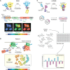
Genetically encoded fluorescent biosensors illuminate kinase signaling in cancer
- PMID: 31434714
- PMCID: PMC6779441
- DOI: 10.1074/jbc.REV119.006177
Genetically encoded fluorescent biosensors illuminate kinase signaling in cancer
Abstract
Protein kinase signaling networks stringently regulate cellular processes, such as proliferation, motility, and cell survival. These networks are also central to the evolution and progression of cancer. Accordingly, genetically encoded fluorescent biosensors capable of directly illuminating the spatiotemporal dynamics of kinase signaling in live cells are being increasingly used to investigate kinase signaling in cancer cells and tumor tissue sections. These biosensors enable visualization of biological processes and events directly in situ, preserving the native biological context and providing detailed insight into their localization and dynamics in cells. Herein, we first review common design strategies for kinase activity biosensors, including signaling targets, biosensor components, and fluorescent proteins involved. Subsequently, we discuss applications of biosensors to study the biology and management of cancer. These versatile molecular tools have been deployed to study oncogenic kinase signaling in living cells and image kinase activities in tumors or to decipher the mechanisms of anticancer drugs. We anticipate that the diversity and precision of genetically encoded biosensors will expand their use to further unravel the dysregulation of kinase signaling in cancer and the modes of actions of cancer-targeting drugs.
Keywords: biosensor; cancer; cell signaling; fluorescence resonance energy transfer (FRET); fluorescent protein; in vivo imaging; kinase signaling; phosphorylation; posttranslational modification.
© 2019 Lin et al.
Conflict of interest statement
The authors declare that they have no conflicts of interest with the contents of this article
Figures


Similar articles
-
Fluorescent biosensors - probing protein kinase function in cancer and drug discovery.Biochim Biophys Acta. 2013 Jul;1834(7):1387-95. doi: 10.1016/j.bbapap.2013.01.025. Epub 2013 Feb 1. Biochim Biophys Acta. 2013. PMID: 23376184 Review.
-
Illuminating the kinome: Visualizing real-time kinase activity in biological systems using genetically encoded fluorescent protein-based biosensors.Curr Opin Chem Biol. 2020 Feb;54:63-69. doi: 10.1016/j.cbpa.2019.11.005. Epub 2020 Jan 3. Curr Opin Chem Biol. 2020. PMID: 31911398 Free PMC article. Review.
-
Multiplexed visualization of dynamic signaling networks using genetically encoded fluorescent protein-based biosensors.Pflugers Arch. 2013 Mar;465(3):373-81. doi: 10.1007/s00424-012-1175-y. Epub 2012 Nov 9. Pflugers Arch. 2013. PMID: 23138230 Free PMC article. Review.
-
Genetically encoded molecular probes to visualize and perturb signaling dynamics in living biological systems.J Cell Sci. 2014 Mar 15;127(Pt 6):1151-60. doi: 10.1242/jcs.099994. J Cell Sci. 2014. PMID: 24634506 Free PMC article. Review.
-
Fluorescent biosensors illuminate the spatial regulation of cell signaling across scales.Biochem J. 2023 Oct 31;480(20):1693-1717. doi: 10.1042/BCJ20220223. Biochem J. 2023. PMID: 37903110 Free PMC article. Review.
Cited by
-
Enhanced kinase translocation reporters for simultaneous real-time measurement of PKA, ERK, and Ca2.bioRxiv [Preprint]. 2024 Oct 2:2024.09.30.615856. doi: 10.1101/2024.09.30.615856. bioRxiv. 2024. Update in: J Biol Chem. 2025 Jan 13:108183. doi: 10.1016/j.jbc.2025.108183 PMID: 39411162 Free PMC article. Updated. Preprint.
-
The Cutting Edge of Disease Modeling: Synergy of Induced Pluripotent Stem Cell Technology and Genetically Encoded Biosensors.Biomedicines. 2021 Aug 5;9(8):960. doi: 10.3390/biomedicines9080960. Biomedicines. 2021. PMID: 34440164 Free PMC article. Review.
-
Drug Screening with Genetically Encoded Fluorescent Sensors: Today and Tomorrow.Int J Mol Sci. 2020 Dec 25;22(1):148. doi: 10.3390/ijms22010148. Int J Mol Sci. 2020. PMID: 33375682 Free PMC article. Review.
-
Expanding the Toolkit of Fluorescent Biosensors for Studying Mitogen Activated Protein Kinases in Plants.Int J Mol Sci. 2020 Jul 28;21(15):5350. doi: 10.3390/ijms21155350. Int J Mol Sci. 2020. PMID: 32731410 Free PMC article.
-
A Novel Single-Color FRET Sensor for Rho-Kinase Reveals Calcium-Dependent Activation of RhoA and ROCK.Sensors (Basel). 2024 Oct 26;24(21):6869. doi: 10.3390/s24216869. Sensors (Basel). 2024. PMID: 39517770 Free PMC article.
References
-
- Wilson L. J., Linley A., Hammond D. E., Hood F. E., Coulson J. M., MacEwan D. J., Ross S. J., Slupsky J. R., Smith P. D., Eyers P. A., and Prior I. A. (2018) New perspectives, opportunities, and challenges in exploring the human protein kinome. Cancer Res. 78, 15–29 10.1158/0008-5472.CAN-17-2291 - DOI - PubMed
-
- Hornbeck P. V., Kornhauser J. M., Latham V., Murray B., Nandhikonda V., Nord A., Skrzypek E., Wheeler T., Zhang B., and Gnad F. (2019) 15 years of PhosphoSitePlus® integrating post-translationally modified sites, disease variants and isoforms. Nucleic Acids Res. 47, D433–D441 10.1093/nar/gky1159 - DOI - PMC - PubMed

