

X chromosome inactivation does not necessarily determine the severity of the phenotype in Rett syndrome patients
- PMID: 31427717
- PMCID: PMC6700087
- DOI: 10.1038/s41598-019-48385-w
X chromosome inactivation does not necessarily determine the severity of the phenotype in Rett syndrome patients
Abstract
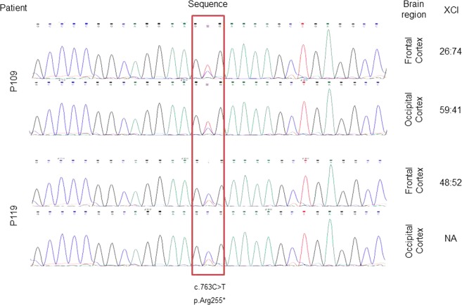
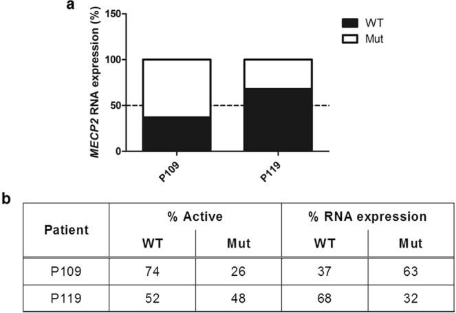
Rett syndrome (RTT) is a severe neurological disorder usually caused by mutations in the MECP2 gene. Since the MECP2 gene is located on the X chromosome, X chromosome inactivation (XCI) could play a role in the wide range of phenotypic variation of RTT patients; however, classical methylation-based protocols to evaluate XCI could not determine whether the preferentially inactivated X chromosome carried the mutant or the wild-type allele. Therefore, we developed an allele-specific methylation-based assay to evaluate methylation at the loci of several recurrent MECP2 mutations. We analyzed the XCI patterns in the blood of 174 RTT patients, but we did not find a clear correlation between XCI and the clinical presentation. We also compared XCI in blood and brain cortex samples of two patients and found differences between XCI patterns in these tissues. However, RTT mainly being a neurological disease complicates the establishment of a correlation between the XCI in blood and the clinical presentation of the patients. Furthermore, we analyzed MECP2 transcript levels and found differences from the expected levels according to XCI. Many factors other than XCI could affect the RTT phenotype, which in combination could influence the clinical presentation of RTT patients to a greater extent than slight variations in the XCI pattern.
Conflict of interest statement
The authors declare no competing interests.
Figures
Similar articles
-
[X chromosome inactivation patterns in patients with Rett syndrome and their mothers and the parental origin of the priority inactive X chromosome].Zhonghua Er Ke Za Zhi. 2006 Sep;44(9):648-52. Zhonghua Er Ke Za Zhi. 2006. PMID: 17217653 Chinese.
-
MECP2 mutations in Czech patients with Rett syndrome and Rett-like phenotypes: novel mutations, genotype-phenotype correlations and validation of high-resolution melting analysis for mutation scanning.J Hum Genet. 2016 Jul;61(7):617-25. doi: 10.1038/jhg.2016.19. Epub 2016 Mar 17. J Hum Genet. 2016. PMID: 26984561
-
X chromosome inactivation in Rett Syndrome and its correlations with MECP2 mutations and phenotype.J Child Neurol. 2008 Jan;23(1):22-5. doi: 10.1177/0883073807307077. J Child Neurol. 2008. PMID: 18184939
-
Two new Rett syndrome families and review of the literature: expanding the knowledge of MECP2 frameshift mutations.Orphanet J Rare Dis. 2011 Aug 30;6:58. doi: 10.1186/1750-1172-6-58. Orphanet J Rare Dis. 2011. PMID: 21878110 Free PMC article. Review.
-
The role of X-chromosome inactivation in the manifestation of Rett syndrome.Brain Dev. 2001 Dec;23 Suppl 1:S182-5. doi: 10.1016/s0387-7604(01)00362-x. Brain Dev. 2001. PMID: 11738869 Review.
Cited by
-
Unraveling Molecular Pathways Altered in MeCP2-Related Syndromes, in the Search for New Potential Avenues for Therapy.Biomedicines. 2021 Feb 3;9(2):148. doi: 10.3390/biomedicines9020148. Biomedicines. 2021. PMID: 33546327 Free PMC article.
-
Rett syndrome.Nat Rev Dis Primers. 2024 Nov 7;10(1):84. doi: 10.1038/s41572-024-00568-0. Nat Rev Dis Primers. 2024. PMID: 39511247 Review.
-
Adenosinergic System and BDNF Signaling Changes as a Cross-Sectional Feature of RTT: Characterization of Mecp2 Heterozygous Mouse Females.Int J Mol Sci. 2023 Nov 13;24(22):16249. doi: 10.3390/ijms242216249. Int J Mol Sci. 2023. PMID: 38003438 Free PMC article.
-
Skewed X-inactivation is associated with retinal dystrophy in female carriers of RPGR mutations.Life Sci Alliance. 2023 Aug 4;6(10):e202201814. doi: 10.26508/lsa.202201814. Print 2023 Oct. Life Sci Alliance. 2023. PMID: 37541846 Free PMC article.
-
Molecular Research in Medical Genetics.Int J Mol Sci. 2022 Jun 14;23(12):6625. doi: 10.3390/ijms23126625. Int J Mol Sci. 2022. PMID: 35743065 Free PMC article.
References
Publication types
MeSH terms
Substances
LinkOut - more resources
Full Text Sources
Medical
