

An anti-inflammatory phenotype in visceral adipose tissue of old lean mice, augmented by exercise
- PMID: 31427677
- PMCID: PMC6700172
- DOI: 10.1038/s41598-019-48587-2
An anti-inflammatory phenotype in visceral adipose tissue of old lean mice, augmented by exercise
Abstract
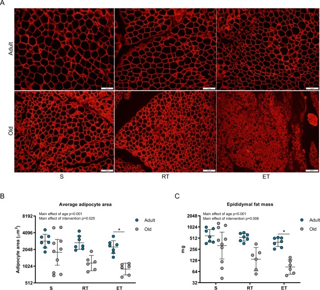
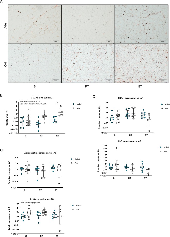
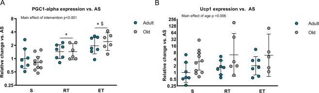
Visceral adipose tissue is an immunogenic tissue, which turns detrimental during obesity by activation of proinflammatory macrophages. During aging, chronic inflammation increases proportional to visceral adipose tissue (VAT) mass and associates with escalating morbidity and mortality. Here, we utilize a mouse model to investigate the inflammatory status of visceral adipose tissue in lean aging mice and assess the effects of exercise training interventions. We randomized adult (11 months; n = 21) and old (23 months; n = 27) mice to resistance training (RT) or endurance training (ET), or to a sedentary control group (S). Strikingly, we observed an anti-inflammatory phenotype in the old mice, consisting of higher accumulation of M2 macrophages and IL-10 expression, compared to the adult mice. In concordance, old mice also had less VAT mass and smaller adipocytes compared to adult mice. In both age groups, exercise training enhanced the anti-inflammatory phenotype and increased PGC1-α mRNA expression. Intriguingly, the brown adipose tissue marker UCP1 was modestly higher in old mice, while remained unchanged by the intervention. In conclusion, in the absence of obesity, visceral adipose tissue possesses a pronounced anti-inflammatory phenotype during aging which is further enhanced by exercise.
Conflict of interest statement
The authors declare no competing interests.
Figures
Similar articles
-
Exercise training inhibits inflammation in adipose tissue via both suppression of macrophage infiltration and acceleration of phenotypic switching from M1 to M2 macrophages in high-fat-diet-induced obese mice.Exerc Immunol Rev. 2010;16:105-18. Exerc Immunol Rev. 2010. PMID: 20839495
-
Fat Quality Matters: Distinct Proteomic Signatures Between Lean and Obese Cardiac Visceral Adipose Tissue Underlie its Differential Myocardial Impact.Cell Physiol Biochem. 2020 Apr 23;54(3):384-400. doi: 10.33594/000000226. Cell Physiol Biochem. 2020. PMID: 32319743
-
The "Big Bang" in obese fat: Events initiating obesity-induced adipose tissue inflammation.Eur J Immunol. 2015 Sep;45(9):2446-56. doi: 10.1002/eji.201545502. Epub 2015 Aug 19. Eur J Immunol. 2015. PMID: 26220361 Review.
-
Adipocyte-secreted exosomal microRNA-34a inhibits M2 macrophage polarization to promote obesity-induced adipose inflammation.J Clin Invest. 2019 Feb 1;129(2):834-849. doi: 10.1172/JCI123069. Epub 2019 Jan 22. J Clin Invest. 2019. PMID: 30667374 Free PMC article.
-
The Immune Landscape of Visceral Adipose Tissue During Obesity and Aging.Front Endocrinol (Lausanne). 2020 May 15;11:267. doi: 10.3389/fendo.2020.00267. eCollection 2020. Front Endocrinol (Lausanne). 2020. PMID: 32499756 Free PMC article. Review.
Cited by
-
The shades of grey in adipose tissue reprogramming.Biosci Rep. 2022 Mar 31;42(3):BSR20212358. doi: 10.1042/BSR20212358. Biosci Rep. 2022. PMID: 35211733 Free PMC article. Review.
-
Peroxisome proliferator-activated receptor gamma coactivator-1 (PGC-1) family in physiological and pathophysiological process and diseases.Signal Transduct Target Ther. 2024 Mar 1;9(1):50. doi: 10.1038/s41392-024-01756-w. Signal Transduct Target Ther. 2024. PMID: 38424050 Free PMC article. Review.
-
Effect of exercise on peritoneal microenvironment and progression of ovarian cancer.Am J Cancer Res. 2021 Oct 15;11(10):5045-5062. eCollection 2021. Am J Cancer Res. 2021. PMID: 34765311 Free PMC article.
-
Modifiable Cardiovascular Risk, Hematopoiesis, and Innate Immunity.Circ Res. 2020 Apr 24;126(9):1242-1259. doi: 10.1161/CIRCRESAHA.120.315936. Epub 2020 Apr 23. Circ Res. 2020. PMID: 32324501 Free PMC article. Review.
-
Analysis of Activity-Dependent Energy Metabolism in Mice Reveals Regulation of Mitochondrial Fission and Fusion mRNA by Voluntary Physical Exercise in Subcutaneous Fat from Male Marathon Mice (DUhTP).Cells. 2020 Dec 16;9(12):2697. doi: 10.3390/cells9122697. Cells. 2020. PMID: 33339143 Free PMC article.
References
Publication types
MeSH terms
LinkOut - more resources
Full Text Sources
Medical
