. 2019 Aug 5;3(8):e00157.
doi: 10.1002/pld3.157. eCollection 2019 Aug.

A tetraspanin gene regulating auxin response and affecting orchid perianth size and various plant developmental processes

Affiliations

A tetraspanin gene regulating auxin response and affecting orchid perianth size and various plant developmental processes

Wei-Hao Chen et al. Plant Direct. .

Abstract

The competition between L (lip) and SP (sepal/petal) complexes in P-code model determines the identity of complex perianth patterns in orchids. Orchid tetraspanin gene Auxin Activation Factor (AAF) orthologs, whose expression strongly correlated with the expansion and size of the perianth after P code established, were identified. Virus-induced gene silencing (VIGS) of OAGL6-2 in L complex resulted in smaller lips and the down-regulation of Oncidium OnAAF. VIGS of PeMADS9 in L complex resulted in the enlarged lips and up-regulation of Phalaenopsis PaAAF. Furthermore, the larger size of Phalaenopsis variety flowers was associated with higher PaAAF expression, larger and more cells in the perianth. Thus, a rule is established that whenever bigger perianth organs are made in orchids, higher OnAAF/PaAAF expression is observed after their identities are determined by P-code complexes. Ectopic expression Arabidopsis AtAAF significantly increased the size of flower organs by promoting cell expansion in transgenic Arabidopsis due to the enhancement of the efficiency of the auxin response and the subsequent suppression of the jasmonic acid (JA) biosynthesis genes (DAD1/OPR3) and BIGPETAL gene during late flower development. In addition, auxin-controlled phenotypes, such as indehiscent anthers, enhanced drought tolerance, and increased lateral root formation, were also observed in 35S::AtAAF plants. Furthermore, 35S::AtAAF root tips maintained gravitropism during auxin treatment. In contrast, the opposite phenotype was observed in palmitoylation-deficient AtAAF mutants. Our data demonstrate an interaction between the tetraspanin AAF and auxin/JA that regulates the size of flower organs and impacts various developmental processes.

Keywords: Arabidopsis thaliana; auxin response; orchids; perianth; tetraspanin.


Conflict of interest statement

The authors declare no conflict of interest associated with the work described in this manuscript.

Figures
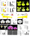
Figure 1
Figure 1
Functional analysis of OnAAF and PaAAF in Oncidium and Phalaenopsis orchids. (a) Total RNA samples isolated from the lips (L), petals (P), dorsal sepals (DS), and lateral sepals (LS) of mature Oncidium flowers were used as templates to detect the expression of OnAAF by quantitative real‐time PCR. (b) Detection of OAGL6‐2 and OnAAF expression in the lips of Oncidium floral bud (FB), open flower (OF), and mature flower (MF). (c) The flowers of Oncidium Lemon Heart peloric mutant (Trilips) with two petals transformed into lips (left), the OAGL6‐2 VIGS flower (right) containing green sepal/petal‐like sectors in the lips (arrow), which are smaller than those in the wild‐type control (Mock, middle). Bar = 10 mm. Top and bottom rows indicate the adaxial and abaxial side of the flower, respectively. (d) Detection of OnAAF expression in L, P, DS, and LS of mature flowers of Oncidium Lemon Heart peloric mutant (Trilips). (e) OnAAF expression was higher and promoted more cell division/expansion in lips than sepals/petals, resulting in the production of larger lips than sepals/petals in Oncidium orchids. (f) Detection of OAGL6‐2 and OnAAF expression in the lips of OAGL6‐2 VIGS (O6‐2‐V‐1 and 2) and wild‐type control (Mock) flowers of Oncidium Lemon Heart. (g) The flower of PeMADS9 (OAGL6‐2 ortholog) VIGS Phalaenopsis Sogo Yukidian “V3” (Pe9‐VIGS, right) contains lips, which are larger and more spread out than in the wild‐type control flower (Mock, left) Bar = 20 mm. (h) Detection of PaAAF expression in lips (Lip) and P of Phalaenopsis Sogo Yukidian “V3” FB and OF. (i) The front lobe (L–f) and side lobe (L‐s) of the lips from (g). Bar = 10 mm. (j) Detection of PeMADS9 (PeM9) and PaAAF expression in lips of PeMADS9 VIGS (PM9‐V) and wild‐type control (Mock) flowers of Phalaenopsis Sogo Yukidian “V3.” (k) The flowers of a Phalaenopsis peloric mutant (Big‐Lip) have much larger sepal/petal‐like L. (l) Detection of PaAAF expression in L, P, DS, and LS from the Big‐Lip mutant flower in (k). (m) Higher PaAAF expression promoted greater cell division/expansion in sepals/petals than in lips, resulting in the production of larger petals than lips in Phalaenopsis orchids. (n) Two varieties of Phalaenopsis orchids with relatively large (P. Red Bell) and small (P. Gold Diamond) mature flower sizes. Bar = 20 mm. (o) The size comparison of the P of P. Red Bell (RB) and P. Gold Diamond (GD) from (n). (p) The comparison of the epidermal cell size in the petals of P. Red Bell (RB) and P. Gold Diamond (GD) from (n). (q, r) SEM of the epidermal cells in the petals of P. Red Bell (q) and P. Gold Diamond (r) from (n). Bar = 100 μm. (s) The comparison of the total cell number in the petals of P. Red Bell (RB) and P. Gold Diamond (GD) from (n). (t) Detection of PaAAF expression in P. Red Bell (RB) and P. Gold Diamond (GD) FB, OF, and MF
Figure 2
Figure 2
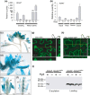
Analysis of the gene expression, protein localization, and palmitoylation for AtAAF in Arabidopsis. (a) The detection of AtAAF expression at 7 days after germination (DAG), 14 DAG, roots (Rt), rosette leaves (RL), cauline leaves (CL), floral buds (FB), and siliques (Sl). (b) The detection of AtAAF expression in sepal, petal, stamen, and carpel of stages 11 and 14 Arabidopsis flowers. (c) GUS staining pattern in floral buds (fb), mature flower (mf), and silique (sil) from an AtAAF::GUS transgenic plant. Bar = 1 mm. (d–f) GUS was detected in sepals (s), petals (p), and anther (an) of stage 10 (d) AtAAF::GUS flowers. GUS activity decreased in s and p and was absent in the anther after stage 12 (e, f). GUS was detected in ovules (o) after pollination (f). c, carpel. Bar = 1 mm. (g, h) Transient expression of 35S::GFP and 35S::AtAAF+GFP in tobacco cells. AtAAF+GFP fusion protein accumulated at the plasma membrane (h) whereas GFP protein accumulated in the cytosol (g). Bar = 30 μm. (i) Assaying palmitoylation of AtAAF and dominant negative mutant AtAAFpalm. Proteins were prepared from the 35S::AtAAF+GFP and 35S::AtAAFpalm +GFP plants by the biotin switch palmitoylation assay. Western blot analysis of proteins with the presence (+) and absence (−) of hydroxylamine treatment using a GFP antibody
Figure 3
Figure 3
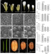
Functional analysis of flower and seed size for 35S::AtAAF and 35S::AtAAFpalm Arabidopsis. (a, b) The flower (a) and petals (b) of 35S::AtAAF (left), wild‐type (middle), and 35S::AtAAFpalm (right) Arabidopsis. Bar = 1 mm. (c) Size comparison of the petals from 35S::AtAAF (AtAAF), WT, and 35S::AtAAFpalm (palm) Arabidopsis. Petals from three flowers for each plant (AtAAF, WT, and palm) were used to measure the average size. The size of the wild‐type petals is set at 100%. (d, e) Comparison of total cell number (d) and the epidermal cell size (e) in petals from 35S::AtAAF (AtAAF), wild‐type (WT), and 35S::AtAAFpalm (palm) Arabidopsis. (f–k) Confocal laser scanning microscopy of the epidermal cells in the petals from 35S::AtAAF (f, g), WT (h, i), and 35S::AtAAFpalm (j, k) flowers. (g), (i), and (k) are close‐ups of (f), (h), and (j), respectively. Bar = 20 μm in (f, h, j) and 5 μm in (g, i, k). (l) The mature siliques of 35S::AtAAF (right) and wild‐type (WT, left) Arabidopsis. Bar = 5 mm. (m–o) The seeds of 35S::AtAAF (m), WT (n), and 35S::AtAAFpalm (o) Arabidopsis. Bar = 0.2 mm. (p) Comparison of the weight for 1,000 seeds from 35S::AtAAF (AtAAF), WT, and 35S::AtAAFpalm (palm) Arabidopsis. (q) Detection of AtAAF expression for one WT and two 35S::AtAAF (−1, −2) plant. The asterisks in (c, d, e, p) indicate a significant difference from the WT value (*p ≤ .05, **p ≤ .01). Statistic analysis was measured by Student's t test
Figure 4
Figure 4
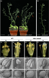
Phenotypic analysis of 35S::AtAAFpalm dominant negative mutant Arabidopsis. (a) Five‐week‐old 35S::AtAAFpalm Arabidopsis (right) produced smaller rosette leaves which showed earlier senescence (arrowed) than the wild‐type plants (left). Bar = 10 mm. (b, c) 35S::AtAAFpalm transgenic plants showed earlier senescence of rosette (b, right) and cauline leaves (c, right, arrow) than the wild‐type (WT, left). Bar = 5 mm. (d) Flowers from a severe 35S::AtAAFpalm inflorescence which showed earlier senescence than WT flowers. Bar = 1 mm. (e) Close‐up of the flowers from a severe 35S::AtAAFpalm inflorescence shown in (d). Bar = 1 mm. (f) Detection of expression for AtAAF, senescence‐related gene (SAG12), and the ethylene response genes (ERF1, EDF1, and EDF2) in floral buds (FB) before stage 12 from one WT plant and two severe 35S::AtAAFpalm plants (palm‐24 and palm‐10). Transcript levels in 35S::AtAAFpalm plants are presented relative to those in the wild‐type plant, which were set to 1. (g) Close‐up of the flowers from a medium–severe 35S::AtAAFpalm‐M inflorescence. The numbers indicate the positions of the open flowers. Bar = 1 mm. (h) Close‐up of the flowers from wild‐type (top row) and 35S::AtAAF (bottom row) inflorescences. The 35S::AtAAF flowers are clearly larger and abscised later than wild‐type flowers. The numbers indicate the positions of the open flowers. Bar = 1 mm. (i) Detection of AtAAF expression in FB and mature flowers (Fl) of one WT, two severe 35S::AtAAFpalm (palm‐10, ‐24), and one medium–severe 35S::AtAAFpalm (palm‐11) plants. (j) Detection of SAG12 expression in WT and two 35S::AtAAF (AtAAF‐14‐1 and AtAAF‐14‐2) plants
Figure 5
Figure 5
Phenotypic analysis of Arabidopsis plants ectopically expressing AtAAF. (a) A 35S::AtAAF plant (right) was sterile and produced short siliques (arrow), whereas wild‐type plants (WT, left) produced long, well‐developed siliques (arrow). Bar = 30 mm. (b) Inflorescences from a 35S::AtAAF plant that showed sterility with short siliques (arrow). Bar = 10 mm. (c–f) Indehiscent anthers (arrow) were observed at stage 12 in WT plants (c), stages 12 (e), and 14 (f) 35S::AtAAF flowers compared with stage 14 wild‐type flower (d), which showed normal anther dehiscence (arrow) and pollen (po) release. Bar = 0.5 mm. (g–j) Close‐up of stage 12 (g) wild‐type, stages 12 (i), and 14 (j) 35S::AtAAF anther compared with stage 14 wild‐type anther (h) by SEM, which showed normal anther dehiscence and pollen (arrow) release. Bar = 200 μm. (k–n) Close‐up of the stage 12 (k), 14 (l) wild‐type and stage 12 (m), 14 (n) 35S::AtAAF pollen grains by SEM. Bar = 10 µm
Figure 6
Figure 6
The analysis of drought and salt tolerance for 35S::AtAAF plants. (a, b) One‐week‐old wild‐type (a) and 35S::AtAAF (b) seedlings were tested for drought tolerance by removing them from MS medium and exposing them to a stream of air for 15, 30, 45, 60, 75, or 90 min. Bar = 5 mm. (c) The 35S::AtAAF (AtAAF) seedlings clearly withered later and lost less weight than wild‐type (WT) seedlings at 15, 30, and 60 min after drought treatment. In contrast, the 35S::AtAAFpalm (palm) seedlings clearly withered earlier and lost more weight than WT seedlings at 15, 30, and 60 min after drought treatment. (d) Comparison of germination for the 35S::AtAAF and WT seedlings grown on MS medium containing 150 mM NaCl. Bar = 10 mm. (e, f) Close‐up of the 35S::AtAAF (e) and WT (f) seedlings grown on MS medium containing 150 mM NaCl. Bar = 5 mm. (g) The survival rate for WT and two lines of 35S::AtAAF (‐1‐4, ‐1‐9) seedlings grown on MS medium containing 150 mM NaCl. (h, i) Detection of AtAAF expression after 15, 30, or 60 min of drought (h) or salt (i) treatment in wild‐type plants
Figure 7
Figure 7
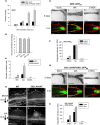
Investigation of the relationship between AtAAF and Auxin response in Arabidopsis. (a) Detection of GFP expression in DR5::GFP and DR5::GFP/35S::AtAAF transgenic Arabidopsis after 1, 2, and 4 hr of external IAA treatment. (b) Detection of the amount of IAA in wild‐type (WT), 35S::AtAAF (AAF), and 35S::AtAAFpalm (palm) flower buds (FB). (c) Number of lateral roots produced in WT and 35S::AtAAF (AtAAF) Arabidopsis seedlings grown on MS medium with or without IAA/2‐NOA (auxin influx inhibitor) treatments. (d) The development of the primordia (arrowhead) for lateral roots of the WT and 35S::AtAAF roots of 14 DAG seedlings 18 hr post‐gravitropic induction with (d‐3 and d‐4) or without (d‐1 and d‐2) IAA treatment. Stage I (d‐1), VII (d‐2), VI (d‐3), and beyond stage VIII (d‐4) primordia of lateral roots were formed. Bar = 50 μm. (e) Root gravitropic assays for DR5::GFP roots with 5 days of a 90‐degree gravitropic stimulus without (mock, e‐1) or with IAA (e‐3) or 2‐NOA (e‐5) treatment. Bar = 0.5 mm. The detection of GFP in the lower (L) and upper (U) parts of the DR5::GFP root tips 5 hr after 90‐degree gravitropic stimulus without (mock, e‐2) or with IAA (e‐4) or 2‐NOA (e‐6) treatment. Bar = 50 μm. (f) The detection of GFP integrated intensity in lower and upper parts of the DR5::GFP root tips from (e‐2), (e‐4) and (e‐6). (g) Root gravitropic assays for 35S::AtAAF/DR5::GFP roots after 5 days of a 90‐degree gravitropic stimulus without mock (g‐1) or with IAA (g‐3) or 2‐NOA (g‐5) treatment. Bar = 0.5 mm. The detection of GFP in lower (L) and upper (U) parts of the 35S::AtAAF/DR5::GFP root tips 5 hr after 90‐degree gravitropic stimulus without (mock, g‐2) or with IAA (g‐4) or 2‐NOA (g‐6) treatment. Bar = 50 μm. (h) The detection of GFP integrated intensity in lower and upper parts of the 35S::AtAAF/DR5::GFP root tips from (g‐2), (g‐4), and (g‐6)
Figure 8
Figure 8
Model for the function of AAF orthologues in regulating auxin response and development in plants. In plants, the targeting of palmitoylated AAF proteins to the plasma membrane promotes auxin uptake and enhances (→) the auxin response. In flowers, AAF is more highly expressed in early than in late developmental stages (gray bar). Thus, it mainly enhances the auxin response and promotes (green arrow) cell division/expansion in the flower organs at an early stage. Ectopic expression of AAF extends its affect to the whole flower and results in an increase in the size of the flower organs. By contrast, a palmitoylation‐deficient mutant of AAF reduces ([formula image]) the auxin response and alters its ability to promote cell division/expansion in early flowering stages resulting in a decrease in flower organ size. In addition, the enhancement of the auxin response by ectopic expression of AAF could suppress anther dehiscence during whole flower development by suppressing the expression of MYB26/NST1/2, DAD1/OPR3, and JA activity (which also causes the suppression of the BPEp and resulted in the expansion of the flower organs). The enhancement of the auxin response by AAF also suppresses ethylene signaling and organ senescence, promotes drought/salt tolerance and lateral root formation, and retains root tip gravitropism in plants. Dominant negative mutation of AAF suppresses auxin response and causes the opposite effect on the processes described above

Similar articles

Cited by

References

    1. Alexander, M. P. (1969). Differential staining of aborted and nonaborted pollen. Stain Technology, 44, 117–122. 10.3109/10520296909063335 - DOI - PubMed
    1. Alonso, J. M. , Stepanova, A. N. , Leisse, T. J. , Kim, C. J. , Chen, H. , Shinn, P. , … Ecker, J. R. (2003). Genome‐wide insertional mutagenesis of Arabidopsis thaliana . Science, 301, 653–657. 10.1126/science.1086391 - DOI - PubMed
    1. Armengot, L. , Marques‐Bueno, M. M. , & Jaillais, Y. (2016). Regulation of polar auxin transport by protein and lipid kinases. Journal of Experimental Botany, 67, 4015–4037. 10.1093/jxb/erw216 - DOI - PMC - PubMed
    1. Azooz, M. M. , Shaddad, M. A. , & Abdel‐Latef, A. A. (2004). Leaf growth and K+/Na+ ratio as an indication of the salt tolerance of three sorghum cultivars grown under salinity stress and IAA treatment. Acta Agronomica Hungarica, 52, 287–296.
    1. Benková, E. , Michniewicz, M. , Sauer, M. , Teichmann, T. , Seifertová, D. , Jürgens, G. , & Friml, J. (2003). Local, efflux‐dependent auxin gradients as a common module for plant organ formation. Cell, 115, 591–602. 10.1016/S0092-8674(03)00924-3 - DOI - PubMed

LinkOut - more resources