. 2019 Aug 12;9(1):11638.
doi: 10.1038/s41598-019-48225-x.

PD-1 of Sigmodon hispidus: Gene identification, characterization and preliminary evaluation of expression in inactivated RSV vaccine-induced enhanced respiratory disease

Affiliations

PD-1 of Sigmodon hispidus: Gene identification, characterization and preliminary evaluation of expression in inactivated RSV vaccine-induced enhanced respiratory disease

Abenaya Muralidharan et al. Sci Rep. .

Abstract

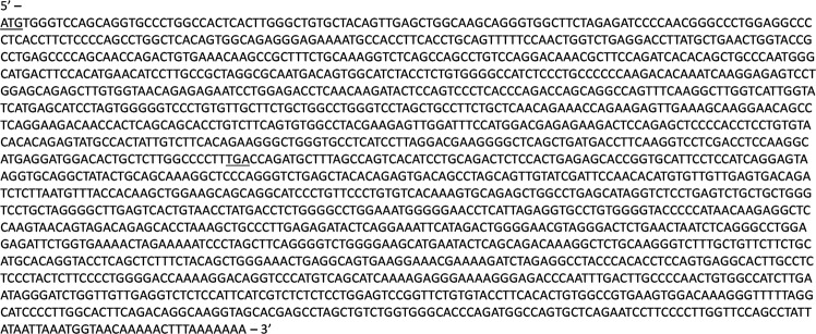
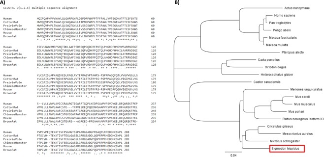
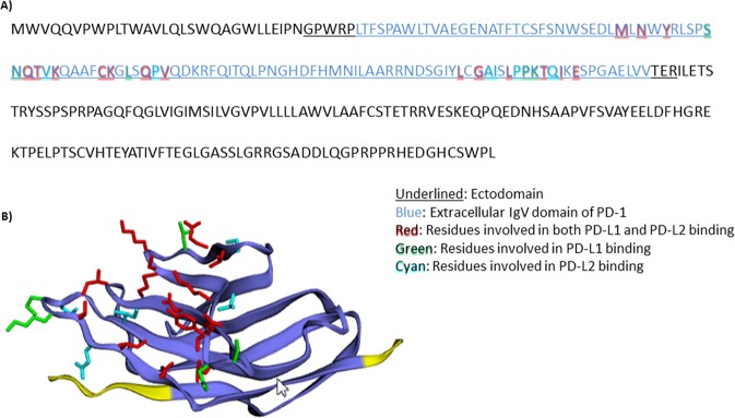
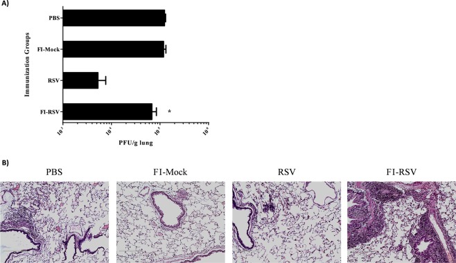
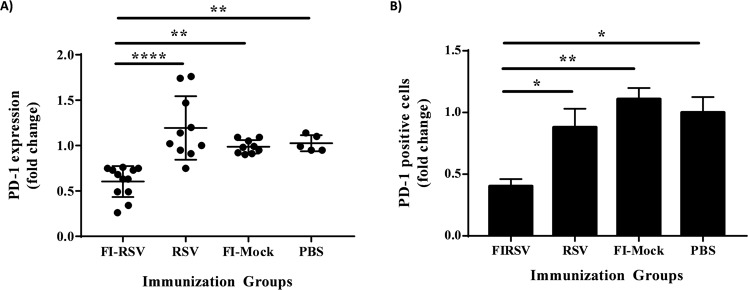
Sigmodon hispidus or cotton rat is an excellent animal model for studying human infections of respiratory viruses including respiratory syncytial virus (RSV), which is the leading cause of hospitalization in infants and causes high rates of infection in the elderly and immunocompromised patient populations. Despite several decades of research, no vaccine has been licensed whereas inactivated vaccines have been shown to induce severe adverse reaction in a clinical trial, with other forms of RSV vaccine also found to induce enhanced disease in preclinical animal studies. While arguably the cotton rat is the best small animal model for evaluation of RSV vaccines and antivirals, many important genes of the immune system remain to be isolated. Programmed cell death-1 (PD-1) plays an integral role in regulating many aspects of immunity by inducing suppressive signals. In this study, we report the isolation of mRNA encoding the cotton rat PD-1 (crPD-1) and characterization of the PD-1 protein. crPD-1 bound to its cognate ligand on dendritic cells and effectively suppressed cytokine secretion. Moreover, using the newly acquired gene sequence, we observed a decreased level of crPD-1 levels in cotton rats with enhanced respiratory disease induced by inactivated RSV vaccine, unraveling a new facet of vaccine-induced disease.


Conflict of interest statement

The authors declare no competing interests.

Figures

Figure 1
Figure 1
Cotton rat (Sigmodon hispidus) PD-1 mRNA sequence. 3′ RACE strategy was used on total RNA extracted from the spleen of a naïve cotton rat to determine the mRNA sequence. The predicted start and stop codon are underlined.
Figure 2
Figure 2
Protein sequence alignment of the cotton rat PD-1. (A) Protein sequence of closely related species and human were aligned with crPD-1 using the Clustal Omega tool from EMBL-EBI. Human (Homo sapiens NCBI Reference Sequence: AAC51773.1), Prairie Vole (Microtus ochrogaster NCBI Reference Sequence: XP_005361412.1), Chinese Hamster (Cricetulus griseus NCBI Reference Sequence: XP_003499314.1), Mouse (Mus musculus NCBI Reference Sequence: NP_032824.1), and Brown Rat (Rattus norvegicus NCBI Reference Sequence: XP_017451871.1). An asterisk (*) indicates positions which have a single, fully conserved residue. A colon (:) indicates conservation between groups of strongly similar properties, scoring >0.5 in the Gonnet PAM 250 matrix. A period (.) indicates conservation between groups of weakly similar properties, scoring = < 0.5 in the Gonnet PAM 250 matrix. (B) A phylogenetic tree was produced using Geneious software.
Figure 3
Figure 3
Identification of putative conserved domains in the cotton rat PD-1. (A) The underlined sequence indicates the putative ectodomain of PD-1; amino acids in blue are the putative extracellular IgV domain of PD-1; putative residues involved in both PD-L1 and PD-L2 binding are highlighted in red; putative residues involved in PD-L1 binding only are highlighted in green and residues involved in PD-L2 binding only in cyan. (B) Predicted structure of the putative ectodomain of the cotton rat PD-1 monomer shown in blue and yellow where the blue region is the putative extracellular IgV domain, residues in red are involved in both PD-L1 and PD-L2 binding, residues in green are involved in PD-L1 binding only and residues in cyan are involved in PD-L2 binding only.
Figure 4
Figure 4
Cotton rat PD-1 protein expression. crPD-1 gene also encoding rat codon optimized secretion signal and ten histidine residues at the 5′-end was synthesized and cloned into pcDNA3.1(+) vector. 293T cells were then transfected for 24 hours, the lysate was collected and his-tag purified. (A) Protein expression was confirmed with western blot using a mouse anti-histidine antibody. The expected size of crPD-1 is 36.4 kDa. A His-tag conjugated truncated recombinant mouse PD-1 (rmPD-1) was used as a positive control and, as expected, migrated from 25 to 45 kDa due to different glycosylation and may have aggregates depending on the reducing conditions. A full-length blot is shown as Supplementary Fig. S1. (B) Mass spectrometry was performed with his-tag purified lipofectamine control and crPD-1 along with rmPD-1. Seven peptides in the newly found sequence were found in the crPD-1 sample at high abundance and two peptides were found at low abundance with total sequence coverage of 41%. No peptides were found in the lipofectamine control, as expected and two peptides were found in the rmPD-1 sample. Mouse PD-1 sequence coverage for the positive control sample (rmPD-1) was 33.33%. (C) Immunofluorescence was also used for protein expression. Cells were permeabilized and stained 24 hours post-transfection. A rabbit anti-mouse PD-1 with Cy2-conjugated anti-rabbit IgG and mouse anti-his tag with Alexa Fluor 555 anti-mouse IgG were used. Representative image of the stained cells at 20X magnification is shown. A merge of the two fluorochromes shows the co-expression of PD-1 and the his-tag, as expected.
Figure 5
Figure 5
crPD-1 binds PD-L1 on dendritic cells in vitro. Purified crPD-1 was added to mouse dendritic cells for 4 hours. Recombinant mouse PD-1 (rmPD-1) and no treatment controls were used. (A) The cells were then stained with a fixable viability dye and PE-conjugated anti-human/mouse PD-L1 blocking antibody for flow cytometry analysis. The schematic displays the strategy used (top left). The mean fluorescence intensity (MFI) of PD-L1 among viable cells is shown. Statistical difference between PD-1 treated and no treatment group is indicated. Data shown is mean ± SEM representative of 2 independent experiments; n = 3 per treatment in each experiment; *p < 0.05 (one-way ANOVA with Bonferroni posttest). (B) The cells were also stained with rabbit anti-mouse PD-1 primary antibody along with a PE-conjugated anti-rabbit secondary antibody for flow cytometry analysis. The schematic displays the strategy used (bottom left). A histogram of the results representative of 2 independent experiments is shown.
Figure 6
Figure 6
FI-RSV immunization of cotton rats results in ineffective viral clearance with pronounced ERD. Cotton rats were immunized twice 21 days apart with FI-RSV, FI-Mock or PBS intramuscularly or wild-type RSV-A2 intranasally. Four weeks following second immunization, the animals were challenged with RSV-A2 intranasally and euthanized 5 days post-challenge for collection of lungs. (A) Lung viral titer determined using plaque assay post challenge. (B) Representative images of H&E stained cotton rat lungs post challenge at 20X magnification. Data shown is mean ± SEM representative of 2 independent experiments; n = 3 per group in each experiment; *p < 0.05 (one-way ANOVA with Bonferroni posttest). FI-RSV: Formaldehyde-inactivated RSV; FI-Mock: Formaldehyde-inactivated cell control; PBS: Phosphate-buffered saline.
Figure 7
Figure 7
PD-1 gene and protein expression is downregulated in FI-RSV immunized cotton rats experiencing ERD. Lungs were collected from twice-immunized and challenged cotton rats. (A) RNA isolated from the lungs were analyzed for PD-1 gene expression using quantitative real-time PCR. CT values were first normalized to β-actin, then, presented as fold change over no immunization control groups, i.e., FI-Mock and PBS that were similar in PD-1 expression levels. (B) Charged slides made from lungs fixed in 10% formalin were trimmed, processed and embedded into paraffin blocks were used for immunohistochemistry analysis of PD-1 protein expression using a mouse PD-1 antibody. The percentage of PD-1 positive cells is presented as fold change over no immunization control groups, i.e., FI-Mock and PBS. Data shown is mean ± SEM representative of 2 independent experiments; n = 10 per group in (A) and n = 3 per group in (B); *p < 0.05, **p < 0.01, ****p < 0.0001 (one-way ANOVA with Bonferroni posttest). FI-RSV: Formaldehyde-inactivated RSV; FI-Mock: Formaldehyde-inactivated cell control; PBS: Phosphate-buffered saline.

Similar articles

References

    1. Jin H-T, Ahmed R, Okazaki T. Role of PD-1 in Regulating T-Cell. Immunity. 2010;350:17–37. - PubMed
    1. Zhang X, et al. Structural and functional analysis of the costimulatory receptor programmed death-1. Immunity. 2004;20:337–47. doi: 10.1016/S1074-7613(04)00051-2. - DOI - PubMed
    1. Nishimura H, Honjo T, Minato N. Facilitation of beta selection and modification of positive selection in the thymus of PD-1-deficient mice. J. Exp. Med. 2000;191:891–8. doi: 10.1084/jem.191.5.891. - DOI - PMC - PubMed
    1. Agata Y, et al. Expression of the PD-1 antigen on the surface of stimulated mouse T and B lymphocytes. Int. Immunol. 1996;8:765–72. doi: 10.1093/intimm/8.5.765. - DOI - PubMed
    1. Nishimura H, et al. Developmentally regulated expression of the PD-1 protein on the surface of double-negative (CD4-CD8-) thymocytes. Int. Immunol. 1996;8:773–80. doi: 10.1093/intimm/8.5.773. - DOI - PubMed

Publication types

MeSH terms