Virophages of Giant Viruses: An Update at Eleven
- PMID: 31398856
- PMCID: PMC6723459
- DOI: 10.3390/v11080733
Virophages of Giant Viruses: An Update at Eleven
Abstract
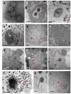
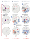
The last decade has been marked by two eminent discoveries that have changed our perception of the virology field: The discovery of giant viruses and a distinct new class of viral agents that parasitize their viral factories, the virophages. Coculture and metagenomics have actively contributed to the expansion of the virophage family by isolating dozens of new members. This increase in the body of data on virophage not only revealed the diversity of the virophage group, but also the relevant ecological impact of these small viruses and their potential role in the dynamics of the microbial network. In addition, the isolation of virophages has led us to discover previously unknown features displayed by their host viruses and cells. In this review, we present an update of all the knowledge on the isolation, biology, genomics, and morphological features of the virophages, a decade after the discovery of their first member, the Sputnik virophage. We discuss their parasitic lifestyle as bona fide viruses of the giant virus factories, genetic parasites of their genomes, and then their role as a key component or target for some host defense mechanisms during the tripartite virophage-giant virus-host cell interaction. We also present the latest advances regarding their origin, classification, and definition that have been widely discussed.
Keywords: coculture; giant virus; host-defense systems; metagenomic; satellite virus; virophage.
Conflict of interest statement
The authors declare no conflict of interest.
Figures


Similar articles
-
Amoebae, Giant Viruses, and Virophages Make Up a Complex, Multilayered Threesome.Front Cell Infect Microbiol. 2018 Jan 11;7:527. doi: 10.3389/fcimb.2017.00527. eCollection 2017. Front Cell Infect Microbiol. 2018. PMID: 29376032 Free PMC article. Review.
-
Virophages: association with human diseases and their predicted role as virus killers.Pathog Dis. 2021 Oct 23;79(8):ftab049. doi: 10.1093/femspd/ftab049. Pathog Dis. 2021. PMID: 34601577 Review.
-
Isolation and Identification of a Large Green Alga Virus (Chlorella Virus XW01) of Mimiviridae and Its Virophage (Chlorella Virus Virophage SW01) by Using Unicellular Green Algal Cultures.J Virol. 2022 Apr 13;96(7):e0211421. doi: 10.1128/jvi.02114-21. Epub 2022 Mar 9. J Virol. 2022. PMID: 35262372 Free PMC article.
-
Polintons, virophages and transpovirons: a tangled web linking viruses, transposons and immunity.Curr Opin Virol. 2017 Aug;25:7-15. doi: 10.1016/j.coviro.2017.06.008. Epub 2017 Jun 30. Curr Opin Virol. 2017. PMID: 28672161 Free PMC article. Review.
-
A virophage cross-species infection through mutant selection represses giant virus propagation, promoting host cell survival.Commun Biol. 2020 May 21;3(1):248. doi: 10.1038/s42003-020-0970-9. Commun Biol. 2020. PMID: 32439847 Free PMC article.
Cited by
-
Virophages Found in Viromes from Lake Baikal.Biomolecules. 2023 Dec 11;13(12):1773. doi: 10.3390/biom13121773. Biomolecules. 2023. PMID: 38136644 Free PMC article.
-
Updated Virophage Taxonomy and Distinction from Polinton-like Viruses.Biomolecules. 2023 Jan 19;13(2):204. doi: 10.3390/biom13020204. Biomolecules. 2023. PMID: 36830574 Free PMC article.
-
Virophages, Satellite Viruses, Virophage Replication and Its Effects and Virophage Defence Mechanisms for Giant Virus Hosts and Giant Virus Defence Systems against Virophages.Int J Mol Sci. 2024 May 28;25(11):5878. doi: 10.3390/ijms25115878. Int J Mol Sci. 2024. PMID: 38892066 Free PMC article. Review.
-
Novel Cell-Virus-Virophage Tripartite Infection Systems Discovered in the Freshwater Lake Dishui Lake in Shanghai, China.J Virol. 2020 May 18;94(11):e00149-20. doi: 10.1128/JVI.00149-20. Print 2020 May 18. J Virol. 2020. PMID: 32188734 Free PMC article.
-
Diverse Trajectories Drive the Expression of a Giant Virus in the Oomycete Plant Pathogen Phytophthora parasitica.Front Microbiol. 2021 Jun 1;12:662762. doi: 10.3389/fmicb.2021.662762. eCollection 2021. Front Microbiol. 2021. PMID: 34140938 Free PMC article.
References
Publication types
MeSH terms
LinkOut - more resources
Full Text Sources

