. 2019 Oct 15;93(21):e01290-19.
doi: 10.1128/JVI.01290-19. Print 2019 Nov 1.

Identification of the Capsid Binding Site in the Herpes Simplex Virus 1 Nuclear Egress Complex and Its Role in Viral Primary Envelopment and Replication

Affiliations

Identification of the Capsid Binding Site in the Herpes Simplex Virus 1 Nuclear Egress Complex and Its Role in Viral Primary Envelopment and Replication

Kosuke Takeshima et al. J Virol. .

Abstract

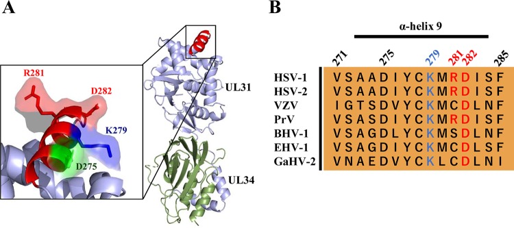
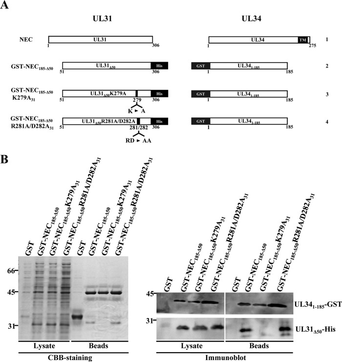
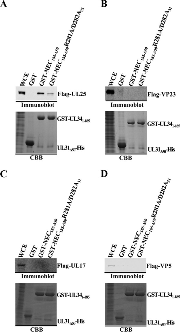
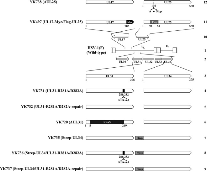
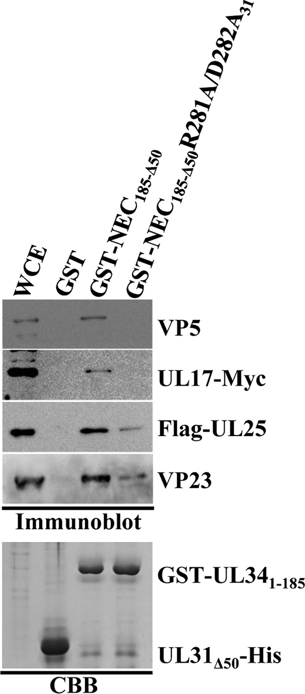
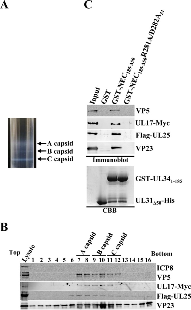
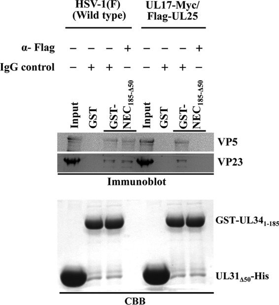
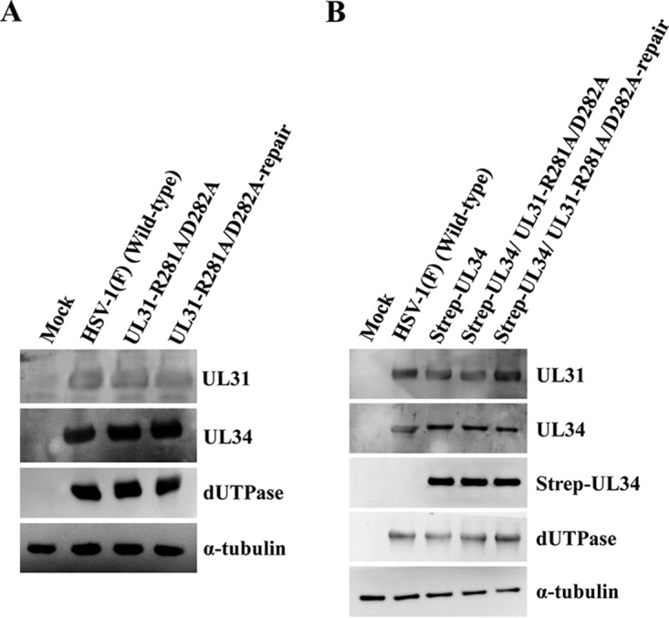
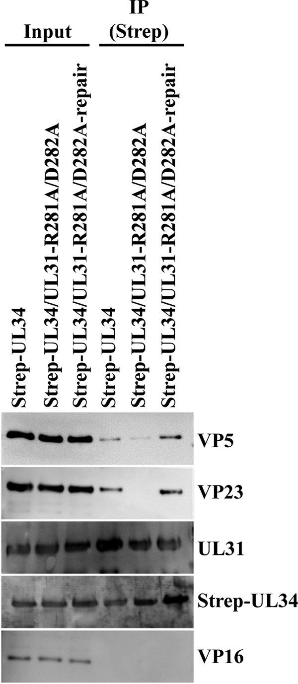
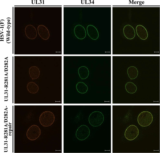
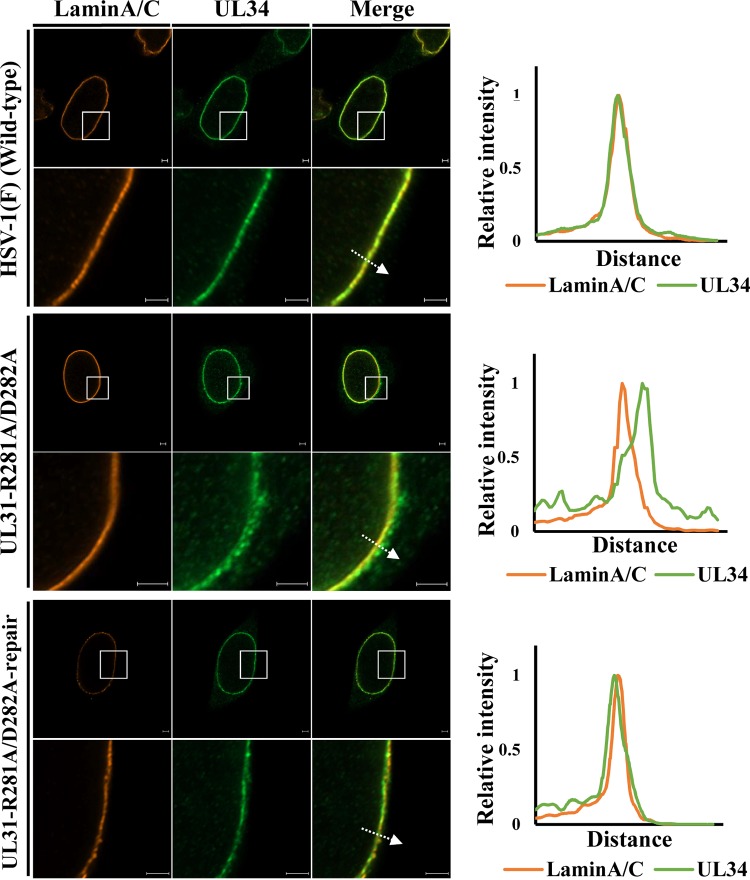
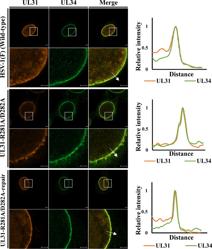
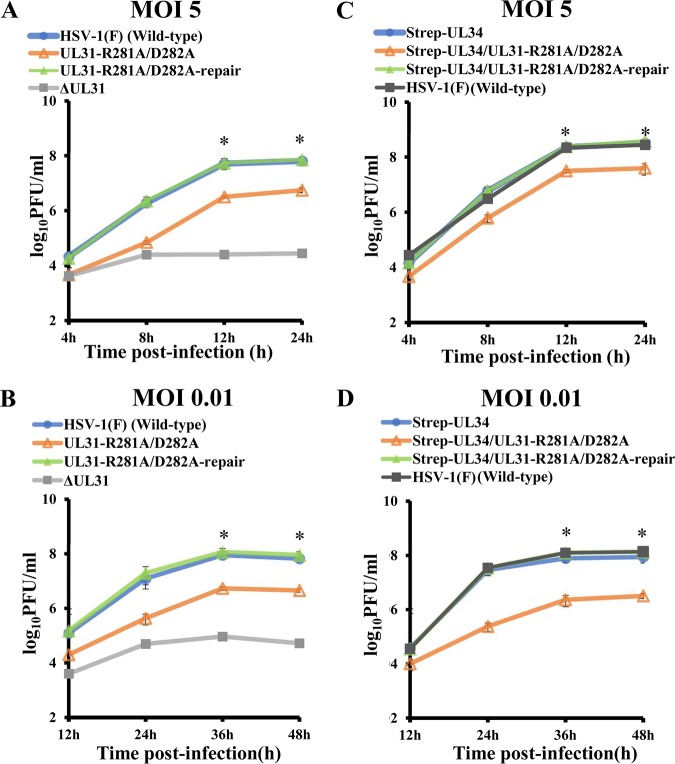
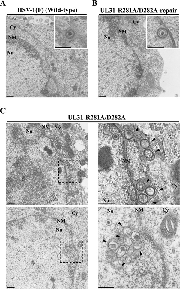
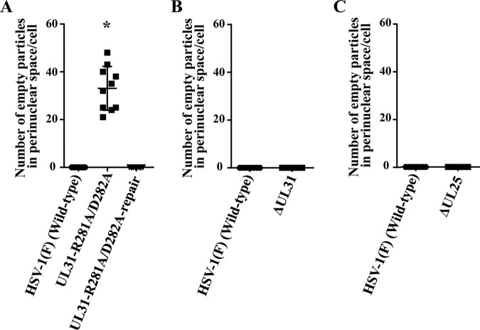
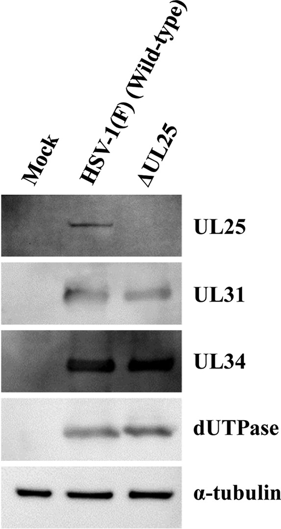
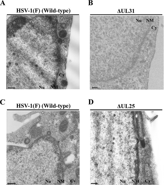
During nuclear egress of nascent progeny herpesvirus nucleocapsids, the nucleocapsids acquire a primary envelope by budding through the inner nuclear membrane of infected cells into the perinuclear space between the inner and outer nuclear membranes. Herpes simplex virus 1 (HSV-1) UL34 and UL31 proteins form a nuclear egress complex (NEC) and play critical roles in this budding process, designated primary envelopment. To clarify the role of NEC binding to progeny nucleocapsids in HSV-1 primary envelopment, we established an assay system for HSV-1 NEC binding to nucleocapsids and capsid proteins in vitro Using this assay system, we showed that HSV-1 NEC bound to nucleocapsids and to capsid protein UL25 but not to the other capsid proteins tested (i.e., VP5, VP23, and UL17) and that HSV-1 NEC binding of nucleocapsids was mediated by the interaction of NEC with UL25. UL31 residues arginine-281 (R281) and aspartic acid-282 (D282) were required for efficient NEC binding to nucleocapsids and UL25. We also showed that alanine substitution of UL31 R281 and D282 reduced HSV-1 replication, caused aberrant accumulation of capsids in the nucleus, and induced an accumulation of empty vesicles that were similar in size and morphology to primary envelopes in the perinuclear space. These results suggested that NEC binding via UL31 R281 and D282 to nucleocapsids, and probably to UL25 in the nucleocapsids, has an important role in HSV-1 replication by promoting the incorporation of nucleocapsids into vesicles during primary envelopment.IMPORTANCE Binding of HSV-1 NEC to nucleocapsids has been thought to promote nucleocapsid budding at the inner nuclear membrane and subsequent incorporation of nucleocapsids into vesicles during nuclear egress of nucleocapsids. However, data to directly support this hypothesis have not been reported thus far. In this study, we have present data showing that two amino acids in the membrane-distal face of the HSV-1 NEC, which contains the putative capsid binding site based on the solved NEC structure, were in fact required for efficient NEC binding to nucleocapsids and for efficient incorporation of nucleocapsids into vesicles during primary envelopment. This is the first report showing direct linkage between NEC binding to nucleocapsids and an increase in nucleocapsid incorporation into vesicles during herpesvirus primary envelopment.

Keywords: capsid; herpes simplex virus; nuclear egress.


Figures

FIG 1
FIG 1
Location of potential capsid binding sites in HSV-1 UL31 and comparison of the amino acid sequences of HSV-1 UL31 helix 9 and the corresponding domains of other alphaherpesvirus UL31 homologs. (A) In the HSV-1 NEC structure (22), UL31 is shown in light blue, and UL34 is shown in pale green. Helix 9 in UL31 is shown in red. The boxed area is enlarged to show the side chains of UL31 helix 9, with the amino acids in green (D275), red (R281 and D282), and blue (K279). Molecular graphics and analyses were performed with the PyMOL molecular graphics system, version 2.0.6 (Schrödinger, LLC). (B) Alignment of the amino acid sequences in HSV-1 UL31 helix 9 and the corresponding domains in alphaherpesvirus UL31 homologs, i.e., HSV-1 (GenBank accession no. CAA32324); herpes simplex virus 2 (HSV-2; GenBank accession no. CAB06756); VZV, varicella-zoster virus (NCBI RefSeq accession no. NP_040150); PrV (GenBank accession no. AFI70796); bovine herpesvirus 1 (BHV-1; NCBI RefSeq accession no. NP_045327); equine herpesvirus 1 (EHV-1; GenBank accession no. AAT67286); and gallid herpesvirus 2 (GaHV-2; GenBank accession no. AAF66766). The mutations investigated in this study are in red (HSV-1 residues R281 and D282) and blue (HSV-1 residue K279).
FIG 2
FIG 2
Purification of recombinant NECs. (A) Schematic diagrams of the wild-type and recombinant HSV-1 UL31 and UL34 viral proteins used in this study. Line 1, wild-type HSV-1 NEC UL31 and UL34; line 2, GST-NEC185-Δ50 fusion proteins; lines 3 and 4, GST-NEC185-Δ50 fusion proteins carrying a single substitution mutation in residue K279 (line 3) or a double mutation in residues R281 and D282 (line 4) of UL31Δ50. (B) GST, GST-NEC185-Δ50, and the two GST-NEC185-Δ50 mutants were expressed in E. coli, lysed, and precipitated using glutathione-Sepharose beads. The lysates and beads were analyzed by electrophoresis in a denaturing gel and then immunoblotted with anti-UL31 and anti-UL34 antisera or stained with CBB.
FIG 3
FIG 3
Effect of the mutations in UL31 R281/D282 on binding of recombinant NEC to capsid proteins from cells expressing each capsid protein. (A to D) 293FT cells were transfected with plasmids expressing either Flag-UL25 (A), Flag-VP23 (B), Flag-UL17 (C), or Flag-VP5 (D) for 24 h. These cells then were lysed and reacted with GST, GST-NEC185-Δ50, or GST-NEC185-Δ50R281A/D282A31 that was immobilized on glutathione-Sepharose beads for 1 h at 4°C. After extensive washing, the beads were divided into two parts. One part was analyzed by electrophoresis in a denaturing gel and immunoblotted with anti-Flag antibody (top gels), and the other part was analyzed by electrophoresis in a denaturing gel and stained with CBB (bottom gels).
FIG 4
FIG 4
Schematic diagrams of the genome structure of wild-type HSV-1(F) and the relevant domains of the recombinant viruses used in this study. Line 1, wild-type HSV-1(F) genome; line 2, domain of the UL30 gene to the UL34 gene; line 3, domains of the UL31 gene and the UL34 gene; lines 4 to 6, recombinant viruses with mutations in UL31; line 7, recombinant virus encoding Strep-tagged UL34, lines 8 and 9; recombinant viruses encoding Strep-tagged UL34 and carrying mutations in UL31; line 10, domains of the UL17 gene and the UL25 gene; line 11, recombinant virus encoding Myc-tagged UL17 and Flag-tagged UL25; and line 12, recombinant virus in which A106 in UL25 was substituted with a stop codon.
FIG 5
FIG 5
Effect of the UL31 R281/D282 mutations on recombinant NEC binding to capsid proteins from HSV-1-infected cells. The GST fusion proteins shown in Fig. 2 were immobilized on glutathione-Sepharose beads and reacted with lysates of Vero cells that had been infected with YK497 (UL17-Myc/Flag-UL25) at an MOI of 5 for 18 h. The beads were washed extensively and divided into two parts. One part was analyzed by electrophoresis in a denaturing gel and immunoblotted with anti-VP5, anti-Myc, anti-Flag, and anti-VP23 antibodies (top gels), and the other part was analyzed by electrophoresis in a denaturing gel and stained with CBB (bottom gel).
FIG 6
FIG 6
Effect of the UL31 R281 and D282 mutations on recombinant NEC binding to nucleocapsids. (A) Vero cells were infected with YK497 (UL17-Myc/Flag-UL25) at an MOI of 3 and harvested at 18 h postinfection. Nuclear lysates were isolated and layered onto 20% to 50% sucrose gradients and ultracentrifuged. The positions of type A, B, and C capsid bands in the sucrose gradient are indicated. (B) Proteins in the gradient fractions, shown in panel A, containing type A, B, or C capsids were analyzed by immunoblotting with the indicated antibodies. (C) Fractions containing C capsids were reacted with GST, GST-NEC185-Δ50, or GST-NEC185-Δ50R281A/D282A31 immobilized on glutathione-Sepharose beads for 1 h at 4°C. Beads were then extensively washed and divided into two parts. One part was analyzed by electrophoresis in a denaturing gel and immunoblotted with anti-VP5, anti-Myc, anti-Flag, and anti-VP23 antibodies (top gels), and the other was analyzed by electrophoresis in a denaturing gel and stained with CBB (bottom gel).
FIG 7
FIG 7
Blocking effect of anti-Flag antibody on recombinant NEC binding to nucleocapsids of the recombinant virus encoding Flag-tagged UL25. (A) Fractions containing C capsids prepared as described in Fig. 6A and B were reacted with GST or GST-NEC185-Δ50 immobilized on glutathione-Sepharose beads in the presence of anti-Flag antibody or an IgG isotype control and analyzed as described Fig. 6C.
FIG 8
FIG 8
Effects of the UL31 R281/D282 mutations and/or the tagging of UL34 with Strep tag on accumulation of viral proteins in HSV-1-infected cells. (A and B) Vero cells were mock infected or infected with wild-type HSV-1(F), YK731 (UL31-R281A/D282A), or YK732 (UL31-R281A/D282A-repair) (A), or mock-infected or infected with wild-type HSV-1(F), YK735 (Strep-UL34), YK736 (Strep-UL34/UL31-R281A/D282A), or YK737 (Strep-UL34/UL31-R281A/D282A-repair) (B), at an MOI of 5 for 18 h. These cells then were analyzed by immunoblotting with the indicated antibodies.
FIG 9
FIG 9
Effect of the UL31 R281/D282 mutations on UL31 interaction with UL34 and capsid proteins in HSV-1-infected cells. Vero cells infected with YK735 (Strep-UL34), YK736 (Strep-UL34/UL31-R281A/D282A), or YK727 (Strep-UL34/UL31-R281A/D282A-repair) at an MOI of 5 for 18 h were lysed, precipitated with Strep-Tactin Sepharose beads, and analyzed by immunoblotting with the indicated antibodies.
FIG 10
FIG 10
Effect of the UL31 R281/D282 mutations on localization of UL31 and UL34 in HSV-1-infected cells examined by confocal microscopy. Vero cells were infected with wild-type HSV-1(F), YK731 (UL31-R281A/D282A), or YK732 (UL31-R281A/D282A-repair) at an MOI of 5 for 18 h and then fixed, permeabilized, stained with anti-UL31 and anti-UL34 antibodies, and examined by confocal microscopy. Scale bars = 5 μm.
FIG 11
FIG 11
Effect of the UL31 R281/D282 mutations on localization of lamin A/C and UL34 in HSV-1-infected cells examined by superresolution confocal microscopy. Vero cells were infected with wild-type HSV-1(F), YK731 (UL31-R281A/D282A), or YK732 (UL31-R281A/D282A-repair) at an MOI of 5 for 18 h and then fixed, permeabilized, stained with anti-lamin A/C and anti-UL34 antibodies, and analyzed with the Airyscan system. Each image in the lower panels is the magnified image of the boxed area in the image in the upper panels. Fluorescence line scans along the dotted lines of the Airyscan images are shown on the right of each image. Scale bars = 2 μm.
FIG 12
FIG 12
Effect of the UL31 R281/D282 mutations on localization of UL31 and UL34 in HSV-1-infected cells examined by superresolution confocal microscopy. Vero cells were infected with wild-type HSV-1(F), YK731 (UL31-R281A/D282A), or YK732 (UL31-R281A/D282A-repair) at an MOI of 5 for 18 h and then fixed, permeabilized, stained with anti-UL31 and anti-UL34 antibodies, and analyzed with the Airyscan system. Each image in the lower panels is the magnified image of the boxed area in the image in the upper panels. Fluorescence line scans along the dotted lines of the Airyscan images are shown on the right of each image. Scale bars = 2 μm.
FIG 13
FIG 13
Effect of the UL31 R281/D282 mutations and/or the tagging of UL34 with Strep tag on HSV-1 growth. (A and B) Vero cells were infected with wild-type HSV-1(F), YK731 (UL31-R281A/D282A), YK732 (UL31-R281A/D282A-repair), or YK720 (ΔUL31) at an MOI of 5 (A) or 0.01 (B). The infected cells were harvested at the indicated times postinfection, and progeny viruses were assayed on UL31-CV1 cells. (C and D) Vero cells were infected with YK735 (Step-UL34), YK736 (Strep-UL34/UL31-R281A/D282A), or YK737 (Strep-UL34/UL31-R281A/D282A-repair) at an MOI of 5 (C) or 0.01 (D). The infected cells were harvested at the indicated times postinfection, and progeny viruses were assayed on Vero cells. Each data point is the mean ± standard error of the results from 5 independent experiments. Statistical analysis was performed by the Student’s t test, and P values of <0.0083 (0.05/6), <0.001 (0.05/5), and <0.00125 (0.05/4) were considered significant after Holm’s sequentially rejective Bonferroni multiple-comparison adjustment. The asterisks indicate statistically significant differences between YK731 (UL31-R281A/D282A) and HSV-1(F), YK731 (UL31-R281A/D282A) and YK732 (UL31-R281A/D282A-repair), YK731 (UL31-R281A/D282A) and YK720 (ΔUL31), YK736 (Strep-UL34/UL31-R281A/D282A) and HSV-1(F), YK736 (Strep-UL34/UL31-R281A/D282A), and YK735 (Strep-UL34), and YK736 (Strep-UL34/UL31-R281A/D282A), and YK737 (Strep-UL34/UL31-R281A/D282A-repair).
FIG 14
FIG 14
Effect of the UL31 R281A/D282A mutations on HSV-1 nuclear egress. (A to C) Vero cells were infected with wild-type HSV-1(F) (A), YK732 (UL31-R281A/D282A-repair) (B), or YK731 (UL31-R281A/D282A) (C) at an MOI of 5. At 18 h postinfection, the cells were fixed, embedded, sectioned, stained, and examined by electron microscopy. (A and B) An enlarged image of an enveloped virion in the perinuclear space is shown in the upper right of each image. (C) Each image on the right is the magnified image of the boxed area on the left. Membranous structures containing empty vesicles formed by evaginations of ONM into the cytoplasm are shown in the top images. Membranous structures containing empty vesicles formed by invaginations of INM into the nucleoplasm are shown on the bottom. Arrowheads indicate empty particles in membranous structures. Nu, nucleus; NM, nuclear membrane; Cy, cytoplasm. Scale bars = 300 nm.
FIG 15
FIG 15
Quantification of empty particles in the perinuclear space of HSV-1-infected cells. (A) Vero cells were infected with wild-type HSV-1(F), YK731 (UL31-R281A/D282A), or YK732 (UL31-R281A/D282A-repair) at an MOI of 5 for 18 h. The infected cells then were examined by electron microscopy as described in Fig. 14, and the numbers of empty particles in the perinuclear spaces of 10 infected cells were quantitated. Statistical analysis was performed by the Student's t test, and P values of <0.0167 (0.05/3), <0.025 (0.05/2), and <0.05 (0.05/1) were sequentially considered significant after Holm’s sequentially rejective Bonferroni multiple-comparison adjustment. The horizontal bars indicate the means ± standard errors. The asterisk indicates a statistical difference between YK731 (UL31-R281A/D282A) and HSV-1(F), and between YK731 (UL31-R281A/D282A) and YK732 (UL31-R281A/D282A-repair). (B and C) Vero cells infected with wild-type HSV-1(F) or YK720 (ΔUL31) (B) or YK738 (ΔUL25) (C) at an MOI of 5 for 18 h. The infected cells were then examined by electron microscopy as described in Fig. 17, and the numbers of empty particles in the perinuclear spaces of 10 infected cells were quantitated.
FIG 16
FIG 16
Effect of the UL25-null mutation on accumulation of viral proteins in HSV-1-infected cells. Vero cells were mock infected or infected with wild-type HSV-1(F) or YK738 (ΔUL25) at an MOI of 5 for 18 h. These cells were then analyzed by immunoblotting with the indicated antibodies.
FIG 17
FIG 17
Effect of the UL31-null or UL25-null mutation on HSV-1 nuclear egress. (A and B) Vero cells were infected with wild-type HSV-1(F) (A) or YK720 (ΔUL31) (B) at an MOI of 5. At 18 h postinfection, the cells were fixed, embedded, sectioned, stained, and examined by electron microscopy. (C and D) Vero cells infected with wild-type HSV-1(F) (C) or YK738 (ΔUL25) (D) at an MOI of 5 for 18 h were analyzed as described in panels A and B. Nu, nucleus; NM, nuclear membrane; Cy, cytoplasm. Scale bars = 300 nm.

Similar articles

Cited by

References

    1. Pellett PE, Roizman B. 2013. Herpesviridae, p 1802–1822. In Knipe DM, Howley PM, Cohen JI, Griffin DE, Lamb RA, Martin MA, Racaniello VR, Roizman B (ed), Fields virology, 6th ed Lippincott-Williams &Wilkins, Philadelphia, PA.
    1. Roizman B, Knipe DM, Whitley RJ. 2013. Herpes simplex viruses, p 1823–1897. In Knipe DM, Howley PM, Cohen JI, Griffin DE, Lamb RA, Martin MA, Racaniello VR, Roizman B (ed), Fields virology, 6th ed. Lippincott-Williams &Wilkins, Philadelphia, PA.
    1. Baines JD. 2011. Herpes simplex virus capsid assembly and DNA packaging: a present and future antiviral drug target. Trends Microbiol 19:606–613. doi:10.1016/j.tim.2011.09.001. - DOI - PubMed
    1. Homa FL, Brown JC. 1997. Capsid assembly and DNA packaging in herpes simplex virus. Rev Med Virol 7:107–122. doi:10.1002/(SICI)1099-1654(199707)7:2<107::AID-RMV191>3.0.CO;2-M. - DOI - PubMed
    1. Cardone G, Heymann JB, Cheng N, Trus BL, Steven AC. 2012. Procapsid assembly, maturation, nuclear exit: dynamic steps in the production of infectious herpesvirions. Adv Exp Med Biol 726:423–439. doi:10.1007/978-1-4614-0980-9_19. - DOI - PMC - PubMed

Publication types

MeSH terms