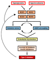
Endothelial Dysfunction: Is There a Hyperglycemia-Induced Imbalance of NOX and NOS?
- PMID: 31382355
- PMCID: PMC6696313
- DOI: 10.3390/ijms20153775
Endothelial Dysfunction: Is There a Hyperglycemia-Induced Imbalance of NOX and NOS?
Abstract
NADPH oxidases (NOX) are enzyme complexes that have received much attention as key molecules in the development of vascular dysfunction. NOX have the primary function of generating reactive oxygen species (ROS), and are considered the main source of ROS production in endothelial cells. The endothelium is a thin monolayer that lines the inner surface of blood vessels, acting as a secretory organ to maintain homeostasis of blood flow. The enzymatic production of nitric oxide (NO) by endothelial NO synthase (eNOS) is critical in mediating endothelial function, and oxidative stress can cause dysregulation of eNOS and endothelial dysfunction. Insulin is a stimulus for increases in blood flow and endothelium-dependent vasodilation. However, cardiovascular disease and type 2 diabetes are characterized by poor control of the endothelial cell redox environment, with a shift toward overproduction of ROS by NOX. Studies in models of type 2 diabetes demonstrate that aberrant NOX activation contributes to uncoupling of eNOS and endothelial dysfunction. It is well-established that endothelial dysfunction precedes the onset of cardiovascular disease, therefore NOX are important molecular links between type 2 diabetes and vascular complications. The aim of the current review is to describe the normal, healthy physiological mechanisms involved in endothelial function, and highlight the central role of NOX in mediating endothelial dysfunction when glucose homeostasis is impaired.
Keywords: NADPH oxidase; ROS; eNOS; endothelium; glucose; hyperglycemia; insulin resistance; obesity; reactive oxygen species; type 2 diabetes; vascular function.
Conflict of interest statement
The authors declare no conflict of interest.
Figures


Similar articles
-
The p47phox- and NADPH oxidase organiser 1 (NOXO1)-dependent activation of NADPH oxidase 1 (NOX1) mediates endothelial nitric oxide synthase (eNOS) uncoupling and endothelial dysfunction in a streptozotocin-induced murine model of diabetes.Diabetologia. 2012 Jul;55(7):2069-79. doi: 10.1007/s00125-012-2557-6. Epub 2012 May 2. Diabetologia. 2012. PMID: 22549734 Free PMC article.
-
β3 Adrenergic Stimulation Restores Nitric Oxide/Redox Balance and Enhances Endothelial Function in Hyperglycemia.J Am Heart Assoc. 2016 Feb 19;5(2):e002824. doi: 10.1161/JAHA.115.002824. J Am Heart Assoc. 2016. PMID: 26896479 Free PMC article.
-
Role of eNOS- and NOX-containing microparticles in endothelial dysfunction in patients with obesity.Obesity (Silver Spring). 2016 Jun;24(6):1305-12. doi: 10.1002/oby.21508. Epub 2016 Apr 30. Obesity (Silver Spring). 2016. PMID: 27130266
-
Reactive oxygen species and endothelial function--role of nitric oxide synthase uncoupling and Nox family nicotinamide adenine dinucleotide phosphate oxidases.Basic Clin Pharmacol Toxicol. 2012 Jan;110(1):87-94. doi: 10.1111/j.1742-7843.2011.00785.x. Epub 2011 Sep 28. Basic Clin Pharmacol Toxicol. 2012. PMID: 21883939 Review.
-
Oxidative stress and diabetic cardiovascular disorders: roles of mitochondria and NADPH oxidase.Can J Physiol Pharmacol. 2010 Mar;88(3):241-8. doi: 10.1139/Y10-018. Can J Physiol Pharmacol. 2010. PMID: 20393589 Review.
Cited by
-
Lipid emulsion attenuates vasodilation by decreasing intracellular calcium and nitric oxide in vascular endothelial cells.Heliyon. 2024 Sep 3;10(17):e37353. doi: 10.1016/j.heliyon.2024.e37353. eCollection 2024 Sep 15. Heliyon. 2024. PMID: 39296045 Free PMC article.
-
Endothelial dysfunction in neuroprogressive disorders-causes and suggested treatments.BMC Med. 2020 Oct 19;18(1):305. doi: 10.1186/s12916-020-01749-w. BMC Med. 2020. PMID: 33070778 Free PMC article. Review.
-
Role of Takeda G protein‑coupled receptor 5 in microvascular endothelial cell dysfunction in diabetic retinopathy (Review).Exp Ther Med. 2022 Sep 15;24(5):674. doi: 10.3892/etm.2022.11610. eCollection 2022 Nov. Exp Ther Med. 2022. PMID: 36237599 Free PMC article. Review.
-
Hemodynamics and Arterial Stiffness in Response to Oral Glucose Loading in Individuals with Type II Diabetes and Controlled Hypertension.High Blood Press Cardiovasc Prev. 2023 Mar;30(2):175-181. doi: 10.1007/s40292-023-00569-2. Epub 2023 Mar 13. High Blood Press Cardiovasc Prev. 2023. PMID: 36913100
-
To the Future: The Role of Exosome-Derived microRNAs as Markers, Mediators, and Therapies for Endothelial Dysfunction in Type 2 Diabetes Mellitus.J Diabetes Res. 2022 Feb 21;2022:5126968. doi: 10.1155/2022/5126968. eCollection 2022. J Diabetes Res. 2022. PMID: 35237694 Free PMC article. Review.
References
-
- Kahn S.E., Prigeon R.L., McCulloch D.K., Boyko E.J., Bergman R.N., Schwartz M.W., Neifing J.L., Ward W.K., Beard J.C., Palmer J.P. Quantification of the relationship between insulin sensitivity and β-cell function in human subjects: Evidence for a hyperbolic function. Diabetes. 1993;42:1663–1672. doi: 10.2337/diab.42.11.1663. - DOI - PubMed
Publication types
MeSH terms
Substances
Grants and funding
LinkOut - more resources
Full Text Sources
Medical

