. 2019 Aug 2;10(1):3493.
doi: 10.1038/s41467-019-11169-x.

Hydrogen peroxide release by bacteria suppresses inflammasome-dependent innate immunity

Affiliations

Hydrogen peroxide release by bacteria suppresses inflammasome-dependent innate immunity

Saskia F Erttmann et al. Nat Commun. .

Abstract

Hydrogen peroxide (H2O2) has a major function in host-microbial interactions. Although most studies have focused on the endogenous H2O2 produced by immune cells to kill microbes, bacteria can also produce H2O2. How microbial H2O2 influences the dynamics of host-microbial interactions is unclear. Here we show that H2O2 released by Streptococcus pneumoniae inhibits inflammasomes, key components of the innate immune system, contributing to the pathogen colonization of the host. We also show that the oral commensal H2O2-producing bacteria Streptococcus oralis can block inflammasome activation. This study uncovers an unexpected role of H2O2 in immune suppression and demonstrates how, through this mechanism, bacteria might restrain the immune system to co-exist with the host.


Conflict of interest statement

The authors declare no competing interests.

Figures
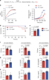
Fig. 1
Fig. 1
Pyruvate oxidase SpxB dampens inflammasome-dependent cytokine response promoting S. pneumoniae survival. a The pyruvate oxidase (SpxB)-mediated H2O2 generation reaction in S. pneumoniae. b H2O2 release by wild-type S. pneumoniae D39 (S.p. WT) and S.pspxB. c In vitro growth rate of S.p. WT (D39) and S.p. ΔspxB. b and c are representative of 3 independent experiments performed in triplicates; data shown as mean ± standard deviation (±s.d.). d Survival of WT mice after intranasal infection (1–2 × 107 cfu/mouse) with S.p. WT (D39) or S.p. ΔspxB analysed by the Kaplan–Meier method, with n = 10 animals per group. P value determined by Gehan-Breslow-Wilcoxon test. e Clinical severity of WT mice infected with S.p. WT (D39) or S.p. ΔspxB (1–2 × 107 cfu/mouse) for 6 h (n = 6 animals per group). P value determined by Mann Whitney test. f S.p. WT (D39) and S.p. ΔspxB counts (cfu) in the lungs of WT mice 12, 24 and 48 h after intranasal infection (1–2 × 107 cfu/mouse). g IL-1β and h TNF-α in the lung fluid of WT mice 12, 24 and 48 h after intranasal infection (1–2 × 107 cfu/mouse) with S.p. WT (D39) or S.p. ΔspxB. Results in f to h are from 2–3 independent experiments with a total of 4–12 animals per group; data shown as the mean ± standard error of the mean (±s.e.m.). P value determined by Mann Whitney test. Source data are provided as a Source Data file
Fig. 2
Fig. 2
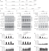
S. pneumoniae impairs NLRP3- and NLRC4-dependent inflammasome activation. a Schematic diagrams of experimental setups used in b to m. Immunoblots of Caspase-1 and IL-1β processing in LPS-primed BMDMs pre-treated with increasing S.p. WT (D39) doses (MOI 1, 10 or 100) for 30 min before stimulation with b ATP for 30 min, c P. aeruginosa (P.a., MOI 20) for 60 min or d nigericin for 30 min. Results are representative of at least 4 independent experiments. ELISA analysis of corresponding supernatants for eg IL-1β, hj IL-18 and km TNF-α. Results are obtained from at least two independent experiments; data are shown as mean ± s.e.m. P values determined by one-way ANOVA followed by Bonferroni post-test. Source data are provided as a Source Data file
Fig. 3
Fig. 3
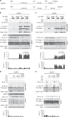
S. pneumoniae blocks multiple inflammasome pathways through its SpxB activity. a Schematic diagram of experimental setups used in (be). b, c Immunoblots of Caspase-1 and IL-1β in cell lysates and supernatants of LPS-primed BMDMs pre-treated with increasing MOIs (1, 10, 100) of S.p. WT (D39) or S.p. ΔspxB and then stimulated with agonists for NLRP3 (ATP, 5 mM, 30 min), or NLRC4 (P.a., MOI 20, 60 min). ELISA for IL-1β secretion and lactate dehydrogenase (LDH) assay in corresponding supernatants. The results are representative of four independent experiments. P values determined by one-way ANOVA followed by Bonferroni post-test. d, e Caspase-1 and IL-1β processing and LDH release of LPS-primed BMDMs pre-treated with S.p. WT (D39), S.p. ΔspxB or S.p. SpxB-complemented ΔspxBspxB + SpxB) (MOI 50) for 30 min before d stimulation with 5 mM ATP for 30 min or e infection with P.a. (MOI 20) for 60 min. The results are representative of four or two independent experiments, respectively. LDH release is depicted as the mean ± s.e.m. Source data are provided as a Source Data file
Fig. 4
Fig. 4
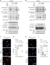
Inflammasome inhibition by S. pneumoniae involves defects in ASC complex formation. a, b Immunoblot analyses of ASC oligomers or Caspase-1 and IL-1β processing in LPS-primed BMDMs pre-treated with S.p. WT or S.p. ΔspxB (D39, MOI 50, 30 min) before stimulation with a ATP for 30 min (NLRP3) or b P.a. (NLRC4, MOI 20) for 60 min. Results are representative of 3 independent experiments. cf Microscopic visualisation of ASC/active Caspase-1 complexes (red/green; overlay yellow) in LPS-primed BMDMs pre-treated with S.p. WT or S.p. ΔspxB (D39, MOI 50) and subsequently stimulated with c, d ATP (5 mM, 30 min) or e, f P.a. (MOI 20, 60 min). Panels d and f depict corresponding percentages of cells in c and e, respectively, containing ASC/active Caspase-1 specks (determined by enumerating at least 100 cells per sample). Scale bar: 50 µm. Results in d and f are from three independent experiments. The data are shown as the mean ± s.e.m. P values determined by one-way ANOVA followed by Bonferroni post-test. Source data are provided as a Source Data file
Fig. 5
Fig. 5
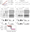
High H2O2-producing bacteria block inflammasome activation whereas low producers do not. a H2O2 production by S. oralis, S. pneumoniae ΔspxB (D39), S. sorbnius, S. suis, S. pyogenes relative to that by S. pneumoniae WT (D39) (100%). Data are representative of two independent experiments performed in duplicates or triplicates presented as the mean ± s.d. b Schematic diagrams of experimental setups used in (ch). c, d Immunoblot analysis of Caspase-1 and IL-1β processing, e, f ELISA analysis of corresponding supernatants for IL-1β secretion and g, h LDH release of LPS-primed BMDMs pre-treated with S. pneumoniae D39 WT (S. p.), S. pyogenes (S. pyo.), S. suis, S. oralis or S. sobrinus (S. sob.) (MOI 40) for 30 min before c, e, g stimulation with ATP (5 mM) for 30 min or d, f, h infection with P. aeruginosa (MOI 20) for 60 min. Data in c and d are representative of two or three independent experiments, respectively. ELISA and LDH release data are from two or three independent experiments and are depicted as the mean ± s.e.m. i Relative inflammasome inhibition or activation by Streptococcus species presented as the mean ± s.e.m. Inhibition depicts decrease in P.a.-induced IL-1β release during co-infection with indicated bacteria. Activation depicts IL-1β release by indicated bacteria relative to that by P.a.. Data are presented as the mean ± s.e.m. of 2–9 experiments. Source data are provided as a Source Data file
Fig. 6
Fig. 6
S. pneumoniae impairs inflammasome activation via SpxB-mediated H2O2 release. a Schematic diagrams of the experimental setups used in (b). b ASC oligomers, Caspase-1 and IL-1β processing in LPS-primed BMDMs pre-treated with S.p. WT (D39, MOI 100) in the presence of catalase (100 U mL−1) for 30 min before ATP stimulation (5 mM, 30 min) or P.a. infection (MOI 20, 60 min). c Schematic diagrams of the experimental setups used in d. d ASC oligomers, Caspase-1 and IL-1β processing in BMDMs pre-treated with 50 µM H2O2 for 10 min prior to stimulation with ATP (5 mM, 30 min) or infection with P.a. (60 min, MOI 20). The data in b and d are representative of three independent experiments. Source data are provided as a Source Data file
Fig. 7
Fig. 7
H2O2 release by S. pneumoniae suppresses inflammasome-dependent bacterial clearance. ac S.p. WT (D39) burden (cfu) in the lungs of WT and ASC−/− mice a 12  h, b 24 h, and c 48 h after intranasal infection (1–2 × 107 cfu/mouse). (df) S.p. ΔspxB (D39) burden in the lungs of WT and ASC−/− mice d 12 h, e 24 h, and f 48 h post infection (1–2 × 107 cfu/mouse). g Clinical severity and h subcutaneous body temperatures of WT and ASC−/− mice after S.p. ΔspxB (D39) infection. Results in a to h are representative of 2 independent experiments with a total of 4–8 animals per group (1–2 × 107 cfu/mouse). i Bacterial burden, j clinical severity, and k subcutaneous body temperature of WT mice co-inoculated with S.p. WT (D39) and active or denatured catalase (500 U per mouse) for 24 h. Results are representative of three independent experiments with a total of 16 animals per group (1 × 107 cfu/mouse). l Bacterial burden, m clinical severity, and n subcutaneous body temperature of WT mice co-inoculated with S.p. ΔspxB (D39) and active or denatured catalase (500 U per mouse) for 24 h. Results are representative of two independent experiments with a total of 10 animals per group (1 × 107 cfu/mouse). The data in a to n are shown as mean ± s.e.m. af, i, l P values determined by Mann Whitney test. g, h, j, k, m, n P values determined by two-way ANOVA followed by Bonferroni post-test. o Proposed model of inflammasome inactivation by high H2O2-producing bacteria. Source data are provided as a Source Data file

Similar articles

Cited by

References

    1. Veal EA, Day AM, Morgan BA. Hydrogen peroxide sensing and signaling. Mol. Cell. 2007;26:1–14. doi: 10.1016/j.molcel.2007.03.016. - DOI - PubMed
    1. Zamocky M, Furtmuller PG, Obinger C. Evolution of catalases from bacteria to humans. Antioxid. Redox Signal. 2008;10:1527–1548. doi: 10.1089/ars.2008.2046. - DOI - PMC - PubMed
    1. Stone JR, Yang S. Hydrogen peroxide: a signaling messenger. Antioxid. Redox Signal. 2006;8:243–270. doi: 10.1089/ars.2006.8.243. - DOI - PubMed
    1. Imlay JA. Cellular defenses against superoxide and hydrogen peroxide. Annu Rev. Biochem. 2008;77:755–776. doi: 10.1146/annurev.biochem.77.061606.161055. - DOI - PMC - PubMed
    1. Nathan C, Cunningham-Bussel A. Beyond oxidative stress: an immunologist’s guide to reactive oxygen species. Nat. Rev. Immunol. 2013;13:349–361. doi: 10.1038/nri3423. - DOI - PMC - PubMed

Publication types

MeSH terms

LinkOut - more resources