. 2019 Aug 1;15(8):e1008294.
doi: 10.1371/journal.pgen.1008294. eCollection 2019 Aug.

Ddc2ATRIP promotes Mec1ATR activation at RPA-ssDNA tracts

Affiliations

Ddc2ATRIP promotes Mec1ATR activation at RPA-ssDNA tracts

Himadri Biswas et al. PLoS Genet. .

Abstract

The DNA damage checkpoint response is controlled by the phosphatidylinositol 3-kinase-related kinases (PIKK), including ataxia telangiectasia-mutated (ATM) and ATM and Rad3-related (ATR). ATR forms a complex with its partner ATRIP. In budding yeast, ATR and ATRIP correspond to Mec1 and Ddc2, respectively. ATRIP/Ddc2 interacts with replication protein A-bound single-stranded DNA (RPA-ssDNA) and recruits ATR/Mec1 to sites of DNA damage. Mec1 is stimulated by the canonical activators including Ddc1, Dpb11 and Dna2. We have characterized the ddc2-S4 mutation and shown that Ddc2 not only recruits Mec1 to sites of DNA damage but also stimulates Mec1 kinase activity. However, the underlying mechanism of Ddc2-dependent Mec1 activation remains to be elucidated. Here we show that Ddc2 promotes Mec1 activation independently of Ddc1/Dpb11/Dna2 function in vivo and through ssDNA recognition in vitro. The ddc2-S4 mutation diminishes damage-induced phosphorylation of the checkpoint mediators, Rad9 and Mrc1. Rad9 controls checkpoint throughout the cell-cycle whereas Mrc1 is specifically required for the S-phase checkpoint. Notably, S-phase checkpoint signaling is more defective in ddc2-S4 mutants than in cells where the Mec1 activators (Ddc1/Dpb11 and Dna2) are dysfunctional. To understand a role of Ddc2 in Mec1 activation, we reconstituted an in vitro assay using purified Mec1-Ddc2 complex, RPA and ssDNA. Whereas ssDNA stimulates kinase activity of the Mec1-Ddc2 complex, RPA does not. However, RPA can promote ssDNA-dependent Mec1 activation. Neither ssDNA nor RPA-ssDNA efficiently stimulates the Mec1-Ddc2 complex containing Ddc2-S4 mutant. Together, our data support a model in which Ddc2 promotes Mec1 activation at RPA-ssDNA tracts.


Conflict of interest statement

The authors have declared that no competing interests exist.

Figures
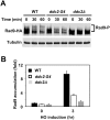
Fig 1
Fig 1. Effect of ddc2-S4 or ddc2Δ mutation on Rad9 phosphorylation after DNA damage.
(A) Wild-type (HB09), ddc2-S4 (HB10) or ddc2Δ (KSC1536) cells expressing Rad9-HA were treated with nocodazole to arrest at G2/M. Cells were then exposed to 0.05% MMS. Cells were collected at the indicated time points, and extracts were subjected to immunoblotting analysis with anti-HA antibodies or anti-tubulin antibodies. (B) Effect of ddc2-S4 or ddc2Δ on Rad9 localization to a HO-induced DSB. Wild-type (HB09), ddc2-S4 (HB10) or ddc2Δ (KSC1536) cells expressing Rad9-HA were transformed with the YCpA-GAL-HO plasmid. Transformed cells were grown in sucrose and treated with nocodazole. After arrest at G2/M, the culture was incubated with galactose for 3 hr to induce HO expression, while half of the culture was maintained in sucrose to repress HO expression. Cells were subjected to chromatin immunoprecipitation with anti-HA antibodies. Association of Rad9 with a HO-induced DSB was analyzed by real-time PCR. Relative enrichment was determined from three independent experiments. The error bars indicate standard deviation.
Fig 2
Fig 2. Effect of ddc2-S4 mutation on DNA end resection and RPA accumulation.
(A) Scheme of the ADH4 locus containing a HO cleavage site. One EcoRI restriction site is located 0.8 kb away from the HO cleavage site whereas another is 5.8 kb away. The black arrows indicate PCR primer pairs to monitor HO or EcoRI cleavage. (B) Effect of ddc2-S4 mutation on DNA end resection. Wild-type (HB01) or ddc2-S4 (HB02) cells carrying YCpA-GAL-HO were grown in sucrose and treated with nocodazole. After arrest at G2/M, the culture was incubated with galactose to induce HO expression. Cells were collected at the indicated times for genomic DNA preparation. Genomic DNA was digested with EcoRI and analyzed by real-time PCR. Experiments were carried out three times. The error bars indicate standard deviation. (C) Effect of ddc2-S4 on Rfa2 localization to a HO-induced DSB. Wild-type (HB01) or ddc2-S4 (HB02) cells were transformed with the YCpA-GAL-HO plasmid. Transformed cells were grown in sucrose and treated with nocodazole. The culture was then incubated with galactose for 3 hr to induce HO expression, while half of the culture was maintained in sucrose to repress HO expression. Cells were subjected to chromatin immunoprecipitation with anti-Rfa2 antibodies. Association of Rfa2 with a HO-induced DSB was analyzed by real-time PCR. Relative enrichment was determined from three independent experiments. The error bars indicate standard deviation.
Fig 3
Fig 3. Effect of ddc2-S4 on Ddc1 phosphorylation and localization in response to DNA damage.
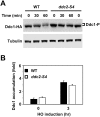
(A) Effect of ddc2-S4 mutation on Ddc1 phosphorylation after DNA damage. Wild-type (HB12) or ddc2-S4 (HB13) cells expressing Ddc1-HA were analyzed as in Fig 1A. (B) Effect of ddc2-S4 mutation on Ddc1 localization to a HO-induced DSB. Wild-type (HB12) or ddc2-S4 (HB13) cells expressing Ddc1-HA were transformed with the YCpA-GAL-HO plasmid. Transformed cells were cultured as in Fig 1B and subjected to ChIP assay to monitor Ddc1 localization. Relative enrichment was determined from three independent experiments. The error bars indicate standard deviation from three independent experiments.
Fig 4
Fig 4. Effect of ddc2-S4 mutation on S-phase checkpoint signaling.
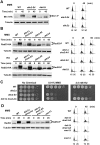
(A) Wild-type (KSC4233), ddc2-S4 (KSC4234) and ddc2Δ (KSC4235) cells expressing Mrc1-HA were arrested with α-factor at G1 and released into medium containing 0.05% MMS. Cells were collected at the indicated time and analyzed as in Fig 1A. Cell cycle progression from G1 to S phase was monitored by DNA flow cytometry. (B) Wild-type (KSC1178), ddc1Δ dna2-AA (KSC4219), ddc2-S4 (KSC3153) and ddc2Δ (KSC1234) cells carrying the YCpT-Rad53-HA plasmid were synchronized with α-factor at G1 and released into medium containing 0.05% MMS or 100 mM HU. Cells were collected at the indicated time (45 min for MMS and 60 min for HU) and analyzed as in Fig 4A. (C) Effect of ddc1Δ dna2-AA or ddc2-S4 on sensitivities to MMS and HU. Wild-type (KSC1178), ddc1Δ dna2-AA (KSC4219), ddc2-S4 (KSC3153) and ddc2Δ (KSC1234) cells were serially diluted and spotted on plates medium containing MMS or HU. (D) Wild-type (KSC1178), ddc1Δ dna2-AA (KSC4219), ddc1Δ dna2-AA mec1Δ (KSC4238) or mec1Δ (KSC1186) cells were synchronized with α-factor at G1 and released into medium containing 0.05% MMS. Cells were collected at the indicated time and subjected to immunoblotting analysis with anti-Rad53 or anti-tubulin antibody. Cell cycle progression from G1 to S phase was monitored by DNA flow cytometry.
Fig 5
Fig 5. Effect of ddc2-S4 mutation on RPA phosphorylation in vivo and in vitro.
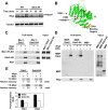
(A) Rfa2 phosphorylation after exposure to MMS. Wild-type (HB01) or ddc2-S4 (HB02) cells were cultured as in Fig 1A and subjected to immunoblotting analysis with anti-Rfa2 antibodies. (B) Position of the ddc2-S4 substitution mutation sites. The putative DNA binding (177KKRK180) and the RPA binding (amino acid 10 to 30) [64] region are highlighted in red and orange, respectively. The side chain of K263 and H382 residues is shown in yellow and blue, respectively. (C) Effect of ddc2-S4 mutation on ssDNA-binding of Mec1-Ddc2. Streptavidin beads were first incubated with RPA (1 pmol) or bio-oligo(dN)80 (5 pmol). Beads were further incubated with Mec1-Ddc2 or Mec1-Ddc2-S4 (0.5 pmol). Captured proteins on beads were detected by immunoblotting with anti-FLAG, anti-HA or anti-Rfa1 antibodies. Note that Mec1 is FLAG-tagged and Ddc2 is HA-tagged. (D) Effect of ddc2-S4 mutation on ssDNA-binding of Ddc2. Streptavidin beads were first incubated with RPA (1 pmol) or bio-oligo(dN)80 (5 pmol). Beads were further incubated with MBP, MBP-Ddc2 or MBP-Ddc2-S4 (0.5 pmol). MBP or MBP-fusion proteins were prepared from E. coli. Captured proteins on beads were analyzed by immunoblotting with anti-MBP or anti-Rfa2 antibodies. (E) Effect of ddc2-S4 mutation on RPA phosphorylation in vitro. Kinase reactions were carried out with Mec1-Ddc2 or Mec1-Ddc2-S4 (5 nM) in the absence or the presence of RPA (10 nM) or bio-oligo(dN)80 (125 nM). Incorporation of 32P into Rfa1 and Rfa2 were normalized to that observed with Rfa1 and Rfa2 alone. The error bars indicate standard deviation from three independent experiments.
Fig 6
Fig 6. Effect of RPA or ssDNA addition on Mec1 activity.
(A) Effect of RPA addition on Mec1 catalytic activity. Kinase reactions were carried out with or without Mec1-Ddc2 (5 nM) using various concentrations of RPA. GST-Rad53 C-terminus fusion (GST-Rad53) was used as a substrate of Mec1. Incorporation of 32P into GST-Rad53 was detected by phosphor imaging. The Rad53 C-terminus does not contain its kinase domain; phosphorylation of GST-Rad53 depends on Mec1-Ddc2. Phosphorylation levels of GST-Rad53 were normalized to that observed with Mec1-Ddc2 alone. The error bars indicate standard deviation from three independent experiments. (B) Effect of ssDNA addition on Mec1 catalytic activity. Kinase reactions were carried out with or without Mec1-Ddc2 (5 nM) using various concentrations of 80-mer biotin-oligo(dN)80. Incorporation of 32P into GST-Rad53 was analyzed as in A. The error bars indicate standard deviation from three independent experiments. (C, D) Effect of length and base-composition of ssDNA on Mec1 activation. Mec1-Ddc2 complex (5 nM) was incubated with various concentrations of oligonucleotides (20, 40, 60, 80-mer oligo(dT), 80-mer bio-oligo(dN)80 (C) or ΦX phage ssDNA (5 kb) (D). Phosphorylation of GST-Rad53 was normalized to that observed without RPA or ssDNA. The error bars indicate standard deviation from three independent experiments. (E) Effect of ddc2-S4 mutation on Mec1 activation by ssDNA. Kinase reactions were carried out with Mec1-Ddc2 or Mec1-Ddc2-S4 (5 nM) using various concentrations of bio-oligo(dN)80. Incorporation of 32P into GST-Rad53 was analyzed as in A. The error bars indicate standard deviation from three independent experiments.
Fig 7
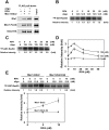
Fig 7. Effects of RPA on ssDNA-dependent Mec1 activation.
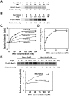
(A) Interaction of Mec1-Ddc2 with RPA in the presence or absence of ssDNA. Mec1-Ddc2 (0.5 pmol) and RPA (1 pmol) were incubated with ANTI-FLAG-M2 agarose in the presence or absence of bio-oligo(dN)80 (5 pmol). Proteins bound to ANTI-FLAG-M2 agarose were analyzed by immunoblotting analysis with anti-FLAG, anti-HA or anti-Rfa1 antibodies. Note that Mec1 is FLAG-tagged and Ddc2 is HA-tagged. (B) Effect of RPA addition on Mec1 activation in the presence of low concentrations of ssDNA. Kinase reactions were carried out with Mec1-Ddc2 (5 nM) using various concentrations of RPA in the absence or presence of bio-oligo(dN)80 (12.5 nM). Incorporation of 32P into GST-Rad53 was analyzed as in Fig 6A. Kinase activities of Mec1-Ddc2, normalized to that observed with Mec1-Ddc2 alone, are shown in comparison with those in the presence of RPA or ssDNA. Experiments were carried out three times and the representative result is shown. (C) Effect of RPA on Mec1 activity in the presence of high concentrations of ssDNA. Kinase reactions were carried out with Mec1-Ddc2 (5 nM) using various concentrations of RPA in the absence or presence of bio-oligo(dN)80 (125 nM). Incorporation of 32P into GST-Rad53 was analyzed as in Fig 6A. Kinase activities of Mec1-Ddc2, normalized to that observed with Mec1-Ddc2 alone, are shown in comparison with those in the presence of RPA or ssDNA. Experiments were carried out three times and the representative result is shown. (D) Effects of RPA concentrations on ssDNA-dependent Mec1 activation. Kinase activities of Mec1-Ddc2 using various concentrations of RPA in the presence of bio-oligo(dN)80 (12.5 nM or 125 nM) were normalized to that observed with Mec1-Ddc2 alone (See Fig 7B or 7C, respectively). Relative kinase activities with various concentrations of RPA in the absence of ssDNA are also included (see Fig 6A). The error bars indicate standard deviation from three independent experiments. (E) Effect of ddc2-S4 mutation on Mec1 activation by RPA and ssDNA. Kinase reactions were carried out with Mec1-Ddc2 or Mec1-Ddc2-S4 (5 nM) using various concentrations of RPA in the absence or the presence of bio-oligo(dN)80 (12.5 nM). Incorporation of 32P into GST-Rad53 was analyzed as in Fig 6A. The error bars indicate standard deviation from three independent experiments.
Fig 8
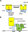
Fig 8. Model for Mec1 activation at RPA-covered ssDNA tracts.
(A) ssDNA binding of Ddc2 increases Mec1 activity. ssDNA binding of Ddc2 triggers conformation changes of the entire Mec1-Ddc2 homodimer, resulting in structural changes of the kinase domain at the C-terminus of Mec1. K263 is involved in homodimerization of the Mec1-Ddc2 heterodimer. The PRD-PRD interface within the kinase domain is also involved in homodimerization of the Mec1-Ddc2 heterodimer. See text for more detail. (B) RPA promotes ssDNA-dependent Mec1 activation. The N-terminus of Ddc2 interacts with the N-terminus of Rfa1 whereas the DNA binding (KKRK) region of Ddc2 is involved in ssDNA binding. RPA alone binds to ssDNA through its own DNA binding domain (DBD). See the text for more detail.

Similar articles

Cited by

References

    1. Ciccia A, Elledge SJ. The DNA damage response: making it safe to play with knives. Mol Cell. 2010;40(2):179–204. 10.1016/j.molcel.2010.09.019 - DOI - PMC - PubMed
    1. Harper JW, Elledge SJ. The DNA damage response: ten years after. Mol Cell. 2007;28(5):739–45. 10.1016/j.molcel.2007.11.015 . - DOI - PubMed
    1. Blackford AN, Jackson SP. ATM, ATR, and DNA-PK: The Trinity at the Heart of the DNA Damage Response. Mol Cell. 2017;66(6):801–17. 10.1016/j.molcel.2017.05.015 . - DOI - PubMed
    1. Yazinski SA, Zou L. Functions, Regulation, and Therapeutic Implications of the ATR Checkpoint Pathway. Annu Rev Genet. 2016;50:155–73. 10.1146/annurev-genet-121415-121658 . - DOI - PubMed
    1. Saldivar JC, Cortez D, Cimprich KA. The essential kinase ATR: ensuring faithful duplication of a challenging genome. Nat Rev Mol Cell Biol. 2017;18(10):622–36. 10.1038/nrm.2017.67 - DOI - PMC - PubMed

Publication types

MeSH terms

Substances