. 2019 Aug 9;8(8):bio045674.
doi: 10.1242/bio.045674.

The ventral epithelium of Trichoplax adhaerens deploys in distinct patterns cells that secrete digestive enzymes, mucus or diverse neuropeptides

Affiliations

The ventral epithelium of Trichoplax adhaerens deploys in distinct patterns cells that secrete digestive enzymes, mucus or diverse neuropeptides

Tatiana D Mayorova et al. Biol Open. .

Abstract

The disk-shaped millimeter-sized marine animal, Trichoplax adhaerens, is notable because of its small number of cell types and primitive mode of feeding. It glides on substrates propelled by beating cilia on its lower surface and periodically pauses to feed on underlying microorganisms, which it digests externally. Here, a combination of advanced electron and light microscopic techniques are used to take a closer look at its secretory cell types and their roles in locomotion and feeding. We identify digestive enzymes in lipophils, a cell type implicated in external digestion and distributed uniformly throughout the ventral epithelium except for a narrow zone near its edge. We find three morphologically distinct types of gland cell. The most prevalent contains and secretes mucus, which is shown to be involved in adhesion and gliding. Half of the mucocytes are arrayed in a tight row around the edge of the ventral epithelium while the rest are scattered further inside, in the region containing lipophils. The secretory granules in mucocytes at the edge label with an antibody against a neuropeptide that was reported to arrest ciliary beating during feeding. A second type of gland cell is arrayed in a narrow row just inside the row of mucocytes while a third is located more centrally. Our maps of the positions of the structurally distinct secretory cell types provide a foundation for further characterization of the multiple peptidergic cell types in Trichoplax and the microscopic techniques we introduce provide tools for carrying out these studies.

Keywords: Digestive system evolution; Gland cell; Mucus; Nervous system evolution; Neuropeptide; Placozoa.


Conflict of interest statement

Competing interestsThe authors declare no competing or financial interests.

Figures
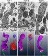
Fig. 1.
Fig. 1.
Three structural types of gland cell in Trichoplax ventral epithelium. Upper row shows gland cells in thin sections in SEM backscatter mode, and lower row shows three-dimensional renderings. Cell body, purple; apical surface, salmon; cilium, green; microvilli, yellow; nucleus, blue; internal granules, red. Gland cells are distinguished by their content of secretory granules (g). Type 1 cells have large granules with a dense content, while Type 2 and 3 cells have smaller and paler granules. Type 1 and 3 cells have an apical cilium. Type 2 cells lack a cilium but have microvilli. Scale bars: 2 µm.
Fig. 2.
Fig. 2.
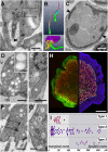
Features and distributions of the three gland cell types and lipophil cells. (A) Thin section from Type 1 gland cell shows its cilium (c) rising from a deep, ovoid pocket, the basal body (b) and the ciliary rootlet (r). Arrowhead indicates coated pit. (B) Three-dimensional reconstructions comparing the cilium and associated structures of a Type 1 gland cell and a VEC. Upper inset shows the short, thin ciliary rootlet (red, right) in a Type 1 cell and the longer and thicker rootlet in a VEC (orange, left), and associated basal bodies (cyan) and cilia (green). Surface renderings made from 12 serial sections. Lower inset shows the apical microvilli (yellow) surrounding the cilium (green) of a Type 1 cell. Surface rendering from 77 serial sections. (C) Thin section from the interior of a Type 1 cell shows distinctive large, grey granules (g) with homogeneous content and markedly dense ER (arrowheads). (D) Section from a Type 2 cell illustrates its clear granules and a microvillus (mv). Inset shows enlarged view of the clear granules. (E) Thin section of the area near the nucleus (n) of a Type 2 shows enlarged ER cistern (asterisk) characteristic of this cell type. (F) Thin section of the apical end of a Type 3 cell shows part of its cilium (c) arising from a shallow cup, the basal body (b) and a segment of the ciliary rootlet (r). (G) Enlarged view of the interior of a Type 3 cell shows the textured content of the granules. (H) Light micrographs showing two views of lipophil cells. Left half: lipophil granules in living animal stained with LipidTox (red). The surface of the animal is stained green with a lectin conjugated to a fluorescent dye (see below). Optical section at the ventral surface. Right half: FISH showing cells expressing phospholipase A (green) and trypsin (red). Nuclei are stained with Hoechst (blue). Maximum projection of a sequence of images comprising the entire animal. (I) Distribution of each type of gland cell mapped on a projection of half of the body of the animal. Map was created from tiled SEM images from one animal. Dark grey area indicates region containing lipophil cells. Scale bars: A,C,D–F: 500 nm; G: 200 nm; inset on D: 100 nm; H: 100 µm.
Fig. 3.
Fig. 3.
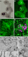
Type 2 gland cells contain and secrete mucus. (A) Projection of a time-lapse bright field image sequence (1.5 h) of a crawling animal. White arrowhead indicates start point and black arrowhead the end point. (B) Trail of mucus left by the crawling animal shown in A after staining with fluorescent WGA (green). Dotted line outlines the animal at one time point. (C) Enlarged view of the mucous clot left at the start point of the trail. (D) Mucus containing cells (mucocytes) labeled with WGA (green) in an en face view of an animal. The image is a maximum intensity projection of a series of optical sections through the animal. Mucocytes are numerous along the rim of the animal and present in moderate numbers further inside, but are absent near the center. (E) Enlarged view of WGA-stained mucocytes located near the rim of an animal. Projection of 38 optical sections. (F–J) TEM images of Types 1, 2 and 3 gland cells in thin sections labeled with WGA-nanogold. (F) Type 1 cell granules (overview and enlarged in inset). Few nanogold particles are present. (G) Numerous nanogold particles congregate in and around clear granules of Type 2 cells. (H) Enlarged view of Type 2 cell showing nanogold-WGA label in and around granules. (I) Type 2 cell Golgi complex labeled with WGA. Asterisk marks ER cistern. (J) Type 3 cell granules (overview and enlarged in inset). Few nanogold particles are present. Scale bars: A,B: 500 µm; C: 50 µm; D: 100 µm; E: 10 µm; F,G,J: 500 nm; H,I, insets on F,J: 100 nm.
Fig. 4.
Fig. 4.
Mucus secretion during feeding. (A) Projection of a time-lapse bright field image sequence (40 min) of a starved animal (gray) placed on a lawn of algae (not visible). White asterisk indicates the start point. After attaching to the substrate, the animal began to crawl but periodically paused to feed. Sites of feeding pauses are labeled 1–3. (B) Mucous trail of the animal shown in A stained with fluorescent-WGA (green). (C) Higher magnification view of the area in the small box in B shows the orientations of mucous strands that accumulated around the edge of the animal during feeding. (D) Mucus surrounding debris from lysed algal cells (magenta) beneath an animal fixed during feeding. (E) Thin section of the apical part of a VEC labeled with nanogold-conjugated WGA. Nanogold particles coat the microvilli (arrowhead) and apical plasma membrane, and label phagosomes (arrow) in the interior. (F) Enlarged view of VEC phagosomes labeled with WGA-nanogold. Scale bars: A,B: 500 µm; C: 20 µm; D: 10 µm; E: 500 nm; F: 200 nm.
Fig. 5.
Fig. 5.
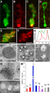
Localization of mucus and endomorphin immunoreactivity in Type 2 gland cells. (A–F) Confocal images from a whole animal stained with fluorescent WGA (green) and immunolabeled for endomorphin 2 (red). (A–C) Color separated and merged images of a Type 2 cell located at the edge of the animal. A cluster of granules near the apex and some granules deeper inside label with both WGA and anti-endomorphin 2. Projections of 25 optical sections. (D) A Type 2 gland cell farther from the edge shows no immunoreactivity for endomorphin 2. (E,F) Single optical section through the middle of a Type 2 gland cell body (E, merged; F, red channel). (G) Profile of green and red channel fluorescence intensity along the white line in E shows varying levels of endomorphin 2 immunoreactivity in WGA stained granules. (H–L) TEM images of thin sections labeled with anti-endomorphin 2 and immunogold-conjugated secondary antibody. (H) Granule in Type 1 cell shows no more than background immunogold labeling. (I) Type 2 cells demonstrate endomorphin immunoreactivity over and surrounding their clear granules. (J) Higher magnification immunolabeled Type 2 gland cell granules. (K) Endomorphin 2 immunogold labeling in the Golgi complex of a Type 2 cell. ER cistern (asterisk) is not labeled. (L) Granules in Type 3 cell show no more than background immunoreactivity. (M) Graph of the density (mean±s.e.m.) of endomorphin-nanogold in three gland cell types (1–3), with grains over granules (g) and cytoplasm (c) shown separately, compared to VEC and background (bg). Nanogold density over Type 2 cell granules (asterisk) is significantly higher than over cytoplasm or granules in the other cell types. Scale bars: A–F: 2 µm; H–L: 200 nm.
Fig. 6.
Fig. 6.
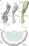
Diagrams showing three gland cell types and their distributions in the ventral epithelium. (A) Type 1 cells are distinguished by large, electron dense secretory granules and a single cilium arising from a deep pit. Type 2 gland cells lack a cilium and have smaller, secretory granules with electron lucent content. Type 3 gland cells have a single cilium and the smallest secretory granules with a textured content. (B) Map of gland cell positions. Type 2 cells are nearest to the edge while Type 1 cells occupy a separate concentric ring further inside. Type 2 and Type 3 cells are intermingled in the lipophil zone but are absent in the center.

Similar articles

Cited by

References

    1. Arendt D., Benito-Gutierrez E., Brunet T. and Marlow H. (2015). Gastric pouches and the mucociliary sole: setting the stage for nervous system evolution. Philos. Trans. R. Soc. B Biol. Sci. 370, 20150286 10.1098/rstb.2015.0286 - DOI - PMC - PubMed
    1. Armon S., Bull M. S., Aranda-Diaz A. and Prakash M. (2018). Ultrafast epithelial contractions provide insights into contraction speed limits and tissue integrity. Proc. Natl. Acad. Sci. USA 115, E10333-E10341. 10.1073/pnas.1802934115 - DOI - PMC - PubMed
    1. Bagby R. M. (1970). The fine structure of pinacocytes in the marine sponge Microciona prolifera (Ellis and Solander). Z. Zellforsch. Mikrosk. Anat. 105, 579-594. 10.1007/BF00335430 - DOI - PubMed
    1. Barker N., Van Es J. H., Kuipers J., Kujala P., Van Den Born M., Cozijnsen M., Haegebarth A., Korving J., Begthel H., Peters P. J. et al. (2007). Identification of stem cells in small intestine and colon by marker gene Lgr5. Nature 449, 1003-1007. 10.1038/nature06196 - DOI - PubMed
    1. Behrendt G. and Ruthmann A. (1986). The cytoskeleton of the fiber cells of Trichoplax adhaerens (Placozoa). Zoomorphology 106, 123-130. 10.1007/BF00312114 - DOI