Review
. 2019 Dec;76(23):4705-4724.
doi: 10.1007/s00018-019-03241-y. Epub 2019 Jul 26.

αKlotho-FGF23 interactions and their role in kidney disease: a molecular insight

Affiliations
Review

αKlotho-FGF23 interactions and their role in kidney disease: a molecular insight

Edward R Smith et al. Cell Mol Life Sci. 2019 Dec.

Abstract

Following the serendipitous discovery of the ageing suppressor, αKlotho (αKl), several decades ago, a growing body of evidence has defined a pivotal role for its various forms in multiple aspects of vertebrate physiology and pathology. The transmembrane form of αKl serves as a co-receptor for the osteocyte-derived mineral regulator, fibroblast growth factor (FGF)23, principally in the renal tubules. However, compelling data also suggest that circulating soluble forms of αKl, derived from the same source, may have independent homeostatic functions either as a hormone, glycan-cleaving enzyme or lectin. Chronic kidney disease (CKD) is of particular interest as disruption of the FGF23-αKl axis is an early and common feature of disease manifesting in markedly deficient αKl expression, but FGF23 excess. Here we critically discuss recent findings in αKl biology that conflict with the view that soluble αKl has substantive functions independent of FGF23 signalling. Although the issue of whether soluble αKl can act without FGF23 has yet to be resolved, we explore the potential significance of these contrary findings in the context of CKD and highlight how this endocrine pathway represents a promising target for novel anti-ageing therapeutics.

Keywords: Cardiovascular disease; Crystallography; Fibroblast growth factor; Kidney disease; Klotho proteins structural biology; Phosphate; Receptors; Therapeutics.


Conflict of interest statement

The authors declare they have no relevant conflicts of interest.

Figures
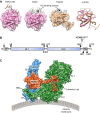
Fig. 1
Fig. 1
Structural basis for the endocrine action of FGF23. a From left to right: Surface representation of prototypical paracrine FGF2 with bound heparan sulphate (HS) octasaccharide (Protein Data Bank, PBD ID 1fq9) [172], aligned FGF2, FGF23 (Tyr25 to Asn170; PBD ID 2p39), and the cartoon structures overlayed [35]. Note the conserved globular core architecture but different conformations of the HS-binding (grey colour). b Cartoon depicting domain structure of 1012 amino acid human αKl. SP, TM, KL1, KL2: denotes signal peptide, transmembrane domain, tandem internal homologous KL repeats 1 and 2. The αKl ectodomain comprising KL1 and KL2 can be cleaved by plasma membrane-tethered ADAM10 or ADAM17 and released into the extracellular fluid. c Surface representation of the FGFR1c–αKl–FGF23 ternary complex from PDB ID 5w2 [48], which depicts D2 and D3 domain of human FGFR1c (blue), KL1 and KL2 domains of the ectodomain of human αKl (green) and human FGF23 (red; Tyr25 to Ser205). Note receptor binding arm (RBA) of KL2, which is necessary to stabilise the FGFR1c–αKl interaction and the tethering of the C-terminal tail of FGF23 through the KL1–KL2 cleft. Crystal structures were visualised in UCSF Chimera ver.1.13.1 [173]. Alignment was performed using the MatchMaker tool and the Needleman-Wunsch algorithm with BLOSUM-62 matrix
Fig. 2
Fig. 2
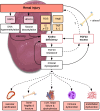
Hypothetical role of αKlotho deficiency in mineral dysregulation and pathological sequelae in CKD. In response to injury, the concerted action of epigenetic and non-epigenetic processes leads to silencing and suppression of αKl expression in the kidney. Renal injury also stimulates increased osteocytic synthesis and secretion of FGF23 with activation of inflammatory and hypoxia signalling pathways strongly implicated as key drivers of this response. FGF23 may suppress αKl expression through direct and indirect mechanisms. Ectopic FGF23 may also be expressed in extra-osseous non-physiological tissues (e.g., heart, liver, kidney) and supplement local and/or systemic levels. αKl deficiency results in end-organ resistance to FGF23 and failure in mineral homeostasis, which may feedback to bone to drive further increases in FGF23. Collectively and/or independently, loss of αKl, FGF23 excess and the consequent mineral dysregulation may promote the dysfunction in cardiovascular, bone and immune systems. Hypothetical pathways awaiting further experimental confirmation are indicated by dashed lines. AngII, angiotensin II; DMTs, dimethyltransferases; FGF23, fibroblast growth factor 23; HDACs, histone deacetylases; IL-1, interleukin-1β; IS, indoxyl sulphate; LVH, left ventricular hypertrophy; p-CS; cresyl sulphate; ROS, reactive oxygen species; TNF, tumour necrosis factor-α
Fig. 3
Fig. 3
Proposed FGF23-dependent and FGF23-independent mechanisms of action of αKl and their links to pathologic signalling in CKD. Flow diagram depicts canonical (left) and non-canonical (right) pathways that may link disturbances in the FGF23:αKl axis with pathological endpoints. Molecular interactions at the cell surface that may underpin these pathways are illustrated underneath. Canonical FGF23:αKl signalling utilising either membrane-tethered αKl (mKl) or the soluble cleaved ectodomain (sKl) in complex with FGFR1c signals via the FRS2a–RAS–MAPK pathway. FGFR3c and FGFR4 may also have minor redundant roles. Over-activity of this pathway may lead to excessive sodium reabsorption, down-regulation of ACE2, hypertension and LVH. On the other hand, after loss of αKl, relative underactivity of this pathway may result in mineral dysregulation characterised by hyperphosphataemia and/or phosphate-related mineral aggregates (calciprotein particles, CPPs [174, 175]) and attendant vascular sequelae. Potentiated by the very high levels in CKD, FGF23 may also signal independently of αKl, directly driving pathology in the heart, kidney and liver. Here αKl-independent signalling is thought to occur through FGFR4 (although alternate co-receptors/co-factors may also be involved) via PLCy-calcium-dependent calcineurin-NFAT pathways. Silencing of αKl in CKD may contribute to pathological signalling through several mechanisms. Firstly, loss of soluble αKl as a decoy receptor potentiating off-target FGF23–FGFR4 or paracrine FGF signalling; secondly loss of direct protective effects of soluble αKl as a TGF-βRII or IGF1 receptor antagonist or via sequestration of Wnt proteins; thirdly, loss of homeostatic functions as a lectin, which binds α2,3-sialyllactose-containing monosialogangliosides (red lipid heads) clustered in lipid rafts and inhibits PI3K-dependent exocytosis of TRPC6. For simplicity the cartoons depict the FGF23–FGFR1c–αKl–HS in 1:1:1:1 stoichiometry rather than the 2:2:2:2 quaternary signalling complex predicted by Chen et al. [53]. D1–D3 denote immunoglobulin-like domains of FGFR. KL1 and KL2 denote tandem internal homologous KL repeats 1 and 2. ACE2, angiotensin I converting enzyme 2; Akt, serine/threonine-protein kinase; CPPs, calciprotein particles; FGF, fibroblast growth factor; FGFR, (FGF) receptor; FRF2a, fibroblast growth factor receptor substrate 2; HS, heparan sulphate; IGF1R, insulin-like growth factor 1 (IGF-1) receptor; LRP, low-density-lipoprotein-related protein; LVH, left ventricular hypertrophy; mKl, membrane αKlotho; MAPK, mitogen-activated protein kinase; mTOR, mammalian target of rapamycin; Na, sodium; NFAT, nuclear factor of activated T-cells; P, phosphate; PI3K, phosphatidylinositol 3-kinases; PLCy, phospholipase C, gamma; RBA, receptor binding arm; sKl, soluble αKlotho; TGF, transforming growth factor; TGFBRII, TGF-β receptor II; TM, transmembrane domain; TRPC, transient receptor potential channel; VC, vascular calcification; Wnt, wingless integrated

Similar articles

Cited by

References

    1. Kuro-o M, Matsumura Y, Aizawa H, Kawaguchi H, Suga T, Utsugi T, Ohyama Y, Kurabayashi M, Kaname T, Kume E, Iwasaki H, Iida A, Shiraki-Iida T, Nishikawa S, Nagai R, Nabeshima YI. Mutation of the mouse klotho gene leads to a syndrome resembling ageing. Nature. 1997;390:45–51. doi: 10.1038/36285. - DOI - PubMed
    1. Tan SJ, Smith ER, Hewitson TD, Holt SG, Toussaint ND. The importance of klotho in phosphate metabolism and kidney disease. Nephrology (Carlton) 2014;19:439–449. doi: 10.1111/nep.12268. - DOI - PubMed
    1. Urakawa I, Yamazaki Y, Shimada T, Iijima K, Hasegawa H, Okawa K, Fujita T, Fukumoto S, Yamashita T. Klotho converts canonical FGF receptor into a specific receptor for FGF23. Nature. 2006;444:770–774. doi: 10.1038/nature05315. - DOI - PubMed
    1. Matsumura Y, Aizawa H, Shiraki-Iida T, Nagai R, Kuro-o M, Nabeshima Y. Identification of the human klotho gene and its two transcripts encoding membrane and secreted klotho protein. Biochem Biophys Res Commun. 1998;242:626–630. doi: 10.1006/bbrc.1997.8019. - DOI - PubMed
    1. Imura A, Iwano A, Tohyama O, Tsuji Y, Nozaki K, Hashimoto N, Fujimori T, Nabeshima Y. Secreted Klotho protein in sera and CSF: implication for post-translational cleavage in release of Klotho protein from cell membrane. FEBS Lett. 2004;565:143–147. doi: 10.1016/j.febslet.2004.03.090. - DOI - PubMed

MeSH terms