. 2019 Sep 2;218(9):2829-2840.
doi: 10.1083/jcb.201904114. Epub 2019 Jul 24.

Direct observation of branching MT nucleation in living animal cells

Affiliations

Direct observation of branching MT nucleation in living animal cells

Vikash Verma et al. J Cell Biol. .

Abstract

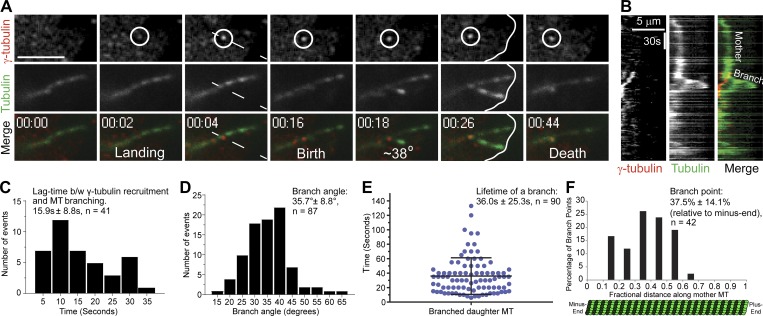
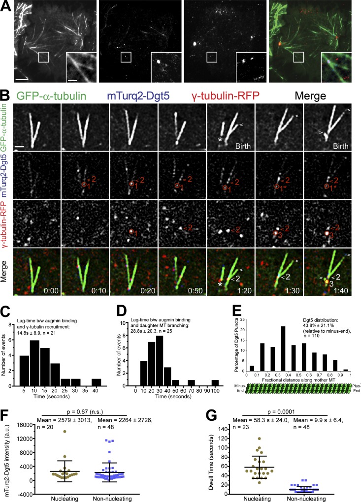
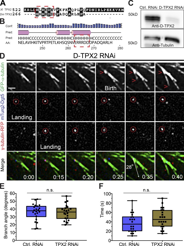
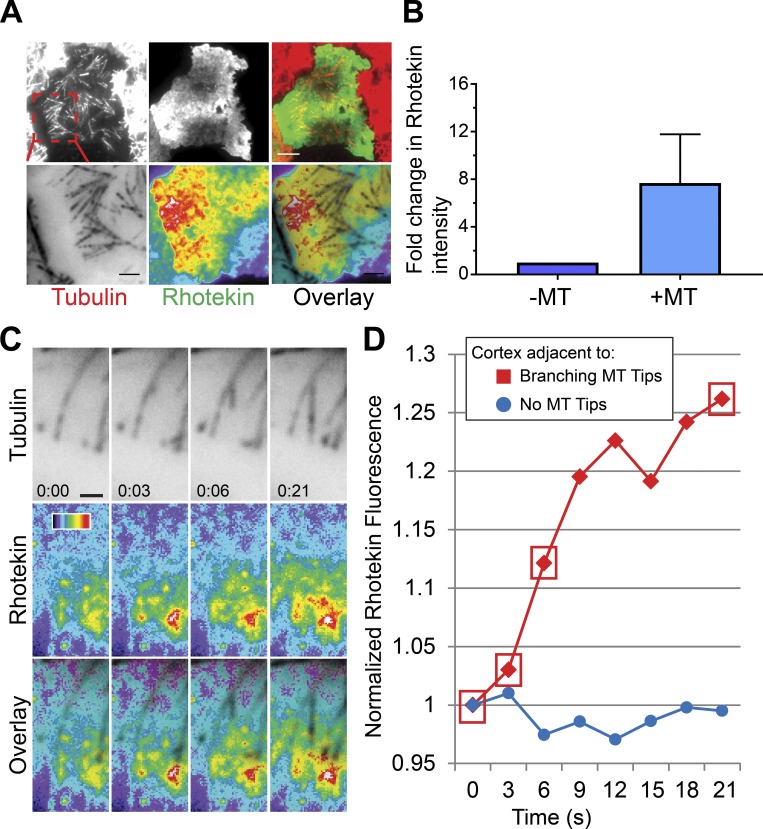
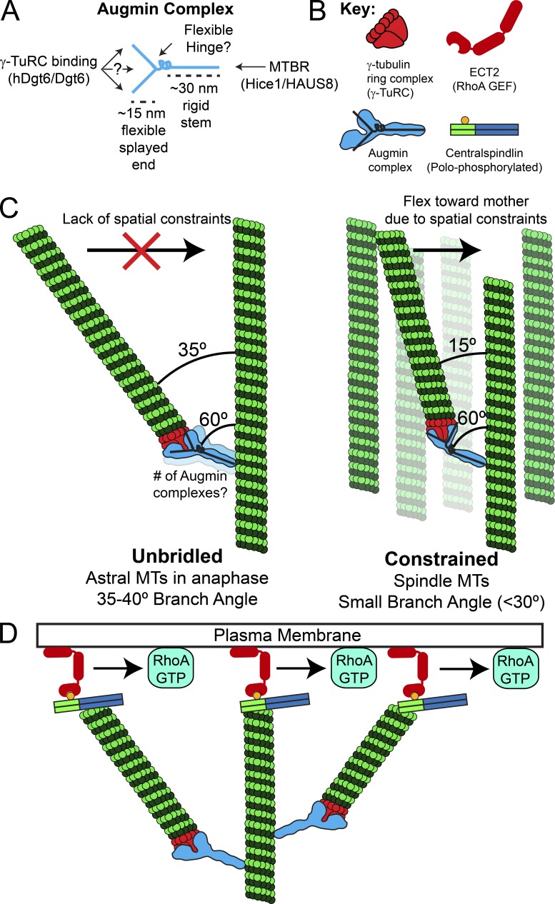
Centrosome-mediated microtubule (MT) nucleation has been well characterized; however, numerous noncentrosomal MT nucleation mechanisms exist. The branching MT nucleation pathway envisages that the γ-tubulin ring complex (γ-TuRC) is recruited to MTs by the augmin complex to initiate nucleation of new MTs. While the pathway is well conserved at a molecular and functional level, branching MT nucleation by core constituents has never been directly observed in animal cells. Here, multicolor TIRF microscopy was applied to visualize and quantitatively define the entire process of branching MT nucleation in dividing Drosophila cells during anaphase. The steps of a stereotypical branching nucleation event entailed augmin binding to a mother MT and recruitment of γ-TuRC after 15 s, followed by nucleation 16 s later of a daughter MT at a 36° branch angle. Daughters typically remained attached throughout their ∼40-s lifetime unless the mother depolymerized past the branch point. Assembly of branched MT arrays, which did not require Drosophila TPX2, enhanced localized RhoA activation during cytokinesis.


Figures

Figure 1.
Figure 1.
Direct visualization of branching MT nucleation in Drosophila S2 cells. (A) Two-color TIRF imaging of an astral MT in a post-anaphase cell coexpressing EGFP-α-tubulin (green) and γ-tubulin-Tag-RFP-T (red). This representative branching event encompasses γ-tubulin landing on the mother MT through disassembly of the daughter MT, both of which contact the cortex (white line at 00:26). (B) Kymograph of the branching event from region (dashed line at 00:04) in A. (C) Histogram of the lag time between (b/w) γ-tubulin recruitment to the mother MT and birth of a daughter MT; n = 41. (D) Distribution of branch angles between mother and daughter MTs. The branch angles were measured 5–10 s after the appearance of daughter MTs; n = 87. (E) Dot plot shows distribution of lifetimes of daughter MTs; n = 90; lifetime is time from birth to death of the daughter MT. (F) Histogram shows distribution of branch origins along the length of mother MTs; n = 42. Time, min:s. Scale bars, 5 µm (A and B). Mean ± SD values are reported in all the histograms. Error bar on the dot plot indicates ± SD.
Figure 2.
Figure 2.
Direct visualization of the key mediators of branching MT nucleation. (A) Representative TIRF micrographs showing expression and localization of γ-tubulin-Tag-RFP-T (red) and mTurquoise2-Dgt5 (blue) in a mid-anaphase Drosophila S2 cell. Inset shows a representative MT branching event where mTurquoise2-Dgt5 and γ-tubulin-Tag-RFP-T colocalized. (B) Still frames from a TIRF time-lapse of MT branching events in a cell coexpressing GFP-α-tubulin (green), mTurquoise2-Dgt5 (blue), and γ-tubulin-TagRFP (red). The events include the following: (a) Localization of two Dgt5 puncta at 10 s on mother MT (one indicated by the red circle and marked 1, and another indicated by the red arrowhead and marked 2 in the mTurquoise2 channel). (b) Dgt5 puncta (1) recruits γ-tubulin (1) (RFP channel, 0:20). The γ-tubulin puncta (1) dissociates from Dgt5 (1) within 30 s without a branch event; although the Dgt5 puncta (1) remains associated with the mother. (c) Dgt5 (2) recruits γ-tubulin (2) (RFP channel, 0:50), and this complex nucleates a daughter within 30 s (1:20). (d) Dgt5 puncta (now denoted 1*) recruits a second γ-tubulin (3) (RFP channel, 1:30), which nucleates a branch within 20 s (1:40). The first daughter MT grows for 20 s (plus end indicated by white arrowheads in GFP channel) while the second daughter MT was born shortly before the mother depolymerizes. The asterisk (GFP channel, 1:20) indicates a rare event in which a daughter with minus end–associated γ-tubulin and Dgt5 dissociates from an intact mother. The unattached daughter depolymerized completely within 20 s. (C) Average lag time between (b/w) mTurquoise2-Dgt5 (augmin complex subunit) binding to the mother MT and recruitment of γ-tubulin-TagRFP-T; n = 21. (D) Histogram of lag time between Dgt5 recruitment to the mother MT and birth of a daughter MT; n = 25. (E) Histogram shows distribution of Dgt5 puncta along the length of mother MTs; n = 110. (F) Dot plot of fluorescence intensities of Dgt5 puncta that supported nucleation of branched MTs (n = 20) versus those that do not support nucleation of branched MTs (n = 48). (G) Dot plot of dwell times of Dgt5 puncta that supported nucleation of branched MTs (n = 23) versus those that do not support nucleation of branched MTs (n = 48). Time: min:s. Scale bars, 5 µm (A), 1 µm (A inset and B). Mean ± SD values are reported. Two-tailed P values of Student’s t tests are reported; n.s., not significant or P > 0.05.
Figure 3.
Figure 3.
Branching MT nucleation is unaffected by D-TPX2 depletion. (A) Protein sequences of Xenopus (Xl) TPX2 and Drosophila (Dm) TPX2 (D-TPX2) were aligned using T-Coffee multiple alignment software (Notredame et al., 2000). Red dashed rectangle indicates a partially conserved CM1/γ-TuNA motif in D-TPX2. (B) Secondary structure prediction of D-TPX2 was generated using PSIPRED bioinformatics software (Buchan and Jones, 2019). Partially conserved CM1/γ-TuNA motif is expected to lie within α-helices (indicated with red dashed square). Conf, confidence; pred, predicted. (C) Western blot showing depletion of endogenous TPX2 with tubulin as a loading control (Ctrl). (D) A representative MT branching event in a D-TPX2–depleted cell coexpressing GFP-α-tubulin (green), mTurquoise2-Dgt5 (blue), and γ-tubulin-TagRFP (red). Time point 00:00 indicates landing of a Dgt5 molecule, which recruits γ-tubulin at 0:15 s. The Dgt5-γ-tubulin complex nucleates a MT branch at 0:35 s. Scale bar, 1 µm. (E) Distribution of branch angles in control and D-TPX2–depleted cells; n = 44. (F) Lifetime of branched daughter MTs in control and D-TPX2–depleted cells; n = 44. Time: min:s. Box plots indicate full range of variation (from minimum to maximum), the interquartile range, and the median. Two-tailed P values of Student’s t tests are reported; n.s., not significant or P > 0.05.
Figure 4.
Figure 4.
Branching MT nucleation amplifies RhoA activation during cytokinesis. (A) The active RhoA reporter Rhotekin is enriched near the MT plus ends of branched MT arrays (top); enlarged view of the branched MT arrays (red dashed box) that activate cortical RhoA (bottom). (B) Fold change in Rhotekin fluorescence intensity near the MT plus ends with respect to a nearby cortical region devoid of astral MTs. Data were pooled from three independent experiments (n = 3). (C) Still frames from a multicolor TIRF time-lapse showing a branching event that locally increases cortical RhoA activation, visualized with Rhotekin. (D) Rhotekin fluorescence increases ∼30% (red) proximal to the mother and daughter MTs during the branching nucleation event shown in C. A nearby cortical region devoid of astral MTs (blue) exhibited no change in Rhotekin fluorescence during the same time period. Red boxes correspond to the time points in C. Error bars indicate SD. Time: min:s. Color wedge, pixel values 150–1,200. Scale bars, 5 µm (A), 1 µm (C).
Figure 5.
Figure 5.
Model of augmin-mediated branching MT nucleation and its role in cytokinesis. (A) Based on negative stain EM of the augmin complex, we propose that the augmin complex contains a rigid ∼30-nm-long stem that binds MTs at one end and an ∼15-nm flexible splayed end that recruits γ-TuRC through Dgt6 and possibly other interfaces. MTBR, MT binding region in Hice 1/HAUS8. (B) Key to molecular schematics in the figure. Note the overlaid “Y” on the augmin complex schematic, which is drawn based on negative stain EM of the complex. (C) A flexible hinge region in the augmin complex and/or flexibility in the splayed Y-end of augmin may allow a daughter MT to sample a broad range of possible branch angles relative to its mother. The branch angle is impacted by the local cellular environment such that “unbridled” daughter MTs with fewer spatial constraints (e.g., astral MTs in anaphase) would have larger branch angles than daughters nucleated in the spatially constrained environment of the spindle and midzone MT array. (D) Branched MT arrays may amplify RhoA activation by generating more MT plus ends that are capable of recruiting cortical ECT2 via direct interaction with plus end–bound (polo-phosphorylated) centralspindlin.

Similar articles

Cited by

References

    1. Alfaro-Aco R., Thawani A., and Petry S.. 2017. Structural analysis of the role of TPX2 in branching microtubule nucleation. J. Cell Biol. 216:983–997. 10.1083/jcb.201607060 - DOI - PMC - PubMed
    1. Basnet N., Nedozralova H., Crevenna A.H., Bodakuntla S., Schlichthaerle T., Taschner M., Cardone G., Janke C., Jungmann R., Magiera M.M., et al. . 2018. Direct induction of microtubule branching by microtubule nucleation factor SSNA1. Nat. Cell Biol. 20:1172–1180. 10.1038/s41556-018-0199-8 - DOI - PMC - PubMed
    1. Bement W.M., Benink H.A., and von Dassow G.. 2005. A microtubule-dependent zone of active RhoA during cleavage plane specification. J. Cell Biol. 170:91–101. 10.1083/jcb.200501131 - DOI - PMC - PubMed
    1. Benink H.A., and Bement W.M.. 2005. Concentric zones of active RhoA and Cdc42 around single cell wounds. J. Cell Biol. 168:429–439. 10.1083/jcb.200411109 - DOI - PMC - PubMed
    1. Brugués J., Nuzzo V., Mazur E., and Needleman D.J.. 2012. Nucleation and transport organize microtubules in metaphase spindles. Cell. 149:554–564. 10.1016/j.cell.2012.03.027 - DOI - PubMed

Publication types

MeSH terms

LinkOut - more resources