M Cells: Intelligent Engineering of Mucosal Immune Surveillance
- PMID: 31312204
- PMCID: PMC6614372
- DOI: 10.3389/fimmu.2019.01499
M Cells: Intelligent Engineering of Mucosal Immune Surveillance
Abstract
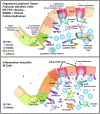
M cells are specialized intestinal epithelial cells that provide the main machinery for sampling luminal microbes for mucosal immune surveillance. M cells are usually found in the epithelium overlying organized mucosal lymphoid tissues, but studies have identified multiple distinct lineages of M cells that are produced under different conditions, including intestinal inflammation. Among these lineages there is a common morphology that helps explain the efficiency of M cells in capturing luminal bacteria and viruses; in addition, M cells recruit novel cellular mechanisms to transport the particles across the mucosal barrier into the lamina propria, a process known as transcytosis. These specializations used by M cells point to a novel engineering of cellular machinery to selectively capture and transport microbial particles of interest. Because of the ability of M cells to effectively violate the mucosal barrier, the circumstances of M cell induction have important consequences. Normal immune surveillance insures that transcytosed bacteria are captured by underlying myeloid/dendritic cells; in contrast, inflammation can induce development of new M cells not accompanied by organized lymphoid tissues, resulting in bacterial transcytosis with the potential to amplify inflammatory disease. In this review, we will discuss our own perspectives on the life history of M cells and also raise a few questions regarding unique aspects of their biology among epithelia.
Keywords: Inflammatory Bowel Disease; endocytosis; epithelium; innate immunity; mucosal immunity.
Figures


Similar articles
-
The gut as a lymphoepithelial organ: the role of intestinal epithelial cells in mucosal immunity.Folia Microbiol (Praha). 1995;40(4):385-91. doi: 10.1007/BF02814746. Folia Microbiol (Praha). 1995. PMID: 8763152 Review.
-
Molecular insights into the mechanisms of M-cell differentiation and transcytosis in the mucosa-associated lymphoid tissues.Anat Sci Int. 2018 Jan;93(1):23-34. doi: 10.1007/s12565-017-0418-6. Epub 2017 Nov 2. Anat Sci Int. 2018. PMID: 29098649 Review.
-
Antigen sampling by epithelial tissues: implication for vaccine design.Behring Inst Mitt. 1997 Feb;(98):24-32. Behring Inst Mitt. 1997. PMID: 9382745 Review.
-
Collaboration of epithelial cells with organized mucosal lymphoid tissues.Nat Immunol. 2001 Nov;2(11):1004-9. doi: 10.1038/ni1101-1004. Nat Immunol. 2001. PMID: 11685223 Review.
-
Commensal bacteria (normal microflora), mucosal immunity and chronic inflammatory and autoimmune diseases.Immunol Lett. 2004 May 15;93(2-3):97-108. doi: 10.1016/j.imlet.2004.02.005. Immunol Lett. 2004. PMID: 15158604 Review.
Cited by
-
Mucosal delivery of nanovaccine strategy against COVID-19 and its variants.Acta Pharm Sin B. 2022 Nov 21;13(7):2897-925. doi: 10.1016/j.apsb.2022.11.022. Online ahead of print. Acta Pharm Sin B. 2022. PMID: 36438851 Free PMC article. Review.
-
Characterization of Canine Peyer's Patches by Multidimensional Analysis: Insights from Immunofluorescence, Flow Cytometry, and Single-Cell RNA Sequencing.Immunohorizons. 2023 Nov 1;7(11):788-805. doi: 10.4049/immunohorizons.2300091. Immunohorizons. 2023. PMID: 38015460 Free PMC article.
-
Identifying Spatial Co-occurrence in Healthy and InflAmed tissues (ISCHIA).Mol Syst Biol. 2024 Feb;20(2):98-119. doi: 10.1038/s44320-023-00006-5. Epub 2024 Jan 15. Mol Syst Biol. 2024. PMID: 38225383 Free PMC article.
-
Understanding human gut diseases at single-cell resolution.Hum Mol Genet. 2020 Sep 30;29(R1):R51-R58. doi: 10.1093/hmg/ddaa130. Hum Mol Genet. 2020. PMID: 32588873 Free PMC article. Review.
-
Intranasal vaccination with lipid-conjugated immunogens promotes antigen transmucosal uptake to drive mucosal and systemic immunity.Sci Transl Med. 2022 Jul 20;14(654):eabn1413. doi: 10.1126/scitranslmed.abn1413. Epub 2022 Jul 20. Sci Transl Med. 2022. PMID: 35857825 Free PMC article.
References
Publication types
MeSH terms
Substances
Grants and funding
LinkOut - more resources
Full Text Sources
Other Literature Sources

