. 2019 Oct;25(10):1377-1392.
doi: 10.1261/rna.071118.119. Epub 2019 Jul 15.

Mutually exclusive amino acid residues of L13a are responsible for its ribosomal incorporation and translational silencing leading to resolution of inflammation

Affiliations

Mutually exclusive amino acid residues of L13a are responsible for its ribosomal incorporation and translational silencing leading to resolution of inflammation

Ravinder Kour et al. RNA. 2019 Oct.

Abstract

Eukaryotic ribosomal protein L13a is a member of the conserved universal ribosomal uL13 protein family. Structurally, L13a is distinguished from its prokaryotic counterparts by the presence of an ∼55 amino acid-long carboxy-terminal α-helical extension. The importance of these evolved residues in the carboxy-terminal extension for mammalian ribosome biogenesis as well as L13a's extraribosomal function in GAIT (γ interferon-activated inhibitor of translation) complex-mediated translation silencing during inflammation is not understood. Here, we present biochemical analyses of L13a mutant variants identifying several mutually exclusive amino acid residues in the eukaryote-specific carboxy-terminal extension of human L13a (Tyr149-Val203) important for ribosomal incorporation and translational silencing. Specifically, we show that mutation of Arg169, Lys170, and Lys171 to Ala abrogate GAIT-mediated translational silencing, but not L13a incorporation into ribosomes. Moreover, we show that the carboxy-terminal helix alone can silence translation of GAIT element-containing mRNAs in vitro. We also show through cellular immunofluorescence experiments that nuclear but not nucleolar localization of L13a is resistant to extensive amino acid alterations, suggesting that multiple complex nuclear import signals are present within this protein. These studies provide new insights into L13a structure and its ribosomal and extraribosomal functions in model human cells.

Keywords: L13a; inflammation; nuclear localization; ribosomal incorporation; translational silencing.


Figures

FIGURE 1.
FIGURE 1.
Putative NLSs, RNA binding sites, and structure of ribosomal protein L13a. (A) Schematic diagram of the human L13a amino acid sequence with predicted NLSs (Arg84 to Met118 and Lys159 to Lys188) and rRNA-binding sites (Lys53 to Ala75 and Arg169 to Lys179) indicated. Arg68, a residue experimentally verified as essential for rRNA-binding and ribosomal incorporation, is also indicated. The eukaryote-specific L13a carboxy-terminal extension spans residues Trp149 to Val203. (B) Human L13a structure depicted as a ribbon diagram based on the CryoEM structure of the human ribosome at 3.6 Å resolution (Protein Data Bank code 5T2C). The conserved globular core domain is shown in dark blue. Predicted NLSs (Arg84 to Met118 and Lys159 to Lys188) are shown in yellow. Predicted RNA-binding sites (Lys53 to Ala75 and Arg169 to Lys179) are shown in red. The eukaryote-specific carboxy-terminal extension (Tyr149 to Val203) is shown in sky blue. Side chains are shown as sticks for Lys53, Lys159, Lys179, Lys188, Ala75, Arg85, Arg169, Met 118, Tyr149, and Val203. (C) Modeling of the interaction between the carboxy-terminal helices of human L13a and L14 proteins. L13a (blue and sky blue) and L14 (red) protein structures are depicted as ribbon diagrams based on the CryoEM structure of the human ribosome at 3.6 Å resolution (Protein Data Bank code 5T2C). Side chains of the L13a residues experimentally determined to affect L13a ribosomal incorporation (Arg68, Lys159, Lys161, Arg169, Lys170, and Lys171) are shown. The van der Waals radius of Arg68 (also essential for L13a ribosomal incorporation) is shown in red. Side chains of L13a residues Val185, Ile189, and Leu196 interacting with the L14 carboxy-terminal helix are shown in yellow.
FIGURE 2.
FIGURE 2.
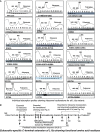
The eukaryotic-specific carboxy-terminal extension of L13a harbors amino acid residues required for its ribosomal incorporation. (A) Density gradient centrifugation cosedimentation analysis of L13a variants. Absorption profiles during ribosome fractionations of individual L13a variants are shown. HA-tagged L13a (wild-type [WT] or mutant variants as indicated at the bottom of each profile) was expressed in HEK 293T cells. Ribosomal fractions (#1–12) were resolved by sucrose density gradient centrifugation (10% to 50%) and analyzed by immunoblotting with anti-HA antibody. The sedimentation of 40S, 60S, and 80S ribosome subunits and polyribosomes is indicated at the top of the figure. (B) Schematic diagram of the human L13a amino acid sequence indicating residues shown to be critical for ribosomal incorporation based on the results shown in A along with other predicted and experimentally determined features.
FIGURE 3.
FIGURE 3.
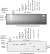
Association of L13a and its mutant variants with 28S rRNA. Lysates prepared from HEK 293T cells expressing HA-tagged L13a (WT or mutant as indicated above each lane) were used for immunoprecipitation with anti-HA-coated agarose beads (or nonantibody-coated blank beads as a control, first lane from left). (Top) Total RNA was extracted from the immunoprecipitates and analyzed by RT-PCR with primers specific for 28S rRNA. RT-PCR products were visualized on an ethidium bromide-stained agarose gel. (Bottom) Equal volumes of the immunoprecipitate from each reaction were run on an SDS-PAGE gel and immunoblotted with anti-HA antibody to confirm the presence of equivalent amounts of HA-tagged protein.
FIGURE 4.
FIGURE 4.
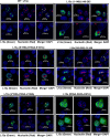
Nuclear and nucleolar localization of L13a and its mutant variants. HEK 293T cells were transfected with constructs directing expression of HA-tagged L13a (WT or deletion mutants [1–148] and [149–203] or triple point mutants Lys159Ala–Arg160Ala–Lys161Ala, Val185Ala–Ile189Ala–Leu196Ala, and Arg59Ala–Lys60Ala–Lys61Ala as indicated above each set of six images). Cells were fixed 24 h post-transfection, stained with anti-HA (green), anti-nucleolin (red; a marker of the nucleolus), and DAPI (blue; to visualize nuclei), and viewed under a fluorescence microscope.
FIGURE 5.
FIGURE 5.
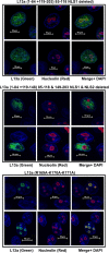
Nuclear and nucleolar localization of L13a variants lacking predicted NLSs or residues important for translational silencing. Nuclear and nucleolar localization of additional L13a variants (1–84 + 119–203; predicted NLS1 deleted), (1–84 + 119–148; predicted NLS1 and NLS2 deleted), and K169A–K170A–K171A (translational silencing incompetent, see Fig. 7) was analyzed by immunofluorescence as described in Figure 4.
FIGURE 6.
FIGURE 6.
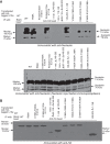
In vivo association of L13a and its mutant variants with nucleolin. (A) Lysates of HEK 293T cells expressing recombinant HA-tagged L13a (WT or mutant as indicated above each lane) were subjected to immunoprecipitation with anti-HA-coated beads (or nonantibody-coated blank beads as a control) as described for Figure 3. The immunoprecipitates were run on SDS-PAGE gels and immunoblotted with antinucleolin antibody (top panel), inputs of nucleolin before immunoprecipitation (bottom panel). (B) Immunoblot with anti-HA antibody.
FIGURE 7.
FIGURE 7.
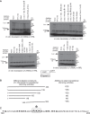
The eukaryote-specific carboxy-terminal extension of L13a harbors amino acid residues required for GAIT element-mediated translational silencing. (A) Effect of L13a variants on in vitro translation of a GAIT element-containing luciferase reporter mRNA and a control mRNA lacking a GAIT element (T7 gene 10). In vitro translation assays were performed as described in Materials and Methods using RRL and purified His-tagged L13a (WT or the indicated deletion or point mutants) produced in a baculovirus expression system. S35-radiolabeled proteins were visualized by 10% SDS-PAGE followed by autoradiography. L13a variants that did not induce GAIT element-dependent translational silencing are marked with a star. The double asterisk at the bottom of each gel shows the front of migration. (B) Schematic illustration of the L13a deletion constructs tested for translational silencing activity and a summary of the results. (C) Amino acid sequence of human L13a from Tyr149 to Val203 with residues tested by point mutations shown in bold font. The three amino acid region identified as essential for translational silencing is indicated by a box with a star.

Similar articles

Cited by

References

    1. Basu A, Poddar D, Robinet P, Smith JD, Febbraio M, Baldwin WM III, Mazumder B. 2014. Ribosomal protein L13a deficiency in macrophages promotes atherosclerosis by limiting translation control-dependent retardation of inflammation. Arterioscler Thromb Vasc Biol 34: 533–542. 10.1161/ATVBAHA.113.302573 - DOI - PMC - PubMed
    1. Basu A, Jain N, Tolbert BS, Komar AA, Mazumder B. 2017. Conserved structures formed by heterogeneous RNA sequences drive silencing of an inflammation responsive post-transcriptional operon. Nucleic Acids Res 45: 12987–13003. 10.1093/nar/gkx979 - DOI - PMC - PubMed
    1. Behrmann E, Loerke J, Budkevich TV, Yamamoto K, Schmidt A, Penczek PA, Vos MR, Bürger J, Mielke T, Scheerer P, et al. 2015. Structural snapshots of actively translating human ribosomes. Cell 161: 845–857. 10.1016/j.cell.2015.03.052 - DOI - PMC - PubMed
    1. Ben-Shem A, Garreau de Loubresse N, Melnikov S, Jenner L, Yusupova G, Yusupov M. 2011. The structure of the eukaryotic ribosome at 3.0 Å resolution. Science 334: 1524–1529. 10.1126/science.1212642 - DOI - PubMed
    1. Boisvert FM, van Koningsbruggen S, Navascués J, Lamond AI. 2007. The multifunctional nucleolus. Nat Rev Mol Cell Biol 8: 574–585. 10.1038/nrm2184 - DOI - PubMed

Publication types

LinkOut - more resources