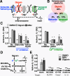
Inflammasome inhibition blocks cardiac glycoside cell toxicity
- PMID: 31300552
- PMCID: PMC6709640
- DOI: 10.1074/jbc.RA119.008330
Inflammasome inhibition blocks cardiac glycoside cell toxicity
Abstract
Chronic heart failure and cardiac arrhythmias have high morbidity and mortality, and drugs for the prevention and management of these diseases are a large part of the pharmaceutical market. Among these drugs are plant-derived cardiac glycosides, which have been used by various cultures over millennia as both medicines and poisons. We report that digoxin and related compounds activate the NLRP3 inflammasome in macrophages and cardiomyocytes at concentrations achievable during clinical use. Inflammasome activation initiates the maturation and release of the inflammatory cytokine IL-1β and the programmed cell death pathway pyroptosis in a caspase-1-dependent manner. Notably, the same fluxes of potassium and calcium cations that affect heart contraction also induce inflammasome activation in human but not murine cells. Pharmaceuticals that antagonize these fluxes, including glyburide and verapamil, also inhibit inflammasome activation by cardiac glycosides. Cardiac glycoside-induced cellular cytotoxicity and IL-1β signaling are likewise antagonized by inhibitors of the NLRP3 inflammasome or the IL-1 receptor-targeting biological agent anakinra. Our results inform on the molecular mechanism by which the inflammasome integrates the diverse signals that activate it through secondary signals like cation flux. Furthermore, this mechanism suggests a contribution of the inflammasome to the toxicity and adverse events associated with cardiac glycosides use in humans and that targeted anti-inflammatories could provide an additional adjunct therapeutic countermeasure.
Keywords: IL-1; cardiac glycoside; cardiomyocyte; caspase 1 (CASP1); cell death; inflammasome; macrophage.
© 2019 LaRock et al.
Conflict of interest statement
The authors declare that they have no conflicts of interest with the contents of this article. The content is solely the responsibility of the authors and does not necessarily represent the official views of the National Institutes of Health
Figures


Similar articles
-
The cardiac glycoside ouabain activates NLRP3 inflammasomes and promotes cardiac inflammation and dysfunction.PLoS One. 2017 May 11;12(5):e0176676. doi: 10.1371/journal.pone.0176676. eCollection 2017. PLoS One. 2017. PMID: 28493895 Free PMC article.
-
NLRP3 inflammasome signaling is activated by low-level lysosome disruption but inhibited by extensive lysosome disruption: roles for K+ efflux and Ca2+ influx.Am J Physiol Cell Physiol. 2016 Jul 1;311(1):C83-C100. doi: 10.1152/ajpcell.00298.2015. Epub 2016 May 11. Am J Physiol Cell Physiol. 2016. PMID: 27170638 Free PMC article.
-
Propofol directly induces caspase-1-dependent macrophage pyroptosis through the NLRP3-ASC inflammasome.Cell Death Dis. 2019 Jul 17;10(8):542. doi: 10.1038/s41419-019-1761-4. Cell Death Dis. 2019. PMID: 31316052 Free PMC article.
-
The role of mitochondria in NLRP3 inflammasome activation.Mol Immunol. 2018 Nov;103:115-124. doi: 10.1016/j.molimm.2018.09.010. Epub 2018 Sep 21. Mol Immunol. 2018. PMID: 30248487 Review.
-
The Role of the Inflammasome in Heart Failure.Front Physiol. 2021 Oct 28;12:709703. doi: 10.3389/fphys.2021.709703. eCollection 2021. Front Physiol. 2021. PMID: 34776995 Free PMC article. Review.
Cited by
-
The Pseudomonas aeruginosa protease LasB directly activates IL-1β.EBioMedicine. 2020 Oct;60:102984. doi: 10.1016/j.ebiom.2020.102984. Epub 2020 Sep 23. EBioMedicine. 2020. PMID: 32979835 Free PMC article.
-
Fibroblast contributions to ischemic cardiac remodeling.Cell Signal. 2021 Jan;77:109824. doi: 10.1016/j.cellsig.2020.109824. Epub 2020 Nov 2. Cell Signal. 2021. PMID: 33144186 Free PMC article. Review.
-
Modeling Nonischemic Genetic Cardiomyopathies Using Induced Pluripotent Stem Cells.Curr Cardiol Rep. 2022 Jun;24(6):631-644. doi: 10.1007/s11886-022-01683-8. Epub 2022 Jun 3. Curr Cardiol Rep. 2022. PMID: 35657495 Free PMC article. Review.
-
Assessing Interleukin-1β Activation During Pyroptosis.Methods Mol Biol. 2023;2641:163-169. doi: 10.1007/978-1-0716-3040-2_13. Methods Mol Biol. 2023. PMID: 37074649
-
Natural toxins and One Health: a review.Sci One Health. 2023 Mar 7;1:100013. doi: 10.1016/j.soh.2023.100013. eCollection 2022 Nov. Sci One Health. 2023. PMID: 39076609 Free PMC article. Review.
References
-
- Tannahill G. M., Curtis A. M., Adamik J., Palsson-McDermott E. M., McGettrick A. F., Goel G., Frezza C., Bernard N. J., Kelly B., Foley N. H., Zheng L., Gardet A., Tong Z., Jany S. S., Corr S. C., et al. (2013) Succinate is an inflammatory signal that induces IL-1β through HIF-1α. Nature 496, 238–242 10.1038/nature11986 - DOI - PMC - PubMed
Publication types
MeSH terms
Substances
Grants and funding
LinkOut - more resources
Full Text Sources
Research Materials

