The oncolytic efficacy and safety of avian reovirus and its dynamic distribution in infected mice
- PMID: 31299861
- PMCID: PMC6879773
- DOI: 10.1177/1535370219861928
The oncolytic efficacy and safety of avian reovirus and its dynamic distribution in infected mice
Abstract
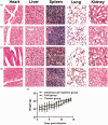
Primary liver cancer is a major public health challenge that ranks as the third most common cause of cancer worldwide despite therapeutic improvement. Reovirus has been emerging as a potential anti-cancer agent and is undergoing multiple clinical trials, and it is reported that reovirus can preferentially cause the cell death of a variety of cancers in a manner of apoptosis. As few studies have reported the efficacy of oncolytic activity and safety profile of avian reovirus, in our study, LDH assay, MTT assay, DAPI staining, and flow cytometry assay were performed to demonstrate the oncolytic effects of avian reovirus against the HepG2 cells, and quantitative real-time PCR (qRT-PCR) and animal experiments were conducted to investigate the dynamic distribution of avian reovirus in infected mice and then illustrate the safety and tissue tropism of avian reovirus. LDH assay, DAPI staining, and flow cytometry assay confirmed the efficacy of the oncotherapeutic effects of avian reovirus, and MTT assay has indicated that avian reovirus suppressed the proliferation of HepG2 cells and decreased their viability significantly. qRT-PCR revealed the dynamic distribution of avian reovirus in infected mice that avian reovirus might replicate better and have more powerful oncolytic activity in liver, kidney, and spleen tissues. Furthermore, histopathological examination clearly supported that avian reovirus appeared non-pathogenic to the normal host, so our study may provide the new insights and rationale for the new strategy of removing liver cancer.
Impact statement: We demonstrated the efficacy of oncolytic activity of avian reovirus (ARV) by LDH assay, MTT assay, DAPI staining, and flow cytometry assay, and also investigated the dynamic distribution of ARV in infected mice and then illustrated the safety and tissue tropism of ARV by quantitative real-time PCR (qRT-PCR) and animal experiments. Collectively, our study may provide the new insights and rationale for the new strategy of removing liver cancer.
Keywords: Hepatocellular carcinoma; avian reovirus; dynamic distribution; oncolytic virus; real-time PCR; safety.
Figures


Similar articles
-
Role of Myeloid Cells in Oncolytic Reovirus-Based Cancer Therapy.Viruses. 2021 Apr 10;13(4):654. doi: 10.3390/v13040654. Viruses. 2021. PMID: 33920168 Free PMC article. Review.
-
Oncolytic avian reovirus-sensitized tumor infiltrating CD8+ T cells triggering immunogenic apoptosis in gastric cancer.Cell Commun Signal. 2024 Oct 21;22(1):514. doi: 10.1186/s12964-024-01888-0. Cell Commun Signal. 2024. PMID: 39434159 Free PMC article.
-
Polymorphisms in the Most Oncolytic Reovirus Strain Confer Enhanced Cell Attachment, Transcription, and Single-Step Replication Kinetics.J Virol. 2020 Jan 31;94(4):e01937-19. doi: 10.1128/JVI.01937-19. Print 2020 Jan 31. J Virol. 2020. PMID: 31776267 Free PMC article.
-
Oncolytic reovirus therapy: Pilot study in dogs with spontaneously occurring tumours.Vet Comp Oncol. 2018 Jun;16(2):229-238. doi: 10.1111/vco.12361. Epub 2017 Oct 27. Vet Comp Oncol. 2018. PMID: 29076241
-
Oncolytic Reoviruses: Can These Emerging Zoonotic Reoviruses Be Tamed and Utilized?DNA Cell Biol. 2023 Jun;42(6):289-304. doi: 10.1089/dna.2022.0561. Epub 2023 Apr 4. DNA Cell Biol. 2023. PMID: 37015068 Review.
Cited by
-
Oncolytic viruses-modulated immunogenic cell death, apoptosis and autophagy linking to virotherapy and cancer immune response.Front Cell Infect Microbiol. 2023 Mar 15;13:1142172. doi: 10.3389/fcimb.2023.1142172. eCollection 2023. Front Cell Infect Microbiol. 2023. PMID: 37009515 Free PMC article. Review.
-
Cell Entry of Avian Reovirus Modulated by Cell-Surface Annexin A2 and Adhesion G Protein-Coupled Receptor Latrophilin-2 Triggers Src and p38 MAPK Signaling Enhancing Caveolin-1- and Dynamin 2-Dependent Endocytosis.Microbiol Spectr. 2023 Jun 15;11(3):e0000923. doi: 10.1128/spectrum.00009-23. Epub 2023 Apr 25. Microbiol Spectr. 2023. PMID: 37097149 Free PMC article.
-
Role of Myeloid Cells in Oncolytic Reovirus-Based Cancer Therapy.Viruses. 2021 Apr 10;13(4):654. doi: 10.3390/v13040654. Viruses. 2021. PMID: 33920168 Free PMC article. Review.
-
p17-Modulated Hsp90/Cdc37 Complex Governs Oncolytic Avian Reovirus Replication by Chaperoning p17, Which Promotes Viral Protein Synthesis and Accumulation of Viral Proteins σC and σA in Viral Factories.J Virol. 2022 Mar 23;96(6):e0007422. doi: 10.1128/jvi.00074-22. Epub 2022 Feb 2. J Virol. 2022. PMID: 35107368 Free PMC article.
-
Oncolytic avian reovirus-sensitized tumor infiltrating CD8+ T cells triggering immunogenic apoptosis in gastric cancer.Cell Commun Signal. 2024 Oct 21;22(1):514. doi: 10.1186/s12964-024-01888-0. Cell Commun Signal. 2024. PMID: 39434159 Free PMC article.
References
-
- Torre LA, Bray F, Siegel RL, Ferlay J, Lortet-Tieulent J, Jemal A. Global cancer statistics. CA Cancer J Clin 2015; 65:87–108 - PubMed
-
- Vucenik I, Zhang ZS, Shamsuddin AM. IP6 in treatment of liver cancer II. Intra-tumoral injection of IP6 regresses pre-existing human liver cancer xenotransplanted in nude mice. Anticancer Res 1998; 18:4091–6 - PubMed
-
- Kowdley KV, Hassanein T, Kaur S, Farrell FJ, Van Thiel DH, Keeffe EB, Sorrell MF, Bacon BR, Weber FJ, Tavill AS. Primary liver cancer and survival in patients undergoing liver transplantation for hemochromatosis. Liver Transpl Surg 1995; 1:237–41 - PubMed
Publication types
MeSH terms
LinkOut - more resources
Full Text Sources
Research Materials

