. 2019 Jul 9;28(2):434-448.e6.
doi: 10.1016/j.celrep.2019.06.027.

SAMHD1 Modulates Early Steps during Human Cytomegalovirus Infection by Limiting NF-κB Activation

Affiliations

SAMHD1 Modulates Early Steps during Human Cytomegalovirus Infection by Limiting NF-κB Activation

Eui Tae Kim et al. Cell Rep. .

Abstract

Cellular SAMHD1 inhibits replication of many viruses by limiting intracellular deoxynucleoside triphosphate (dNTP) pools. We investigate the influence of SAMHD1 on human cytomegalovirus (HCMV). During HCMV infection, we observe SAMHD1 induction, accompanied by phosphorylation via viral kinase UL97. SAMHD1 depletion increases HCMV replication in permissive fibroblasts and conditionally permissive myeloid cells. We show this is due to enhanced gene expression from the major immediate-early (MIE) promoter and is independent of dNTP levels. SAMHD1 suppresses innate immune responses by inhibiting nuclear factor κB (NF-κB) activation. We show that SAMHD1 regulates the HCMV MIE promoter through NF-κB activation. Chromatin immunoprecipitation reveals increased RELA and RNA polymerase II on the HCMV MIE promoter in the absence of SAMHD1. Our studies reveal a mechanism of HCMV virus restriction by SAMHD1 and show how SAMHD1 deficiency activates an innate immune pathway that paradoxically results in increased viral replication through transcriptional activation of the HCMV MIE gene promoter.

Keywords: HCMV; NF-κB; SAMHD1; human cytomegalovirus; virus restriction.


Conflict of interest statement

DECLARATION OF INTERESTS

The authors declare no competing interests.

Figures

Figure 1.
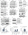
Figure 1.. HCMV Infection Induces SAMHD1 Expression and Phosphorylation
THP-1 or HFF cells were mock infected (0 or M) or infected with HCMV at an MOI of 1. (A) Total RNAs were prepared at the indicated time points, and the levels of SAMHD1 transcripts were measured by qRT-PCR. The results are shown as a.u. relative to the level in uninfected THP-1 cells and represented as the mean values and SD of three independent experiments. The p values were corrected for multiple comparisons using the Benjamini-Hochberg procedure. NS, not significant. (B) Protein level of SAMHD1 was examined over a time course of HCMV infection in THP-1 or HFF cells. Italic numbers beneath the upper blots represent relative fold changes in densities of the bands when compared with uninfected (M) cells after normalization with the loading control. (C) HFF cells were mock infected (M) or infected at an MOI of 3 with either the intact TB40/E strain of HCMV or UV-inactivated HCMV. Cell lysates were prepared at the indicated time points and immunoblotted with the indicated antibodies. (D) THP-1 cell lysates from mock or PMA-treated cells and/or cells infected with HCMV at an MOI of 10 were separated on a Phos-tag acrylamide or on classical acrylamide gels and analyzed by immunoblot using indicated antibodies. (E) HFF cells were infected with HCMV (AD169 strain) wild type (WT) or UL97 kinase active site dead mutant K355Q at an MOI of 1 or 5 for 24 h. Total cell lysates were prepared and immunoblotted with the indicated antibodies. (F) In vitro kinase assays using recombinant proteins as indicated, together with ATP (1 mM) and maribavir (MBV; 1 mM). Reactions were visualized by immunoblotting using anti-phospho-T592-SAMHD1-specific antibody or by silver staining reagents.
Figure 2.
Figure 2.. Depletion of SAMHD1 Increases HCMV Replication and MIE Gene Expression in Primary Fibroblasts
(A) HFF cells were stably transduced with lentiviruses containing non-target control shRNA (NT) or two independent shRNAs to SAMHD1 (SAMHD1#1 and SAMHD1#2). (B) HFF cells were infected with HCMV harboring GFP as a reporter. Cells infected at an MOI of 0.1 were monitored for the spread of GFP signal. GFP images merged with phase-contrast images were collected at 7 days post-infection. Scale bar, 400 μm. (C) Mean fluorescence intensity (MFI) of GFP signals per cell (n = 100). The percentages of GFP-positive cells are indicated above. (D) DNA was extracted at 72 hpi from the SAMHD1 stable knockdown HFF cells infected with HCMV at an MOI of 3. The relative number of viral genomes was determined by qPCR using primers to the UL123 gene region. Data expressed as fold increase over control are shown with mean ± SD replicates of a representative experiment (n = 3). (E) Control and SAMHD1 stable knockdown HFF cells were infected with HCMV at an MOI of 3. Total RNAs were prepared at 24 hpi, and the level of UL123 transcripts (IE1) was measured by qRT-PCR. The results shown are fold increase over control cells. Data are expressed as mean ± SD replicates of a representative experiment (n = 3). (F) HFF cells transiently transfected with scramble siRNA or two independent siRNAs to SAMHD1 were infected with HCMV at an MOI of 3. Total RNAs were prepared at 24 hpi, and the level of UL123 transcripts (IE1) was measured by qRT-PCR. The results are shown as fold increase over control cells. Data are expressed as mean ± SD replicates of a representative experiment (n = 3). (G) HFF cells transfected with siRNAs were infected with HCMV at an MOI of 3. At 24 hpi, total cell lysates were prepared and analyzed by immunoblotting. (H) Control and SAMHD1 stable knockdown HFF cells were mock infected or infected with HCMV at an MOI of 3. Total cell lysates were prepared at the indicated time points, and immunoblot analysis was carried out with the indicated antibodies. (I) Levels of SAMHD1 protein in proliferating and quiescent cell conditions. Immunoblots are of SAMHD1 from THP-1 and HFF cells cultured under proliferating or quiescent conditions and when switched from quiescent to proliferating (Q/P) conditions. (J) Proliferating or quiescent stage HFF cells stably transduced with shRNAs of control or SAMHD1 were mock infected or infected with HCMV at an MOI of 0.1. At 7 days post-infection, total cell lysates were prepared and analyzed by immunoblotting. (K) HCMV was infected at an MOI of 3 for 12 or 24 h into proliferating or quiescent stage HFF cells stably transduced with shRNAs. At 12 or 24 hpi, total cell lysates were prepared and analyzed by immunoblotting.
Figure 3.
Figure 3.. Depletion of SAMHD1 Increases HCMV Replication and MIE Gene Expression in Macrophage-like THP-1
(A) THP-1 cells were stably transduced with lentiviruses containing control shRNA or shRNA to SAMHD1. SAMHD1 protein levels were analyzed by immunoblot. (B) PMA-treated THP-1 cells were infected at an MOI of 5 with HCMV expressing GFP as a reporter. Cells were analyzed by flow cytometry at 72 hpi. (C) PMA-treated THP-1 cells were infected with HCMV at an MOI of 0.2, 1, or 5. At 72 hpi, the relative number of viral genomes was determined by qPCR using primers to the UL123 gene. Data are expressed as mean ± SD replicates of a representative experiment (n = 3). (D) Cell culture supernatants from cells infected with HCMV at an MOI of 5 were analyzed at 10 days post-infection for progeny virus titers using infectious center assays in HFF cells. Data are expressed as mean ± SD replicates of a representative experiment (n = 3). (E) PMA-treated THP-1 cells were infected with HCMV at an MOI of 5. Total RNAs were prepared at the indicated time points, and the levels of UL123 transcripts were measured by qRT-PCR. Data expressed as fold change compared with 12 hpi in control cells are shown as mean ± SD replicates of a representative experiment (n = 3). (F) PMA-treated control THP-1 and cells expressing shRNA for SAMHD1 were mock infected or infected with HCMV at an MOI of 5. Total cell lysates were prepared at the indicated time points, and immunoblot analysis was carried out with indicated antibodies. (G) SAMHD1 protein expression in THP-1 control (Cas9) and KO cells (KO pool and selected monoclonal cells KO#1 and KO#2) confirms efficient knockout (KO) of the SAMHD1 gene. (H) PMA-treated control THP-1 and SAMHD1 KO cells were mock infected or infected with HCMV at an MOI of 5. Total cell lysates were analyzed by immunoblot for viral protein expression. (I) PMA-treated THP-1 cells were infected at an MOI of 5 in the absence or presence of VPA (1 mM) analyzed for protein expression by immunoblot.
Figure 4.
Figure 4.. HCMV Quiescence Is Impaired in SAMHD1-Depleted Cells
(A) Kasumi-3 cells were infected with HCMV (TB40/E-mCherry:IE2-EGFP) at an MOI of 5. Fluorescence microscopy reveals mCherry-positive (virus-infected) and EGFP-positive (IE2-expressing) cells at the indicated time points. Scale bar, 100 μm. (B) Control and SAMHD1 stable knockdown Kasumi-3 cells were infected with HCMV at an MOI of 5. Total RNAs were prepared 10 days post-infection, and the levels of UL122 (IE2) transcripts were measured by qRT-PCR. The results shown are fold increase over parental cells. IE, immediate-early. (C) Levels of UL32 (pp150) transcripts were measured by qRT-PCR as in (B). L, late. (D) Kasumi-3 cells were infected with HCMV (TB40/E-mCherry:UL99-EGFP) at an MOI of 5 for 8 days to model latent infection. Samples were divided and treated with DMSO or 20 μM PMA for 48 h. Kasumi-3 cells were then washed and cocultured in serial dilutions with NUFF cells for 48 h. Kasumi-3 cells were removed, and plaques in the fibroblast monolayer were monitored for an additional 7 days. ELDA was used to calculate frequency of reactivation. Red lines represent cultures treated with 20 μM PMA; black lines represent cultures treated with DMSO as a solvent control. Solid lines are the log-active cell fraction; dashed lines are the 90% confidence interval. Circles represent data points of the log proportion of negative cultures; down-pointing triangles represent data with zero negative response. Scale bar, 100 μm.
Figure 5.
Figure 5.. Ectopic Expression of SAMHD1 Inhibits MIE Gene Expression
(A) U937 cells were stably transduced with control lentivirus (LPCX), lentiviruses expressing wild-type SAMHD1 (WT), and SAMHD1 mutants (HDAA, T592D, and T592V). SAMHD1 protein levels were immunoblotted with anti-FLAG antibody. (B) Untreated and PMA-treated differentiated U937 cells were infected with HCMV at an MOI of 5. Total RNAs were prepared at 24 hpi, and the levels of UL123 transcripts were measured by qRT-PCR. Data expressed as fold change compared with LPCX control cells are shown as mean ± SD replicates of a representative experiment (n = 3). NS, not significant. (C) U937 cells were infected with HCMV at an MOI of 5. Total RNAs were prepared at 24 hpi, and the levels of UL123 transcripts were measured by qRT-PCR. (D–F) Expression of MIE transcript was not affected by dNs treatment. (D) U937 cells were infected with HCMV at an MOI of 5 for 24 h in cells incubated with different doses of dNs. Total RNAs were prepared, and the levels of UL123 transcripts were measured by qRT-PCR. (E) U937 cells were infected with HCMV at an MOI of 5 in cells incubated with 2 mM dNs. UL123 transcripts were analyzed at indicated time points by qRT-PCR. (F) Control or SAMHD1 KO THP-1 cells were infected with HCMV at an MOI of 5 for 24 h in cells incubated with 2 mM dNs. Total RNAs were prepared, and the levels of UL123 transcripts were measured by qRT-PCR. Data expressed as fold change compared with Cas9 control cells without dNs are shown as mean ± SD replicates of a representative experiment (n = 3).
Figure 6.
Figure 6.. SAMHD1 Downregulates NF-κB
(A) Control THP-1 and SAMHD1 KO cells were mock infected or infected with HCMV at an MOI of 5. At 24 hpi, total cell lysates were analyzed by immunoblot using indicated antibodies. (B) HFF cells transfected with siRNAs were mock infected or infected with HCMV at and MOI of 3. At 24 hpi, total cell lysates were prepared and analyzed by immunoblotting. (C) Control THP-1 and SAMHD1 KO cells were mock infected and infected with HCMV at an MOI of 5. At 24 hpi, nuclear and cytoplasmic fractions were prepared and immunoblot analysis for RELA was performed. CTCF and GAPDH were probed to ensure protein loading, as well as to rule out cross-contamination of nuclear and cytoplasmic fractions. (D) Control THP-1 and SAMHD1 KO cells were infected with HCMV at an MOI of 5. At 24 hpi, nuclear and cytoplasmic fractions were prepared and immunoblot analysis for NF-κB1 (p105 and p50) was performed. (E) U937 cells transduced with empty vector or expressing SAMHD1 (WT or HDAA) were infected with HCMV at an MOI of 5. At 24 hpi, total cell lysates were prepared and analyzed by immunoblotting. (F) HFF cells with endogenous SAMHD1 silenced by shRNA were transduced with adenovirus vectors expressing control (LacZ), wild-type SAMHD1, or mutant SAMHD1 (HDAA). At 3 days post-transduction, cells were infected with HCMV at an MOI of 3. Total cell lysates were prepared at 24 hpi, and immunoblot analysis was carried out with antibodies for indicated proteins. Antibody to the V5 epitope detected transgene expression. (G) U937 cells with empty vector, wild-type SAMHD1, or SAMHD1 mutants were infected with HCMV at an MOI of 5. At 24 hpi, total cell lysates were prepared and analyzed by immunoblotting. (H) Control THP-1 and SAMHD1 KO cells were infected with HCMV at an MOI of 5 in the absence or presence of JSH-23 (30 mM), an inhibitor of NF-κB nuclear translocation. At 24 hpi, nuclear and cytoplasmic fractions were prepared and immunoblot analysis for RELA was performed. (I) Control THP-1 and SAMHD1 KO cells were infected with HCMV at an MOI of 5 in the absence or presence of JSH-23. At 24 hpi, total RNAs were prepared and the levels of UL123 transcripts were measured by qRT-PCR. Data expressed as fold change compared with levels in untreated Cas9 control cells are shown as mean ± SD replicates of a representative experiment (n = 3). (J) ChIP assays were performed with THP-1 control and SAMHD1 KO cells at 24 hpi. Cross-linked cell extracts from THP-1 cells with HCMV at an MOI of 5 were immunoprecipitated with RELA, RNA polymerase II (POLR2A), and control immunoglobulin G (IgG) antibodies and analyzed for the presence of a DNA fragment of the MIE promoter region by real-time PCR. ChIP DNA signals were normalized to the input DNA signal as a percentage. Data are expressed as mean ± SD replicates of a representative experiment (n = 3). (K) ChIP assays were performed with histone H3 antibody, as described in (J).

Similar articles

Cited by

References

    1. Ablasser A, Hemmerling I, Schmid-Burgk JL, Behrendt R, Roers A, and Hornung V (2014). TREX1 deficiency triggers cell-autonomous immunity in a cGAS-dependent manner. J. Immunol 192, 5993–5997. - PubMed
    1. Adler M, Tavalai N, Müller R, and Stamminger T (2011). Human cytomegalovirus immediate-early gene expression is restricted by the nuclear domain 10 component Sp100. J. Gen. Virol 92, 1532–1538. - PubMed
    1. Ahn JH, and Hayward GS (2000). Disruption of PML-associated nuclear bodies by IE1 correlates with efficient early stages of viral gene expression and DNA replication in human cytomegalovirus infection. Virology 274, 39–55. - PubMed
    1. Antonucci JM, St Gelais C, de Silva S, Yount JS, Tang C, Ji X, Shepard C, Xiong Y, Kim B, and Wu L (2016). SAMHD1-mediated HIV-1 restriction in cells does not involve ribonuclease activity. Nat. Med 22, 1072–1074. - PMC - PubMed
    1. Antonucci JM, Kim SH, St Gelais C, Bonifati S, Li TW, Buzovetsky O, Knecht KM, Duchon AA, Xiong Y, Musier-Forsyth K, and Wu L (2018). SAMHD1 impairs HIV-1 gene expression and negatively modulates reactivation of viral latency in CD4+ T cells. J. Virol 92, e00292–18. - PMC - PubMed

Publication types

MeSH terms

Substances