. 2019 Jul 9;10(4):e00875-19.
doi: 10.1128/mBio.00875-19.

Positional Dynamics and Glycosomal Recruitment of Developmental Regulators during Trypanosome Differentiation

Affiliations

Positional Dynamics and Glycosomal Recruitment of Developmental Regulators during Trypanosome Differentiation

Balázs Szöőr et al. mBio. .

Abstract

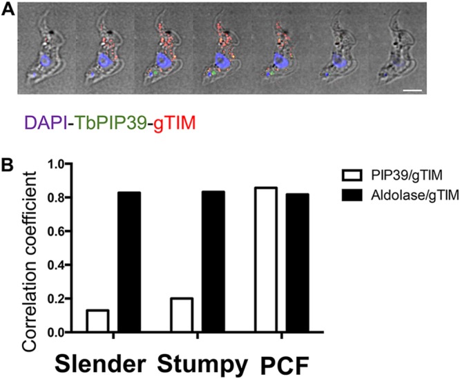
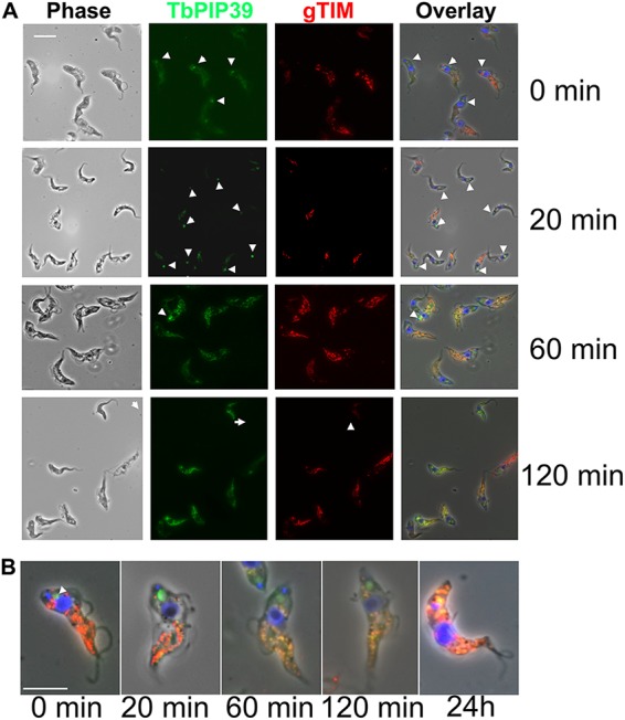
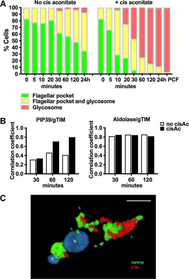
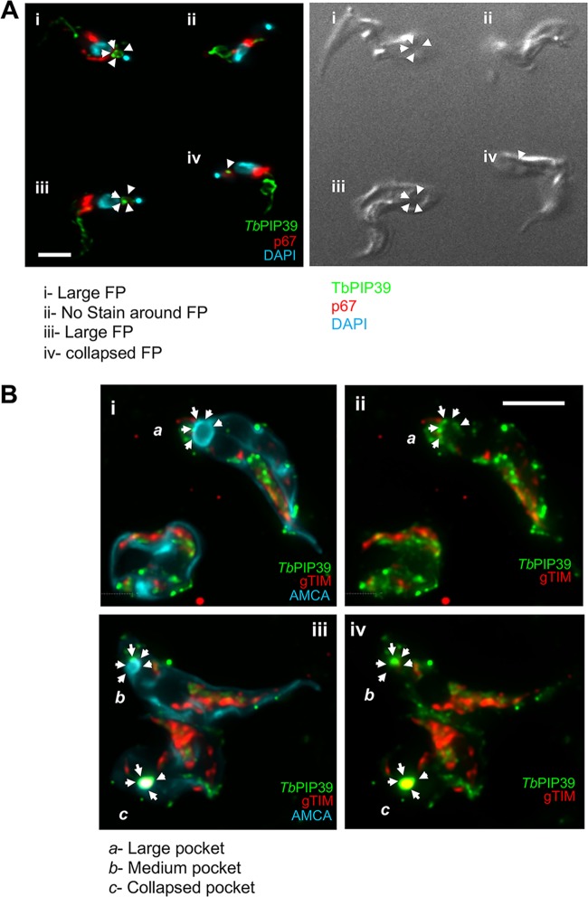
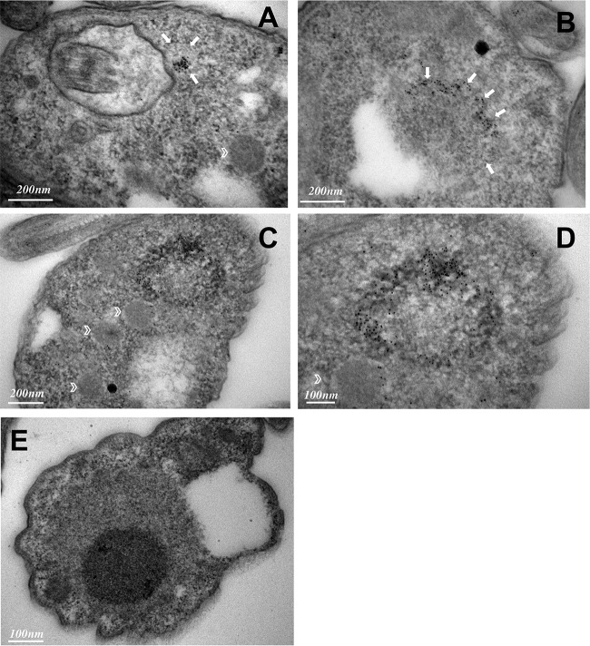
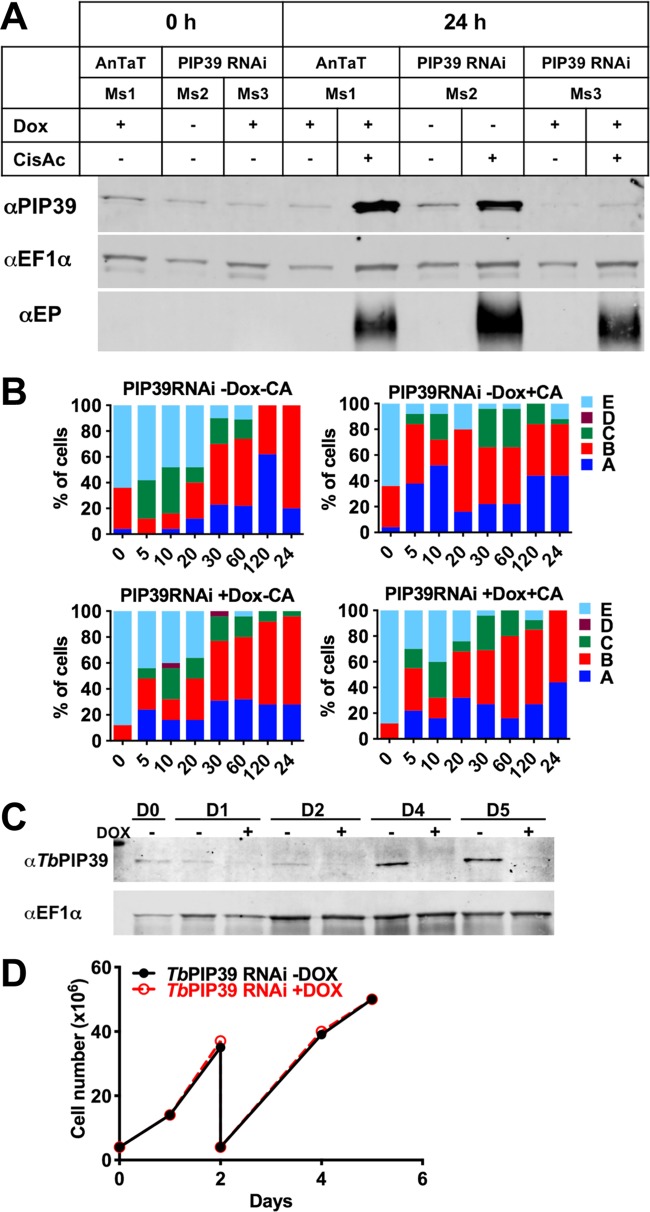
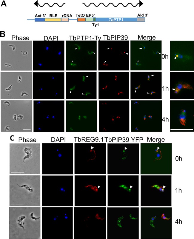
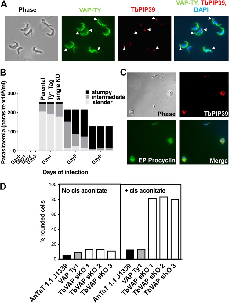
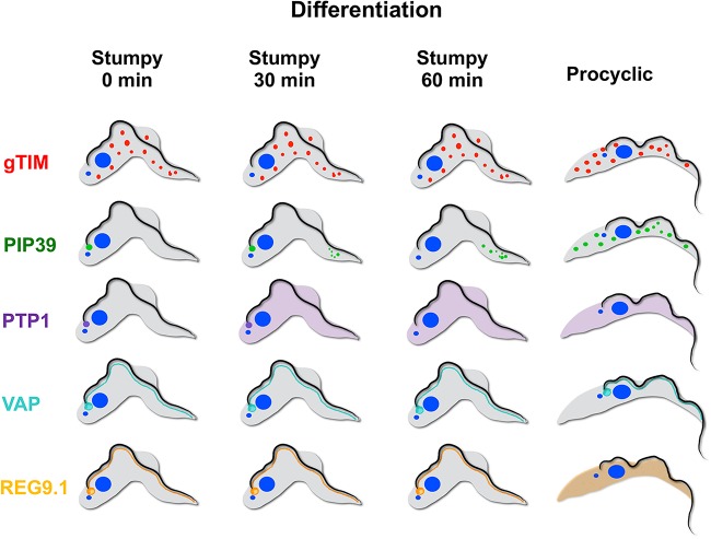
Glycosomes are peroxisome-related organelles that compartmentalize the glycolytic enzymes in kinetoplastid parasites. These organelles are developmentally regulated in their number and composition, allowing metabolic adaptation to the parasite's needs in the blood of mammalian hosts or within their arthropod vector. A protein phosphatase cascade regulates differentiation between parasite developmental forms, comprising a tyrosine phosphatase, Trypanosoma brucei PTP1 (TbPTP1), which dephosphorylates and inhibits a serine threonine phosphatase, TbPIP39, which promotes differentiation. When TbPTP1 is inactivated, TbPIP39 is activated and during differentiation becomes located in glycosomes. Here we have tracked TbPIP39 recruitment to glycosomes during differentiation from bloodstream "stumpy" forms to procyclic forms. Detailed microscopy and live-cell imaging during the synchronous transition between life cycle stages revealed that in stumpy forms, TbPIP39 is located at a periflagellar pocket site closely associated with TbVAP, which defines the flagellar pocket endoplasmic reticulum. TbPTP1 is also located at the same site in stumpy forms, as is REG9.1, a regulator of stumpy-enriched mRNAs. This site provides a molecular node for the interaction between TbPTP1 and TbPIP39. Within 30 min of the initiation of differentiation, TbPIP39 relocates to glycosomes, whereas TbPTP1 disperses to the cytosol. Overall, the study identifies a "stumpy regulatory nexus" (STuRN) that coordinates the molecular components of life cycle signaling and glycosomal development during transmission of Trypanosoma bruceiIMPORTANCE African trypanosomes are parasites of sub-Saharan Africa responsible for both human and animal disease. The parasites are transmitted by tsetse flies, and completion of their life cycle involves progression through several development steps. The initiation of differentiation between blood and tsetse fly forms is signaled by a phosphatase cascade, ultimately trafficked into peroxisome-related organelles called glycosomes that are unique to this group of organisms. Glycosomes undergo substantial remodeling of their composition and function during the differentiation step, but how this is regulated is not understood. Here we identify a cytological site where the signaling molecules controlling differentiation converge before the dispersal of one of them into glycosomes. In combination, the study provides the first insight into the spatial coordination of signaling pathway components in trypanosomes as they undergo cell-type differentiation.

Keywords: development; differentiation; glycosome; organelle; parasite; trypanosome.


Figures

FIG 1
FIG 1
(A) Serial 0.3-μm Z stack slices through a stumpy-form trypanosome cell stained to localize the differentiation regulator TbPIP39 (green) or glycosomal TIM (red). The cell nucleus and kinetoplast are shown in blue. The TbPIP39 is located close to, but slightly anterior of, the kinetoplast and is not colocated with the glycosomal marker. DAPI, 4,6′-diamidino-2-phenylindole. Bar = 5 μm. (B) Pearson coefficient of colocalization between TbPIP39 and glycosomal TIM or between aldolase and glycosomal TIM in bloodstream slender and stumpy forms or in procyclic forms (PCF). Colocalization values were calculated using Volocity software based on captured confocal images. The threshold was set according to the background of images.
FIG 2
FIG 2
(A) Representative images of trypanosomes undergoing differentiation from stumpy forms to procyclic forms. Samples were taken at the time points indicated after the initiation of differentiation by cis-aconitate. Cells were labeled for the location of TbPIP39 (green) or the glycosomal marker gTIM (red), with the nucleus and kinetoplast being labeled with DAPI (4′,6-diamidino-2-phenylindole [blue]). Phase-contrast images are shown on the left-hand side, and merged images are shown on the right. Bar = 10 μm. (B) Selected fields of cells stained for TbPIP39, gTIM, and DAPI at time points after the initiation of differentiation. The location of TbPIP39 proximally to the flagellar pocket region of the cell is highlighted with arrowheads. Bar = 12 μm.
FIG 3
FIG 3
(A) Quantitation of the distribution of TbPIP39 between the periflagellar pocket location only (green), at the periflagellar pocket and in glycosomes (yellow), or exclusively in glycosomes (red). At each time point and under each condition (with or without cis-aconitate, to initiate differentiation), 250 cells were scored. In each case, cells remained in HMI-9 at 37°C to retain the viability of undifferentiated bloodstream-form parasites. (B) Pearson’s coefficient of colocalization between TbPIP39 and glycosomal TIM or between aldolase and glycosomal TIM at time points after the exposure of stumpy forms to cis-aconitate. Colocalization values were calculated using Volocity software based on captured confocal images. The threshold was set according to the background of images. n = 250 cells per condition/time point. (C) Three-dimensional reconstruction of a cell 30 min after exposure to cis-aconitate and stained for TbPIP39 (green) and glycosomal gTIM (red). The cell nucleus (N) and kinetoplast (K) are labeled blue. TbPIP39 is concentrated around the flagellar pocket of the cell but also shows labeling at a dispersed glycosomal location more anterior in the cell, coincident with the distribution of glycosomal gTIM. The labeling is exclusive so that merged staining associated with the coincident location of gTIM and TbPIP39 is not visible. Bar = 5 μm.
FIG 4
FIG 4
(A) Colabeling of stumpy-form cells (without exposure to cis-aconitate) stained for TbPIP39 (green) and the lysosomal marker p67 (red). The nucleus and kinetoplast are stained in blue. The TbPIP39 labeling is distinct from the lysosome, being positioned unevenly around the flagellar pocket (FP [cell i]) or at a tight focus in cells where the flagellar pocket is collapsed (cells iii and iv). Cell ii has no staining detected at the flagellar pocket region. Bar = 5 μm. (B) Colabeling of stumpy-form cells (without exposure to cis-aconitate) stained for TBPIP39 (green) and the glycosomal gTIM (red). The flagellar pocket and cell surface membrane are labeled blue with aminomethylcoumarin (AMCA). Arrows indicate the distribution of the TbPIP39 signal unevenly around the flagellar pocket (Cells a and b [images i and ii]), or at a tight focus associated with a collapsed flagellar pocket (cell c [images iii and iv]). Bar = 5 μm.
FIG 5
FIG 5
(A to D) Immunoelectron micrographs taken of thin sections of the flagellar pocket region of stumpy cells immunogold labeled for TbPIP39. Arrows indicate the boundary of membrane-bound vesicles containing TbPIP39 signal; glycosomes are indicated by chevrons in panels A, C, and D. (E) Immunoelectron micrographs of the flagellar pocket region of stumpy cells in the absence of primary antibody.
FIG 6
FIG 6
(A) RNAi depletion of TbPIP39. T. brucei EATRO 1125 AnTat1.1 90:13 TbPIP39 RNAi cells or T. brucei EATRO 1125 AnTat1.1 90:13 cells were grown in mice, with or without doxycycline induction. The generated stumpy cells were then exposed, or not, to cis-aconitate to initiate differentiation (with doxycycline remaining in the RNAi-induced samples), and TbPIP39 protein was detected at 24 h. TbPIP39 levels increase during differentiation, but this is greatly reduced in induced RNAi samples. EF1α provides a loading control, this being a little higher in cells exposed to cis-aconitate due to their replication as differentiated procyclic forms. EP procyclin staining shows relative differentiation in the presence or absence of cis-aconitate. (B) Distribution of the glycosomal marker aldolase and the lysosomal marker p67 during differentiation between stumpy and procyclic forms. The panel shows the prevalence of different categories of lysosomal/glycosomal staining (as defined by Herman et al. [23]) at times through differentiation when TbPIP39 was depleted by RNAi or not. The cytological profiles were as follows: A, enlarged lysosomal signal; B, lysosome enlarged but separated into distinct smaller vesicles; C, normal lysosomal size, with glycosomal colocalization; D, no lysosome observed; E, lysosome normal, with no colocalization with glycosomes. (C) Western blot of TbPIP39 in cells differentiated to proliferative procyclic forms with TbPIP39 RNAi induced or not. EF1α shows the loading control. The lower panel shows that by days 4 and 5, the cells remain proliferative (the cells were passaged at day 2) despite expressing significantly less TbPIP39.
FIG 7
FIG 7
(A) Schematic representation of the construct used to N-terminally tag TbPTP1 with the Ty1 epitope tag. (B) Colocalization of TbPTP1-Ty1 (green), and TbPIP39 (red) in stumpy forms (0 h) and after 1 and 4 h of exposure to cis-aconitate. The TbPTP1 signal is detected as an ectopically expressed copy incorporating an N-terminal Ty1 epitope tag to allow its detection. The TbPIP39 signal is detected using an antibody recognizing that protein. The cell nucleus and kinetoplast are labeled with DAPI (blue). TbPTP1 colocalized with TbPIP39 in stumpy forms (arrowheads), but after 1 h, TbPIP39 has relocated to glycosomes, while TbPTP1 has become more diffuse, albeit some remains concentrated in the periflagellar pocket region (arrowheads). At 4 h, TbPIP39 is glycosomal, while TbPTP1 is diffuse throughout the cell body. Bar = 10 μm. Enlarged images on the right show selected cells from the merged panel to highlight signal at the periflagellar pocket region (chevron). (C) Detection of YFP-tagged TbPIP39 (green) in stumpy-form cells and its location with respect to REG9.1 (red). DAPI (blue) denotes the position of the cell nucleus and kinetoplast. Bar = 15 μm.
FIG 8
FIG 8
(A) Detection of Ty1 epitope-tagged TbVAP in stumpy-form cells and its location with respect to TbPIP39. The Ty1-tagged TbVAP (green) is detected at the flagellar pocket region, along the flagellum attachment zone, and in the cell body. TbPIP39 (red) is closely proximal at the flagellar pocket region but not precisely coincident. DAPI (blue) denotes the position of the cell nucleus and kinetoplast. Arrowheads indicate the region of TbPIP39. Bar = 15 μm. (B) Proportion of slender, intermediate, and stumpy forms in TbVAP single-knockout cells versus parental cells or epitope-tagged TbVAP cells. (C) TbVAP single-allele-replacement mutants 24 h after the initiation of differentiation to procyclic forms. The differentiating cells (indicated by their expression of EP procyclin [green]) are swollen and lose integrity. TbPIP39 is punctate throughout the cell, indicating a glycosomal rather than periflagellar pocket distribution. The merged panel shows TbPIP39 (red), EP procyclin (green), and DAPI (blue) revealing the nucleus and kinetoplast. Bar = 25 μm. (D) Quantitation of cells that exhibit a rounded morphology. Parental T. brucei AnTat1.1 J1339 cells, TbVAP1-mNeonGreen-Ty1-expressing cells, or three TbVAP single-allele-deletion mutants (sKO) are shown. In each case, the percentage of rounded cells is shown after 24 h in either the absence or presence of cis-aconitate as a differentiation stimulus. At least 250 cells were scored for each cell line and each condition.
FIG 9
FIG 9
Schematic representation of the distribution of gTIM (representing glycosomes) TbPIP39, TbPTP1, TbVAP, and REG9.1 in stumpy forms and at 30 and 60 min after exposure to cis-aconitate and in procyclic forms.

Similar articles

Cited by

References

    1. Wu H, Carvalho P, Voeltz GK. 2018. Here, there, and everywhere: the importance of ER membrane contact sites. Science 361:eaan5835. doi:10.1126/science.aan5835. - DOI - PMC - PubMed
    1. Eden ER, White IJ, Tsapara A, Futter CE. 2010. Membrane contacts between endosomes and ER provide sites for PTP1B-epidermal growth factor receptor interaction. Nat Cell Biol 12:267–272. doi:10.1038/ncb2026. - DOI - PubMed
    1. Allmann S, Bringaud F. 2017. Glycosomes: a comprehensive view of their metabolic roles in T. brucei. Int J Biochem Cell Biol 85:85–90. doi:10.1016/j.biocel.2017.01.015. - DOI - PubMed
    1. Bauer S, Morris MT. 2017. Glycosome biogenesis in trypanosomes and the de novo dilemma. PLoS Negl Trop Dis 11:e0005333. doi:10.1371/journal.pntd.0005333. - DOI - PMC - PubMed
    1. Kennedy PG. 2013. Clinical features, diagnosis, and treatment of human African trypanosomiasis (sleeping sickness). Lancet Neurol 12:186–194. doi:10.1016/S1474-4422(12)70296-X. - DOI - PubMed

Publication types

MeSH terms

Substances

LinkOut - more resources