. 2019 Sep 6:17:323-336.
doi: 10.1016/j.omtn.2019.06.006. Epub 2019 Jun 20.

Selective miRNA Modulation Fails to Activate HIV Replication in In Vitro Latency Models

Affiliations

Selective miRNA Modulation Fails to Activate HIV Replication in In Vitro Latency Models

María Rosa López-Huertas et al. Mol Ther Nucleic Acids. .

Abstract

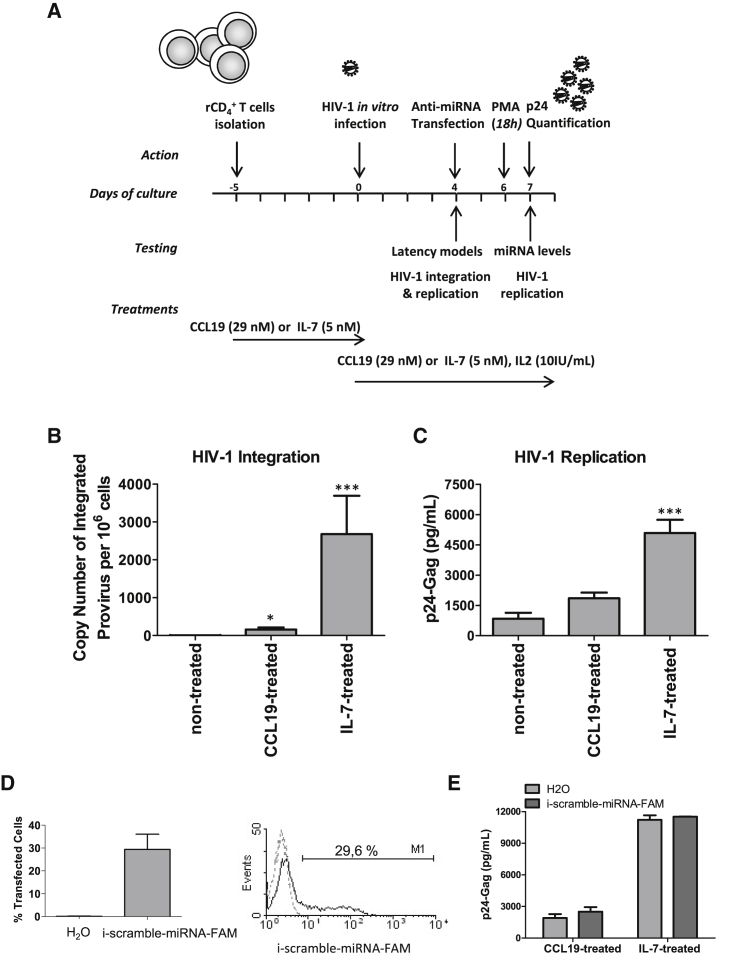
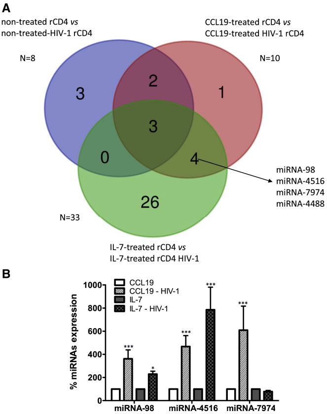
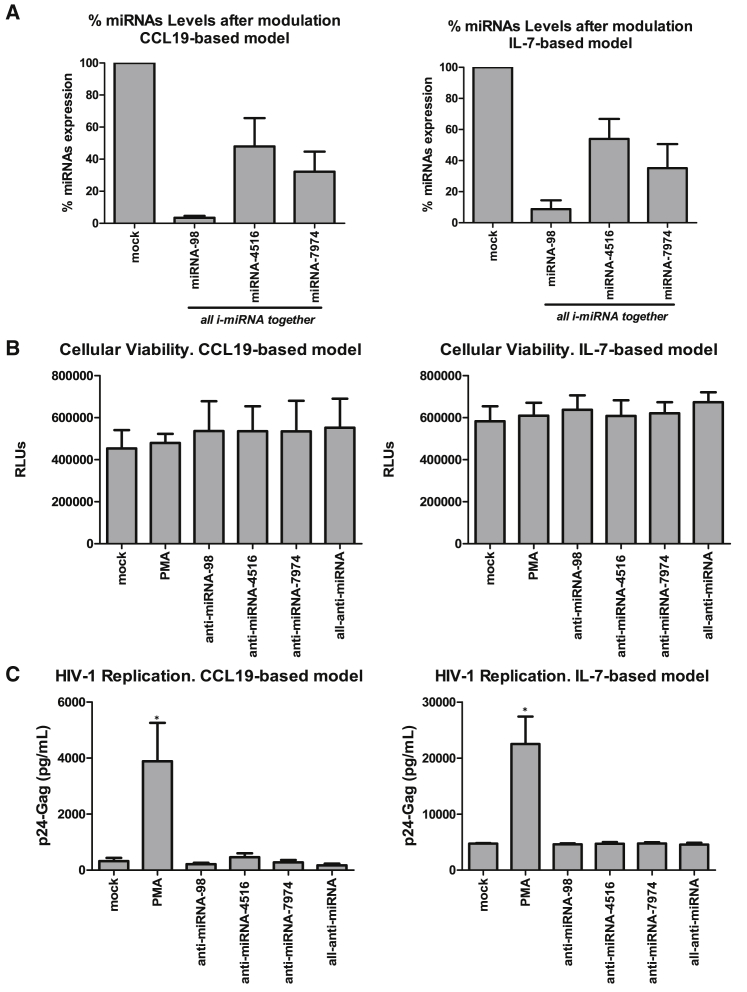
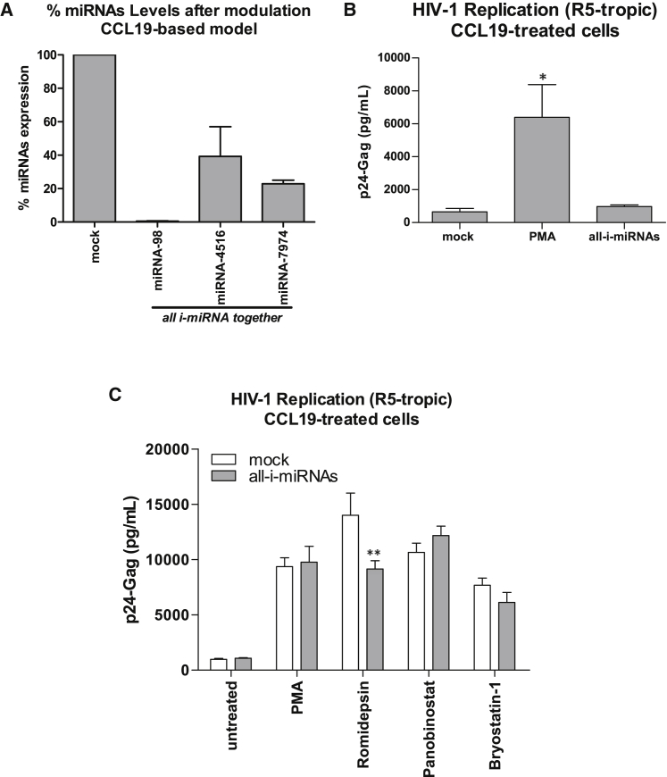
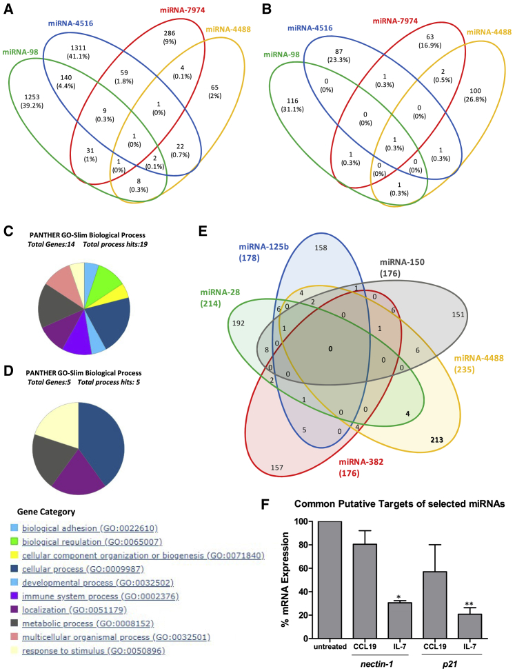
HIV remains incurable because of viral persistence in latent reservoirs that are inaccessible to antiretroviral therapy. A potential curative strategy is to reactivate viral gene expression in latently infected cells. However, no drug so far has proven to be successful in vivo in reducing the reservoir, and therefore new anti-latency compounds are needed. We explored the role of microRNAs (miRNAs) in latency maintenance and their modulation as a potential anti-latency strategy. Latency models based on treating resting CD4 T cells with chemokine (C-C motif) ligand 19 (CCL19) or interleukin-7 (IL7) before HIV infection and next-generation sequencing were used to identify the miRNAs involved in HIV latency. We detected four upregulated miRNAs (miRNA-98, miRNA-4516, miRNA-4488, and miRNA-7974). Individual or combined inhibition of these miRNAs was performed by transfection into cells latently infected with HIV. Viral replication, assessed 72 h after transfection, did not increase after miRNA modulation, despite miRNA inhibition and lack of toxicity. Furthermore, the combined modulation of five miRNAs previously associated with HIV latency was not effective in these models. Our results do not support the modulation of miRNAs as a useful strategy for the reversal of HIV latency. As shown with other drugs, the potential of miRNA modulation as an HIV reactivation strategy could be dependent on the latency model used.

Keywords: CCL19; HIV latency; HIV latency model; IL-7; LRA; latency-reversing agent; miRNA modulation.


Figures

Figure 1
Figure 1
Establishment and Characterization of HIV-1 Latency Models Based on CCL19 or IL-7 Treatment rCD4 lymphocytes were treated with CCL19 (29 nM) or IL-7 (5 nM) for 5 days, infected with X4-tropic NL4-3 HIV-1 strain, and cultured for an additional 4 days before use in modulating expression of cellular miRNAs. (A) A schematic of the HIV-1 primary-cell latency model. (B) qPCR analysis of HIV-1 proviral integration. (C) p24/gag levels in the supernatant of non-activated HIV-1-infected cells were assessed to measure basal replication. Statistical significance was determined by Kruskal-Wallis test with Dunn’s Multiple Comparison post-test analysis (*p < 0.05 and ***p < 0.001). Data in (A) and (B) are the mean ± SEM from eight independent experiments. Comparison are referred to non-treated cells. (D) A vector expressing a non-relevant, scrambled anti-miRNA and the reporter FAM (i-scrambled-miRNA-FAM) was transfected as a control of transfection efficiency. Data are the mean ± SEM (graphs) or a representative experiment (histogram). (E) HIV-1 replication was measured as p24/gag levels in the supernatant of HIV-1 latently infected cells after the expression of i-scrambled-miRNA-FAM vector. Data are the mean ± SEM of two independent experiments.
Figure 2
Figure 2
Differentially Expressed miRNAs Are Shared by Both CCL19- and IL-7-Based HIV-1 Latency Models (A) A Venn diagram showing the differentially expressed miRNAs between all cell conditions studied. A subset of four deregulated miRNAs (miRNA-4488, miRNA-7974, miRNA-98-5p, and miRNA-4516) is shared by the CCL19- and IL-7-treated, HIV-1-infected rCD4 T lymphocytes. (B) The expression levels of miRNA-98, miRNA-4516, and miRNA-7974 were analyzed by qRT-PCR in CCL19- and IL-7-treated, HIV-1-infected T cells. Levels of miRNA-4488 could not be measured, as the commercial oligonucleotides were not accurate. 5S was used as the housekeeping gene. Data are the mean ± SEM and are expressed as the relative percentage of miRNAs levels. Comparisons were performed between HIV-1-infected and uninfected cells within the same treatment (CCL19 versus CCL19-HIV cells or IL-7 versus IL-7-HIV-1 cells). Statistical significance was calculated by two-way ANOVA, followed by the Bonferroni post-test analysis (*p < 0.05, ***p < 0.001).
Figure 3
Figure 3
Modulation of Shared miRNAs in HIV-1-Infected CCL19- or IL-7-Treated rCD4 T Lymphocytes Primary rCD4 T lymphocytes were treated with CCL19 or IL-7 for 5 days, infected with X4-tropic NL4-3 HIV-1, and cultured for an additional 4 days. Then, miRNA-98, miRNA-4516, and miRNA-7974 were inhibited by the transfection of specific anti-miRNA-98, anti-miRNA-4516, and anti-miRNA-7974. Cells were cultured for 3 days and PMA was added for the last 18 h. (A) Expression levels of miRNA-98, miRNA-4516, and miRNA-7974 were measured by qRT-PCR 72 h after transfection of all the anti-miRNAs combined. Data are the mean ± SEM and are expressed as the percentage of change, with 5S levels used as the reference. (B) Cell viability was measured in HIV-1 latently infected rCD4 T cells 3 days after the inhibition of miRNA-98, miRNA-4516, and miRNA-7974. Data are represented as absolute values of relative light units (RLUs). (C) HIV-1 replication was assessed by quantifying p24/gag levels in the culture supernatant 72 h after miRNA modulation or 18 h after PMA activation. Statistical significance was determined by using the Kruskal-Wallis test with Dunn’s multiple comparison post-test analysis (*p < 0.05). Mock cells served as the reference. Data from four independent experiments are shown.
Figure 4
Figure 4
Modulation of Shared miRNAs in rCD4 T Lymphocytes Treated with CCL19 and Infected with R5-Tropic HIV Primary rCD4 T lymphocytes were treated with CCL19 for 5 days, infected with R5-tropic JR-CSF HIV-1, and cultured for an additional 4 days. Then, miRNA-98, miRNA-4516, and miRNA-7974 were altogether inhibited by the transfection of specific anti-miRNA-98, anti-miRNA-4516, and anti-miRNA-7974. The cells were cultured for 3 days, and PMA or the indicated latency-reversing agent (LRA) was added for the last 18 h. (A) Expression levels of miRNA-98, miRNA-4516, and miRNA-7974 were measured by qRT-PCR 72 h after transfection of the combined anti-miRNAs. Data are the mean ± SEM and are expressed as the percentage of change, using 5S levels as the reference. (B) HIV-1 replication was assessed in the same cells by quantifying p24/gag levels in the culture supernatant 72 h after miRNA modulation or 18 h after PMA activation. Statistical significance was estimated using the Kruskal-Wallis test with Dunn’s multiple-comparison post-test analysis (*p < 0.05). Mock cells served as the reference. (C) HIV-1 replication was assessed by quantifying p24/gag levels in the culture supernatant 72 h after miRNA modulation. PMA or the indicated LRA was added for the last 18 h to mock-transfected cells and to cells transfected with the miRNA inhibitors (all-i-miRNAs). The LRAs used were romidepsin (40 nM), panobinostat (30 nM), and bryostatin-1 (10 nM). Statistical significance was studied using two-way ANOVA with the Bonferroni post-test analysis; differences between mock-transfected cells and cells transfected with the miRNA inhibitors were not significant. Data are from four independent experiments.
Figure 5
Figure 5
Modulation in the CCL19 and IL7 Models of miRNAs Previously Described as Being Associated with Latency Primary rCD4 T lymphocytes were treated with CCL19 or IL-7 for 5 days, infected with X4-tropic NL4-3 HIV-1, and cultured for an additional 4 days. Then, miRNA-28, miRNA-125b, miRNA-150, miRNA-223, and miRNA-382 were inhibited by the transfection of specific anti-miRNA-28, anti-miRNA-125b, anti-miRNA-150, anti-miRNA-223, and anti-miRNA-382. Cells were cultured for 3 days, and PMA was added for the last 18 h. (A) Expression levels of miRNA-28, miRNA-125b, miRNA-150, miRNA-223, and miRNA-382 were measured by qRT-PCR 72 h after transfecting all the anti-miRNAs in combination. Data are the mean ± SEM and are expressed as the percentage of change, using 5S levels as the reference. (B) Cell viability was measured 72 h after modulation of the miRNAs or 18 h after PMA activation. Data are absolute values of RLUs. (C) HIV-1 replication was assessed by quantifying p24/gag levels in the culture supernatant 72 h after miRNA modulation or 18 h after PMA activation. Statistical significance was determined by using the Kruskal-Wallis test with Dunn’s multiple-comparison post-test analysis (*p < 0.05). Mock cells served as the reference. Data are from three independent experiments.
Figure 6
Figure 6
Functional Implications of miRNAs Associated with Latency Based on Prediction of Targets Predicted targets for miRNAs deregulated in CCL19- and IL-7-based models performed with (A) miRDB or (B) MiRTarBase databases. Common targets are shown in Venn diagrams. The intersection of the four sets indicates one common target for each database: nectin-1 or p21, respectively, for the miRDB or MiRTarBase. (C and D) Functional enrichment in the biological process of the common targets identified in (A) and (B), respectively, and studied using the Panther-Go terms program. (E) Venn diagrams of predicted targets for miRNAs previously described as being associated with targets identified by the MiRTarBase database. (F) Expression of mRNA encoding for nectin-1 and p21 was analyzed by qRT-PCR in CCL19- or IL-7-treated cells, infected with the HIV NL4.3 strain. Non-treated cells were used as a negative control. 5S was used as the housekeeping gene. Media and SEM from three independent studies are shown. Statistical significance was estimated by using the Kruskal-Wallis test with Dunn’s multiple comparison post-test analysis (*p < 0.05 and **p < 0.01).

Similar articles

Cited by

References

    1. UNAIDS . 2017. Fact sheet 2015.https://www.aidsdatahub.org/sites/default/files/publication/UNAIDS_fact_...
    1. Siliciano J.D., Kajdas J., Finzi D., Quinn T.C., Chadwick K., Margolick J.B., Kovacs C., Gange S.J., Siliciano R.F. Long-term follow-up studies confirm the stability of the latent reservoir for HIV-1 in resting CD4+ T cells. Nat. Med. 2003;9:727–728. - PubMed
    1. Coiras M., López-Huertas M.R., Pérez-Olmeda M., Alcamí J. Understanding HIV-1 latency provides clues for the eradication of long-term reservoirs. Nat. Rev. Microbiol. 2009;7:798–812. - PubMed
    1. Eisele E., Siliciano R.F. Redefining the viral reservoirs that prevent HIV-1 eradication. Immunity. 2012;37:377–388. - PMC - PubMed
    1. Buzon M.J., Sun H., Li C., Shaw A., Seiss K., Ouyang Z., Martin-Gayo E., Leng J., Henrich T.J., Li J.Z. HIV-1 persistence in CD4+ T cells with stem cell-like properties. Nat. Med. 2014;20:139–142. - PMC - PubMed