Effect of neuron-derived neurotrophic factor on rejuvenation of human adipose-derived stem cells for cardiac repair after myocardial infarction
- PMID: 31287219
- PMCID: PMC6714174
- DOI: 10.1111/jcmm.14456
Effect of neuron-derived neurotrophic factor on rejuvenation of human adipose-derived stem cells for cardiac repair after myocardial infarction
Abstract
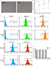
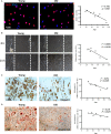
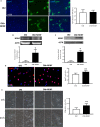
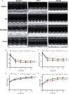
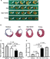
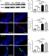
The decline of cell function caused by ageing directly impacts the therapeutic effects of autologous stem cell transplantation for heart repair. The aim of this study was to investigate whether overexpression of neuron-derived neurotrophic factor (NDNF) can rejuvenate the adipose-derived stem cells in the elderly and such rejuvenated stem cells can be used for cardiac repair. Human adipose-derived stem cells (hADSCs) were obtained from donors age ranged from 17 to 92 years old. The effects of age on the biological characteristics of hADSCs and the expression of ageing-related genes were investigated. The effects of transplantation of NDNF over-expression stem cells on heart repair after myocardial infarction (MI) in adult mice were investigated. The proliferation, migration, adipogenic and osteogenic differentiation of hADSCs inversely correlated with age. The mRNA and protein levels of NDNF were significantly decreased in old (>60 years old) compared to young hADSCs (<40 years old). Overexpression of NDNF in old hADSCs significantly improved their proliferation and migration capacity in vitro. Transplantation of NDNF-overexpressing old hADSCs preserved cardiac function through promoting angiogenesis on MI mice. NDNF rejuvenated the cellular function of aged hADSCs. Implantation of NDNF-rejuvenated hADSCs improved angiogenesis and cardiac function in infarcted mouse hearts.
Keywords: NDNF; ageing; cardiac injury repair; human adipose-derived stem cells; rejuvenation.
© 2019 The Authors. Journal of Cellular and Molecular Medicine published by John Wiley & Sons Ltd and Foundation for Cellular and Molecular Medicine.
Conflict of interest statement
The authors confirm that there are no conflicts of interest.
Figures

Similar articles
-
Aged Human Multipotent Mesenchymal Stromal Cells Can Be Rejuvenated by Neuron-Derived Neurotrophic Factor and Improve Heart Function After Injury.JACC Basic Transl Sci. 2017 Nov 29;2(6):702-716. doi: 10.1016/j.jacbts.2017.07.014. eCollection 2017 Dec. JACC Basic Transl Sci. 2017. PMID: 30062183 Free PMC article.
-
Host pre-conditioning improves human adipose-derived stem cell transplantation in ageing rats after myocardial infarction: Role of NLRP3 inflammasome.J Cell Mol Med. 2020 Nov;24(21):12272-12284. doi: 10.1111/jcmm.15403. Epub 2020 Oct 6. J Cell Mol Med. 2020. PMID: 33022900 Free PMC article.
-
Intramyocardial injection of human adipose-derived stem cells ameliorates cognitive deficit by regulating oxidative stress-mediated hippocampal damage after myocardial infarction.J Mol Med (Berl). 2021 Dec;99(12):1815-1827. doi: 10.1007/s00109-021-02135-6. Epub 2021 Oct 11. J Mol Med (Berl). 2021. PMID: 34633469 Free PMC article.
-
Local activation of cardiac stem cells for post-myocardial infarction cardiac repair.J Cell Mol Med. 2012 Nov;16(11):2549-63. doi: 10.1111/j.1582-4934.2012.01589.x. J Cell Mol Med. 2012. PMID: 22613044 Free PMC article. Review.
-
Embryonic stem cells: differentiation into cardiomyocytes and potential for heart repair and regeneration.Coron Artery Dis. 2005 Mar;16(2):111-6. doi: 10.1097/00019501-200503000-00006. Coron Artery Dis. 2005. PMID: 15735404 Review.
Cited by
-
Senescent mesenchymal stem/stromal cells and restoring their cellular functions.World J Stem Cells. 2020 Sep 26;12(9):966-985. doi: 10.4252/wjsc.v12.i9.966. World J Stem Cells. 2020. PMID: 33033558 Free PMC article. Review.
-
Human MuStem Cell Grafting into Infarcted Rat Heart Attenuates Adverse Tissue Remodeling and Preserves Cardiac Function.Mol Ther Methods Clin Dev. 2020 Jun 15;18:446-463. doi: 10.1016/j.omtm.2020.06.009. eCollection 2020 Sep 11. Mol Ther Methods Clin Dev. 2020. PMID: 32695846 Free PMC article.
-
Inhibition of miR-199a-5p rejuvenates aged mesenchymal stem cells derived from patients with idiopathic pulmonary fibrosis and improves their therapeutic efficacy in experimental pulmonary fibrosis.Stem Cell Res Ther. 2021 Feb 25;12(1):147. doi: 10.1186/s13287-021-02215-x. Stem Cell Res Ther. 2021. PMID: 33632305 Free PMC article.
-
Utilization of Human Induced Pluripotent Stem Cells for Cardiac Repair.Front Cell Dev Biol. 2020 Jan 31;8:36. doi: 10.3389/fcell.2020.00036. eCollection 2020. Front Cell Dev Biol. 2020. PMID: 32117968 Free PMC article. Review.
-
Cellular Therapeutics for Chronic Wound Healing: Future for Regenerative Medicine.Curr Drug Targets. 2022;23(16):1489-1504. doi: 10.2174/138945012309220623144620. Curr Drug Targets. 2022. PMID: 35748548 Review.
References
-
- Mendis S, Davis S, Norrving B. Organizational update: the world health organization global status report on noncommunicable diseases 2014; one more landmark step in the combat against stroke and vascular disease. Stroke. 2015;46:e121‐e122. - PubMed
-
- Task Force on the management of STseamiotESoC , Steg PG, James SK, et al. Guidelines for the management of acute myocardial infarction in patients presenting with ST‐segment elevation. Eur Heart J. 2012;33:2569‐2619. - PubMed
-
- Reinsch M, Weinberger F. Stem cell‐based cardiac regeneration after myocardial infarction. Herz. 2018;43:109‐114. - PubMed
Publication types
MeSH terms
Substances
LinkOut - more resources
Full Text Sources
Medical

