. 2019 Aug 28;93(18):e00241-19.
doi: 10.1128/JVI.00241-19. Print 2019 Sep 15.

Immune-Mediated Control of a Dormant Neurotropic RNA Virus Infection

Affiliations

Immune-Mediated Control of a Dormant Neurotropic RNA Virus Infection

Katelyn D Miller et al. J Virol. .

Abstract

Genomic material from many neurotropic RNA viruses (e.g., measles virus [MV], West Nile virus [WNV], Sindbis virus [SV], rabies virus [RV], and influenza A virus [IAV]) remains detectable in the mouse brain parenchyma long after resolution of the acute infection. The presence of these RNAs in the absence of overt central nervous system (CNS) disease has led to the suggestion that they are viral remnants, with little or no potential to reactivate. Here we show that MV RNA remains detectable in permissive mouse neurons long after challenge with MV and, moreover, that immunosuppression can cause RNA and protein synthesis to rebound, triggering neuropathogenesis months after acute viral control. Robust recrudescence of viral transcription and protein synthesis occurs after experimental depletion of cells of the adaptive immune response and is associated with a loss of T resident memory (Trm) lymphocytes within the brain. The disease associated with loss of immune control is distinct from that seen during the acute infection: immune cell-depleted, long-term-infected mice display severe gait and motor problems, in contrast to the wasting and lethal disease that occur during acute infection of immunodeficient hosts. These results illuminate the potential consequences of noncytolytic, immune-mediated viral control in the CNS and demonstrate that what were once considered "resolved" RNA viral infections may, in fact, induce diseases later in life that are distinct from those caused by acute infection.IMPORTANCE Viral infections of neurons are often not cytopathic; thus, once-infected neurons survive, and viral RNAs can be detected long after apparent viral control. These RNAs are generally considered viral fossils, unlikely to contribute to central nervous system (CNS) disease. Using a mouse model of measles virus (MV) neuronal infection, we show that MV RNA is maintained in the CNS of infected mice long after acute control and in the absence of overt disease. Viral replication is suppressed by the adaptive immune response; when these immune cells are depleted, viral protein synthesis recurs, inducing a CNS disease that is distinct from that observed during acute infection. The studies presented here provide the basis for understanding how persistent RNA infections in the CNS are controlled by the host immune response, as well as the pathogenic consequences of noncytolytic viral control.

Keywords: RNA virus; T resident memory cells; central nervous system; measles virus; neuron; viral persistence.


Figures

FIG 1
FIG 1
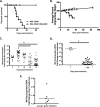
NSE-CD46+ mice survive MV challenge but fail to clear viral RNA. (A) NSE-CD46+ (n = 10) and NSE-CD46+ RAG2 KO (n = 8) mice were challenged i.c. with 1 × 104 PFU of MV-Ed and monitored daily for survival. (B) Weights of all mice from panel A were obtained throughout the infection time course and compared to baseline. Percent weight gain or loss was then calculated. (C) NSE-CD46+ mice were challenged i.c. with 1 × 104 PFU of MV-Ed and were sacrificed at the indicated day postinfection. RNA was purified from perfused brains and analyzed by RT-qPCR (using random hexamers to synthesize cDNA). (D) MV nucleoprotein mRNA in NSE-CD46+ mice challenged i.c. with MV-Ed was detected using the approach outlined above, substituting oligo(dT) primers to generate cDNA. Data are represented using the ΔΔCT method. Results are representative of at least 3 independent experiments with 5 to 10 mice per group. (E) NSE-CD46+ (n = 6) mice were challenged i.c. with 1 × 104 PFU of MV-Ed and were sacrificed at 2 years postinfection. RNA was purified from perfused brains and analyzed by RT-qPCR (using random hexamers to synthesize cDNA). Data are represented using the ΔΔCT method. *, P < 0.05, Mann-Whitney U test.
FIG 2
FIG 2
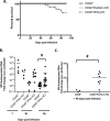
Immune-mediated control of a persistent viral infection. (A) NSE-CD46+, NSE-CD46+ perforin KO, and NSE-CD46+ IFN-γ KO mice were challenged i.c. with 1 × 104 PFU of MV Edmonston. Mice were monitored daily. Percent survival is indicated. Data represent at least 2 individual experiments using NSE-CD46+, NSE-CD46+ perforin KO, and NSE-CD46+ IFN-γ KO mice. (B and C) RNA was collected from whole-brain tissue of mice of the indicated genotypes. RT-qPCR data were generated using random-hexamer priming for cDNA generation, followed by qPCR using primers specific for the MV nucleoprotein and cyclophilin B as a standard. Data were analyzed using the ΔΔCT method. #, P < 0.05, Mann-Whitney U test of NSE-CD46+ controls compared to immunodeficient mice.
FIG 3
FIG 3
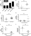
CD8+ T resident memory cells are abundant in the CNS during long-term infection and maintain an effector phenotype. (A) Lymphocytes were purified from whole-brain single-cell suspensions of perfused mice at the indicated days postinfection and subjected to flow cytometric analysis. Black bars, total numbers of CD3+ CD8+ T cells; white bars, total numbers of CD3+ CD8+ CD103+ T cells. #, P < 0.05, Mann-Whitney U test for the proportion of CD3+ CD8+ CD103+ T cells to CD3+ CD8+ T cells (n = 5 to 15/group). (B) Lymphocytes were purified from perfused whole brains of mice infected for 7 or ≥90 days and immunostained for CD103. The percentage of CD103+ cells of the CD3+ CD8+ population is shown. (C) Lymphocytes were purified from perfused whole brains of mice infected for ≥90 days; the percentage of CD69+ cells among CD3+ CD8+ CD103+ T cells in the brains of uninfected or persistently infected NSE-CD46+ mice is shown. (D) Lymphocytes purified from whole-brain tissue of mock-infected mice (PBS), mice inoculated with inactivated virus (UV MV), or mice infected with replication-competent MV, collected at 90 dpi, and subjected to flow cytometric analysis. CD103+ CD3+ CD8+ cells are shown as a percentage of total CD3+ CD8+ T cells (n ≥ 6/group). The percentage of CD3+ CD8+ CD103+ (Trm) cells among CD3+ CD8+ T cells in brains of mice infected for ≥90 days (n = 5 to 8/group) is also shown. (E and F) Lymphocytes purified from perfused whole brains of mice infected for ≥90 days. (E) Percentages of IFN-γ+ CD3+ CD8+ CD103+ and CD3+ CD8+ CD103 cells (n = 8/group). (F) Percentages of granzyme B+ CD3+ CD8+ CD103+ and CD3+ CD8+ CD103 cells (n = 8/group). #, P < 0.05, Mann-Whitney U test. Data are representative of those from at least 2 independent experiments.
FIG 4
FIG 4
Sublethal irradiation leads to increased detection of MV RNA and mRNA. NSE-CD46+ mice were challenged i.c. with 1 × 104 PFU of MV Edmonston, and sublethally irradiated (6.5 gy) at least 90 days later. RNA expression levels from perfused brains were determined by RT-qPCR using random-hexamer or oligo(dT) priming for cDNA generation, followed by qPCR with primers specific for the MV nucleoprotein and cyclophilin B as a standard. Data were analyzed using the ΔΔCT method from at least 2 independent experiments (n = 8 to 12/group). #, P < 0.05, Mann-Whitney U Test. (A) cDNA generated using random-hexamer priming. (B) cDNA generated using oligo(dT) priming. (C) RNA was purified from perfused brains at 14 days post-sublethal irradiation, at the peak of viral RNA reemergence. Data are shown as percent decrease from mock-infected controls (dashed line). dpr, days postirradiation.
FIG 5
FIG 5
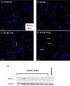
Sublethal irradiation leads to increased expression of MV protein. (A to D) Immunofluorescence of whole-brain tissue obtained from uninfected mice (A), immunocompetent persistently infected (134 days postinfection) mice (B), persistently infected mice sublethally irradiated for 7 days (C), and persistently infected mice sublethally irradiated for 14 days (D). Nuclear staining (Hoechst [blue]) and MV staining (human polyclonal MV antibody [red]) were used. Arrows indicate MV-positive cells. Scale bar = 50 μm. (E) NSE-CD46+ mice were challenged i.c. with 1 × 104 PFU of MV-Ed for at least 90 dpi, followed by 14 days of sublethal irradiation. Shown is Western blot analysis of protein purified from whole-brain tissue of individual mice probed for MV nucleoprotein and GAPDH.
FIG 6
FIG 6
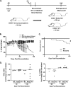
Loss of adaptive immunity after clearance of acute infection leads to pathogenesis. Bone marrow chimeras were generated using NSE-CD46+ mice that had been infected with 1 × 104 PFU of MV-Ed at least 90 days previously. Infected mice were reconstituted with the indicated bone marrow (WT or RAG2 KO) and monitored daily. (A) Schematic of bone marrow chimera generation. (B) Average weight gain or loss of indicated bone marrow recipients. (C) Survival of reconstituted mice (n = 14 to 18/group). (D) RNA was purified from perfused brains of bone marrow recipients (both wild type and RAG2 KO) at 50 and 100 days postreconstitution, and levels of tumor necrosis factor alpha (TNF-α) and IL-6 RNAs were determined. Data are shown as percent change (increase or decrease) compared to mock-infected controls (dashed line). Log rank (Mantel-Cox) test for significance was used. #, P < 0.05.
FIG 7
FIG 7
Loss of adaptive immunity after clearance of acute infection leads to viral reactivation. Bone marrow chimeras were generated using NSE-CD46+ mice that had been infected with 1 × 104 PFU of MV-Ed at least 90 days previously. Infected mice were reconstituted with the indicated bone marrow (WT or RAG2 KO) and monitored daily. (A) RT-qPCR analysis of whole-brain tissue collected from reconstituted mice at the indicated times postreconstitution (n = 3 to 6/group). Data are representative of those from 2 or 3 independent experiments. (B) Western blot analysis of protein purified from whole-brain tissue of individual, reconstituted mice probed for MV nucleoprotein and GAPDH.

Similar articles

Cited by

References

    1. Jarius S, Eichhorn P, Franciotta D, Petereit HF, Akman-Demir G, Wick M, Wildemann B. 2017. The MRZ reaction as a highly specific marker of multiple sclerosis: re-evaluation and structured review of the literature. J Neurol 264:453–466. doi:10.1007/s00415-016-8360-4. - DOI - PubMed
    1. Hottenrott T, Dersch R, Berger B, Rauer S, Huzly D, Stich O. 2017. The MRZ reaction in primary progressive multiple sclerosis. Fluids Barriers CNS 14:2. doi:10.1186/s12987-016-0049-7. - DOI - PMC - PubMed
    1. Li W, Lee M-H, Henderson L, Tyagi R, Bachani M, Steiner J, Campanac E, Hoffman DA, Geldern von G, Johnson K, Maric D, Morris HD, Lentz M, Pak K, Mammen A, Ostrow L, Rothstein J, Nath A. 2015. Human endogenous retrovirus-K contributes to motor neuron disease. Sci Transl Med 7:307ra153. doi:10.1126/scitranslmed.aac8201. - DOI - PMC - PubMed
    1. Jurgens HA, Amancherla K, Johnson RW. 2012. Influenza infection induces neuroinflammation, alters hippocampal neuron morphology, and impairs cognition in adult mice. J Neurosci 32:3958–3968. doi:10.1523/JNEUROSCI.6389-11.2012. - DOI - PMC - PubMed
    1. Brot MD, Rail GF, Oldstone MB, Koob GF, Gold LH. 1997. Deficits in discriminated learning remain despite clearance of long-term persistent viral infection in mice. J Neurovirol 3:265–273. doi:10.3109/13550289709029467. - DOI - PubMed

Publication types

LinkOut - more resources