. 2019 Aug 28;93(18):e00322-19.
doi: 10.1128/JVI.00322-19. Print 2019 Sep 15.

Newcastle Disease Virus V Protein Degrades Mitochondrial Antiviral Signaling Protein To Inhibit Host Type I Interferon Production via E3 Ubiquitin Ligase RNF5

Affiliations

Newcastle Disease Virus V Protein Degrades Mitochondrial Antiviral Signaling Protein To Inhibit Host Type I Interferon Production via E3 Ubiquitin Ligase RNF5

Yingjie Sun et al. J Virol. .

Abstract

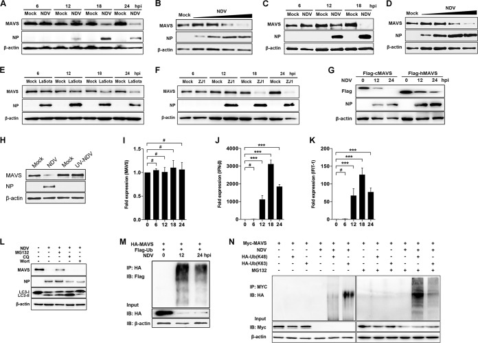
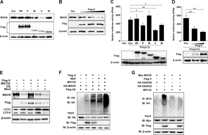
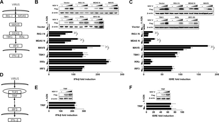
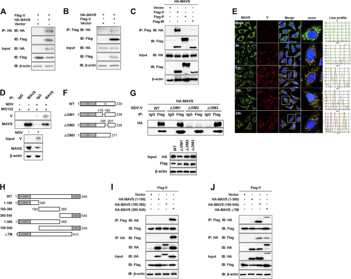
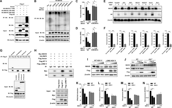
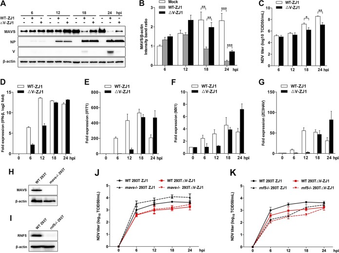
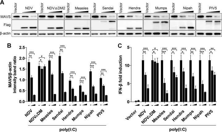
Paramyxovirus establishes an intimate and complex interaction with the host cell to counteract the antiviral responses elicited by the cell. Of the various pattern recognition receptors in the host, the cytosolic RNA helicases interact with viral RNA to activate the mitochondrial antiviral signaling protein (MAVS) and subsequent cellular interferon (IFN) response. On the other hand, viruses explore multiple strategies to resist host immunity. In this study, we found that Newcastle disease virus (NDV) infection induced MAVS degradation. Further analysis showed that NDV V protein degraded MAVS through the ubiquitin-proteasome pathway to inhibit IFN-β production. Moreover, NDV V protein led to proteasomal degradation of MAVS through Lys362 and Lys461 ubiquitin to prevent IFN production. Further studies showed that NDV V protein recruited E3 ubiquitin ligase RNF5 to polyubiquitinate and degrade MAVS. Compared with levels for wild-type NDV infection, V-deficient NDV induced attenuated MAVS degradation and enhanced IFN-β production at the late stage of infection. Several other paramyxovirus V proteins showed activities of degrading MAVS and blocking IFN production similar to those of NDV V protein. The present study revealed a novel role of NDV V protein in targeting MAVS to inhibit cellular IFN production, which reinforces the fact that the virus orchestrates the cellular antiviral response to its own benefit.IMPORTANCE Host anti-RNA virus innate immunity relies mainly on the recognition by retinoic acid-inducible gene I and melanoma differentiation-associated protein 5 and subsequently initiates downstream signaling through interaction with MAVS. On the other hand, viruses have developed various strategies to counteract MAVS-mediated signaling. The mechanism for paramyxoviruses regulating MAVS to benefit their infection remains unknown. In this article, we demonstrate that the V proteins of NDV and several other paramyxoviruses target MAVS for ubiquitin-mediated degradation through E3 ubiquitin ligase RING-finger protein 5 (RNF5). MAVS degradation leads to the inhibition of the downstream IFN-β pathway and therefore benefits virus proliferation. Our study reveals a novel mechanism of NDV evading host innate immunity and provides insight into the therapeutic strategies for the control of paramyxovirus infection.

Keywords: MAVS; NDV; RNF5; degradation; paramyxovirus.


Figures

FIG 1
FIG 1
NDV infection induced MAVS degradation. (A) HeLa cells were mock treated or infected with NDV at an MOI of 1. Cells were harvested at 6, 12, 18, and 24 hpi and detected using immunoblot (IB) analysis with anti-MAVS, anti-NP, or anti-β-actin antibody. (B) HeLa cells were mock treated or infected with NDV at an MOI of 0.01, 0.1, 1, or 5 (wedge). Cells were harvested at 18 hpi and detected using immunoblot analysis with anti-MAVS, anti-NP, or anti-β-actin antibody. (C) A549 cells were mock treated or infected with NDV Herts/33 strain at an MOI of 5. Cells were harvested at 6, 12, and 18 hpi and detected using immunoblot analysis with anti-MAVS, anti-NP, or anti-β-actin antibody. (D) A549 cells were mock treated or infected with NDV Herts/33 strain at an MOI of 0.01, 0.1, 1, or 5 (wedge). Cells were harvested at 18 hpi and detected using immunoblot analysis with anti-MAVS, anti-NP, or anti-β-actin antibody. (E and F) HeLa cells were mock treated or infected with NDV LaSota (E) or ZJ1 (F) strain at an MOI of 1. Cells were harvested at 6, 12, 18, and 24 hpi and detected using immunoblot analysis with anti-MAVS, anti-NP, or anti-β-actin antibody. (G) HEK-293T was transfected with Flag-tagged chicken MAVS (cMAV) and human MAVS (hMAVS). At 24 h after transfection, cells were infected with NDV at an MOI of 1. Cells were harvested at 12 and 24 hpi and detected using immunoblot analysis with anti-Flag, anti-NP, or anti-β-actin antibody. (H) HeLa cells were mock treated or infected with NDV (MOI of 1) or UV-treated NDV (MOI of 10). Cells were harvested at 18 hpi and detected with anti-MAVS, anti-NP, or anti-β-actin antibody. (I to K) HeLa cells were mock treated or infected with NDV at an MOI of 1. Cells were harvested at 6, 12, 18, and 24 hpi and detected using qRT-PCR with MAVS (I), IFN-β (J), or IFIT1 (K) primers. (L) HeLa cells were mock infected or infected with NDV at an MOI of 1 and maintained in the presence or absence of the lysosome inhibitor CQ (50 μM) or the autophagy inhibitor wortmannin (Wort; 100 nM) for 12 h. Cells were harvested and detected using immunoblot analysis with anti-MAVS, anti-NP, anti-LC3, or anti-β-actin antibody. For proteasome inhibition assay, the infected cells were treated with MG132 (20 μM) for 6 h prior to immunoblot analysis. (M) HEK-293T cells were cotransfected with HA-MAVS and Flag-ubiquitin. At 24 h after transfection, cells were infected with NDV at an MOI of 1. At 12 and 24 hpi, cells were harvested, immunoprecipitated (IP) with anti-HA antibody, and further detected using immunoblot analysis with anti-Flag antibody. Expression levels of the proteins were analyzed by immunoblot analysis of the lysates with anti-HA or anti-β-actin antibody. (N) HEK-293T cells were cotransfected with Myc-MAVS and either HA-ubiquitin (K48) or HA-ubiquitin (K63). At 24 h after transfection, cells were infected with NDV at an MOI of 1 and maintained in the presence or absence of the proteasome inhibitor MG132 (20 μM, 6 h prior to immunoprecipitation). At 18 hpi, cells were harvested, immunoprecipitated with anti-Myc antibody, and further detected using immunoblot analysis with anti-HA antibody. Expression levels of the proteins were analyzed by immunoblot analysis of the lysates with anti-Myc or anti-β-actin antibody. Data are presented as means from three independent experiments. #, P > 0.05; ***, P < 0.001.
FIG 2
FIG 2
NDV V protein triggers MAVS degradation through ubiquitin-proteasome pathway. (A) HeLa cells were transfected with either empty vector (Vec) or Flag-tagged NP, P, M, V, or W. At 12 h posttransfection (hpt), cells were transfected with poly(I·C) (20 μg/ml). Cells were harvested at 12 hpt with poly(I·C) and detected using immunoblot analysis with anti-MAVS, anti-Flag, or anti-β-actin antibody. (B) HeLa cells were transfected with either empty vector or Flag-V (0.5, 1, or 2 μg/ml; wedge). At 12 hpt, cells were transfected with poly(I·C) (20 μg/ml). Cells were harvested at 12 hpt with poly(I·C) and detected using immunoblot analysis with anti-MAVS, anti-Flag, or anti-β-actin antibody. (C) HEK-293T cells were cotransfected with p-125Luc, PRL-TK, and either empty vector or Flag-tagged NP, P, M, V, or W. At 12 hpt, cells were mock treated or transfected with poly(I·C) (20 μg/ml). Cells were harvested at 12 hpt and assessed for luciferase activity. The results are presented as relative luciferase activity. Expression levels of expressed proteins were analyzed by immunoblot analysis of the lysates with anti-Flag or anti-β-actin antibody. (D) HEK-293T cells were cotransfected with p-125Luc, PRL-TK, and either empty vector or Flag-V (0, 0.5, or 1 μg/ml; wedge). At 12 hpt, cells were transfected with poly(I·C) (20 μg/ml). Cells were harvested at 12 hpt and assessed for luciferase activity. The results are presented as relative luciferase activity. Expression levels of expressed proteins were analyzed by immunoblot analysis of the lysates with anti-Flag or anti-β-actin antibody. (E) HeLa cells were transfected with either empty vector or Flag-V and maintained in the presence or absence of the proteasome inhibitor MG132 (20 μM, 6 h prior to immunoblot analysis), the lysosome inhibitor CQ (50 μM), or the autophagy inhibitor wortmannin (100 nM) for 12 h. Cells were harvested and detected using immunoblot analysis with anti-MAVS, anti-NP, or anti-β-actin antibody. (F) HEK-293T cells were cotransfected with HA-MAVS, Flag-ubiquitin (Ub), and either empty vector or Flag-V and maintained in the presence or absence of the proteasome inhibitor MG132 (20 μM, 6 h prior to immunoprecipitation). At 24 hpt, cells were harvested, immunoprecipitated with anti-HA antibody, and further detected using immunoblot analysis with anti-ubiquitin MAbs. For the NDV infection group, HEK-293T cells were cotransfected with HA-MAVS and Flag-ubiquitin. At 24 hpt, cells were infected with NDV at an MOI of 1 and maintained in the presence or absence of MG132 (20 μM, 6 h prior to immunoprecipitation). At 12 hpi, cells were harvested for immunoprecipitation assay. Expression levels of the proteins were analyzed by immunoblot analysis of the lysates with anti-HA, anti-Flag, or anti-β-actin antibody. (G) HEK-293T cells were cotransfected with Myc-MAVS, HA-ubiquitin (K48), or HA-ubiquitin (K63) and either empty vector or Flag-V and maintained in the presence of the proteasome inhibitor MG132 (20 μM, 6 h prior to immunoprecipitation). At 24 hpt, cells were harvested, immunoprecipitated with anti-Myc antibody, and further detected using immunoblot analysis with anti-HA antibody. Expression levels of the proteins were analyzed by immunoblot analysis of the lysates with anti-Myc, anti-Flag, or anti-β-actin antibody. Data are presented as means from three independent experiments. *, P < 0.05; **, P < 0.01; ***, P < 0.001.
FIG 3
FIG 3
NDV V protein suppresses MAVS-mediated IFN-β signaling transduction. (A) Schematic diagram of RIG-I/MDA5-mediated IFN-β signaling pathway. (B and C) HEK-293T cells were cotransfected with PRL-TK, expression plasmid RIG-I N, MDA5 N, MAVS, TBK1, IKKε, or IRF3-5D or empty plasmid (vector), and either empty vector (−) or Flag-V (1 or 2 μg/ml; wedge) as well as p-125Luc (B) or pISRE-luc (C). At 24 hpt, cells were harvested and assessed for luciferase activity. The results are presented as relative luciferase activity. Expression levels of expressed proteins were analyzed by immunoblot analysis of the lysates with anti-Flag or anti-β-actin antibody. (D) Schematic diagram of TLR3-mediated signaling pathway. (E and F) HEK-293T cells were cotransfected with PRL-TK, TRIF, and either empty vector or Flag-V (1 or 2 μg/ml; wedge) as well as p-125Luc (E) or pISRE-luc (F). At 24 hpt, cells were harvested and assessed for luciferase activity. The results are presented as relative luciferase activity. Expression levels of expressed proteins were analyzed by immunoblot analysis of the lysates with anti-Flag or anti-β-actin antibody. Data are presented as means from three independent experiments. **, P < 0.01; ***, P < 0.001.
FIG 4
FIG 4
NDV V interacts with MAVS. (A) HEK-293T cells were cotransfected with Flag-V and either empty vector or HA-MAVS. At 24 hpt, cells were harvested, immunoprecipitated with anti-HA antibody, and further detected using immunoblot analysis with anti-HA or anti-Flag antibody. Expression levels of the proteins were analyzed by immunoblot analysis of the lysates with anti-HA, anti-Flag, or anti-β-actin antibody. (B) HEK-293T cells were cotransfected with HA-MAVS and either empty vector or Flag-V. At 24 hpt, cells were harvested, immunoprecipitated with anti-HA antibody, and further detected using immunoblot analysis with anti-HA or anti-Flag antibody. Expression levels of the proteins were analyzed by immunoblot analysis of the lysates with anti-HA, anti-Flag, or anti-β-actin antibody. (C) HEK-293T cells were cotransfected with HA-MAVS and either empty vector or Flag-V, -P, or -W. At 24 hpt, cells were harvested, immunoprecipitated with anti-Flag antibody, and further detected using immunoblot analysis with anti-HA or anti-Flag antibody. Expression levels of the proteins were analyzed by immunoblot analysis of the lysates with anti-HA, anti-Flag, or anti-β-actin antibody. (D) HeLa cells were mock treated or infected with NDV at an MOI of 1. At 24 hpt, cells were harvested, immunoprecipitated with anti-IgG or MAVS antibody, and further detected using immunoblot analysis with anti-V or anti-MAVS antibody. Expression levels of the proteins were analyzed by immunoblot analysis of the lysates with anti-V, anti-MAVS, or anti-β-actin antibody. (E) HeLa cells were mock treated or infected with NDV at an MOI of 1. Cells were harvested at 6, 12, 18, and 24 hpi and detected using IF assay with anti-MAVS or anti-V antibody. Shown is an intensity profile of the linear region of interest (ROI) across the HeLa cell costained with V and MAVS. (F) Schematics of a series of Flag-tagged truncated V constructs. (G) HEK-293T cells were cotransfected with HA-MAVS and either empty vector, WT, or truncated Flag-V (ΔDM1, ΔDM2, and ΔDM3). At 24 hpt, cells were harvested, immunoprecipitated with anti-IgG or anti-Flag antibody, and further detected using immunoblot analysis with anti-HA or anti-Flag antibody. Expression levels of the proteins were analyzed by immunoblot analysis of the lysates with anti-HA, anti-Flag, or anti-β-actin antibody. (H) Schematics of a series of HA-tagged truncated MAVS constructs. (I and J) HEK-293T cells were cotransfected with Flag-V and either empty vector or truncated HA-MAVS aa 1 to 180, aa 180 to 360, and aa 360 to 540 (I) or aa 1 to 360, aa 180 to 540, and ΔTM (J). At 24 hpt, cells were harvested, immunoprecipitated with anti-Flag or anti-HA antibody, and further detected using immunoblot analysis with anti-HA or anti-Flag antibody. Expression levels of the proteins were analyzed by immunoblot analysis of the lysates with anti-HA, anti-Flag, or anti-β-actin antibody.
FIG 5
FIG 5
Characterization of RNF5 as the E3 ubiquitin ligase for V-mediated MAVS degradation. (A) HEK-293T cells were cotransfected with Flag-V, Flag-ubiquitin, and either empty vector or truncated HA-MAVS aa 1 to 180, 180 to 360, and 360 to 540. At 24 hpt, cells were harvested, immunoprecipitated with anti-HA antibody, and further detected using immunoblot analysis with anti-ubiquitin or anti-HA antibody. Expression levels of the proteins were analyzed by immunoblot analysis of the lysates with anti-HA, anti-Flag, or anti-β-actin antibody. (B) HEK-293T cells were cotransfected with Flag-V, Flag-ubiquitin, and either empty vector, WT, or mutated HA-MAVS K362A, K371A, K420A, K461A, K500A, and K362/461A and maintained in the presence of the proteasome inhibitor MG132 (20 μM, 6 h prior to immunoprecipitation). At 24 hpt, cells were harvested, immunoprecipitated with anti-HA antibody, and further detected using immunoblot analysis with anti-ubiquitin antibody. Expression levels of the proteins were analyzed by immunoblot analysis of the lysates with anti-HA, anti-Flag, or anti-β-actin antibody. (C and D) HEK-293T cells were cotransfected with PRL-TK, HA-MAVS, or K362/461A MAVS and either empty vector (−) or Flag-V (1 or 2 μg/ml; wedge) as well as p-125Luc (C) or pISRE-luc (D). At 24 hpt, cells were harvested and assessed for luciferase activity. The results are presented as relative luciferase activity. (E) HeLa cells were transfected with either scrambled siRNA or specific siRNA targeting AIP4, RNF5, RNF125, MARCH5, Smurf1, or MUL1. At 48 h after transfection, cells were infected with NDV at an MOI of 1. Cells were harvested at 18 hpi and detected using immunoblot analysis with anti-MAVS, anti-NP, or anti-β-actin antibody. (F) The transfection experiments were performed as described for panel A. Cells were harvested and detected using qRT-PCR with primers for AIP4, RNF5, RNF125, MARCH5, Smurf1, or MUL1. (G) HEK-293T cells were cotransfected with Flag-V and HA-tagged RNF5, MARCH5, Smurf1, or Smurf2. At 24 hpt, cells were harvested, immunoprecipitated with anti-IgG or anti-Flag antibody, and further detected using immunoblot analysis with anti-HA or anti-Flag antibody. Expression levels of the proteins were analyzed by immunoblot analysis of the lysates with anti-HA, anti-Flag, or anti-β-actin antibody. (H) HEK-293T cells were cotransfected with Myc-MAVS, HA-RNF5, and either WT or ΔDM2 Flag-V vector. At 24 hpt, cells were harvested, immunoprecipitated with anti-IgG or anti-HA antibody, and further detected using immunoblot analysis with anti-Myc or anti-HA antibody. Expression levels of the proteins were analyzed by immunoblot analysis of the lysates with anti-HA, anti-Myc, anti-Flag, or anti-β-actin antibody. (I) HeLa cells were transfected with either WT or ΔDM2 Flag-V vector (1 or 2 μg/ml; wedge). At 12 hpt, cells were transfected with poly(I·C) (20 μg/ml). Cells were harvested at 12 hpt with poly(I·C) and detected using immunoblot analysis with anti-MAVS, anti-Flag, or anti-β-actin antibody. (J) WT and rnf5−/− HeLa cells were transfected with either vector or Flag-V (1 or 2 μg/ml; wedge). At 12 hpt, cells were transfected with poly(I·C) (20 μg/ml). Cells were harvested at 12 hpt with poly(I·C) and detected using immunoblot analysis with anti-MAVS, anti-RNF5, anti-Flag, or anti-β-actin antibody. (K) WT and rnf5−/− HeLa cells were cotransfected with p-125Luc, PRL-TK, and either vector or Flag-V (1 or 2 μg/ml; wedge). At 12 hpt, cells were transfected with poly(I·C) (20 μg/ml). Cells were harvested at 12 hpt with poly(I·C) and assessed for luciferase activity. The results are presented as relative luciferase activity. (L to N) The transfection experiments were performed as described for panel D. Cells were harvested at 12 hpt with poly(I·C) and detected using qRT-PCR with IFIT1 (L), MX1 (M), or ZC3HAV (N) primers.
FIG 6
FIG 6
Depletion of V attenuated NDV-induced MAVS degradation. (A) HeLa cells were mock treated or infected with WT ZJ1 or ΔV ZJ1 at an MOI of 1. Cells were harvested at 6, 12, 18, and 24 hpi and detected using immunoblot analysis with anti-MAVS, anti-NP, anti-V, or anti-β-actin antibody. (B) The intensity band ratio of MAVS to β-actin. Representative results are shown with graphs representing the ratio of MAVS to β-actin normalized to the control condition. Data are presented as means from three independent experiments. (C) Extracellular virus yields in WT ZJ1 or ΔV ZJ1 infection group. (D to G) Virus infection experiments were performed as described for panel A. Cells were harvested and detected using qRT-PCR with IFN-β (D), IFIT-1(E), MX1 (F), or ZC3HAV (G) primers. (H and I) WT and either mavs−/− (H) or rnf5−/− (I) 293T cells were seeded in 6-well plates (1 × 106 cells/well) and collected for immunoblot analysis with anti-MAVS, anti-RNF5, or anti-β-actin antibody. (J) WT and mavs−/− HEK-293T cells were infected with WT ZJ1 or ΔV ZJ1, respectively, at an MOI of 1. Cells were harvested at 6, 12, 18, and 24 hpi. The extracellular virus yields in the WT ZJ1 or ΔV ZJ1 infection group were detected as TCID50 on DF-1 cells. (K) WT and rnf5−/− HEK-293T cells were infected with WT ZJ1 or ΔV ZJ1, respectively, at an MOI of 1. Cells were harvested at 6, 12, 18, and 24 hpi. The extracellular virus yields in the WT ZJ1 or ΔV ZJ1 infection group were detected as TCID50 on DF-1 cells. Data are presented as means from three independent experiments. *, P < 0.05; **, P < 0.01; ***, P < 0.001.
FIG 7
FIG 7
Paramyxovirus V proteins trigger MAVS degradation. (A) HeLa cells were transfected with either empty vector or Flag-tagged V of NDV, NDV ΔDM2, measles virus, Sendia virus, Hendra virus, mumps, Nipah virus, or PIV5. At 12 hpt, cells were transfected with poly(I·C) (20 μg/ml). Cells were harvested at 12 hpt with poly(I·C) and detected using immunoblot analysis with anti-MAVS, anti-Flag, or anti-β-actin antibody. (B) The intensity band ratio of MAVS to β-actin. Representative results are shown with graphs representing the ratio of MAVS to β-actin normalized to the control condition. Data are presented as means from three independent experiments. (C) HeLa cells were cotransfected with p-125Luc, PRL-TK, and either empty vector or Flag-tagged V of NDV, NDV ΔDM2, measles virus, Sendai virus, Hendra virus, mumps virus, Nipah virus, or PIV5. At 12 hpt, cells were mock treated or transfected with poly(I·C) (20 μg/ml). Cells were harvested at 12 hpt and assessed for luciferase activity. The results are presented as relative luciferase activity. Data are presented as means from three independent experiments. #, P > 0.05; *, P < 0.05; **, P < 0.01; ***, P < 0.001.

Similar articles

Cited by

References

    1. Akira S, Uematsu S, Takeuchi O. 2006. Pathogen recognition and innate immunity. Cell 124:783–801. doi:10.1016/j.cell.2006.02.015. - DOI - PubMed
    1. Yoneyama M, Fujita T. 2009. RNA recognition and signal transduction by RIG-I-like receptors. Immunol Rev 227:54–65. doi:10.1111/j.1600-065X.2008.00727.x. - DOI - PubMed
    1. Kato H, Takeuchi O, Sato S, Yoneyama M, Yamamoto M, Matsui K, Uematsu S, Jung A, Kawai T, Ishii KJ, Yamaguchi O, Otsu K, Tsujimura T, Koh CS, Reis e Sousa C, Matsuura Y, Fujita T, Akira S. 2006. Differential roles of MDA5 and RIG-I helicases in the recognition of RNA viruses. Nature 441:101–105. doi:10.1038/nature04734. - DOI - PubMed
    1. Kawai T, Takahashi K, Sato S, Coban C, Kumar H, Kato H, Ishii KJ, Takeuchi O, Akira S. 2005. IPS-1, an adaptor triggering RIG-I- and Mda5-mediated type I interferon induction. Nat Immunol 6:981–988. doi:10.1038/ni1243. - DOI - PubMed
    1. Meylan E, Curran J, Hofmann K, Moradpour D, Binder M, Bartenschlager R, Tschopp J. 2005. Cardif is an adaptor protein in the RIG-I antiviral pathway and is targeted by hepatitis C virus. Nature 437:1167–1172. doi:10.1038/nature04193. - DOI - PubMed

Publication types

MeSH terms

LinkOut - more resources