

Vascular Cognitive Impairment and Dementia: JACC Scientific Expert Panel
- PMID: 31248555
- PMCID: PMC6719789
- DOI: 10.1016/j.jacc.2019.04.034
Vascular Cognitive Impairment and Dementia: JACC Scientific Expert Panel
Abstract
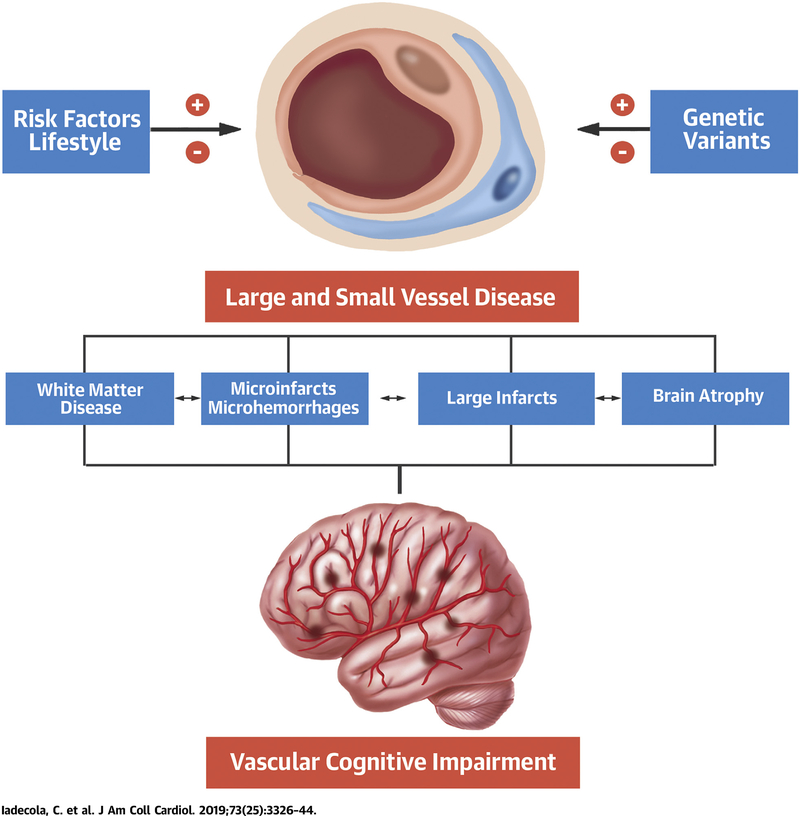
Cognitive impairment associated with aging has emerged as one of the major public health challenges of our time. Although Alzheimer's disease is the leading cause of clinically diagnosed dementia in Western countries, cognitive impairment of vascular etiology is the second most common cause and may be the predominant one in East Asia. Furthermore, alterations of the large and small cerebral vasculature, including those affecting the microcirculation of the subcortical white matter, are key contributors to the clinical expression of cognitive dysfunction caused by other pathologies, including Alzheimer's disease. This scientific expert panel provides a critical appraisal of the epidemiology, pathobiology, neuropathology, and neuroimaging of vascular cognitive impairment and dementia, and of current diagnostic and therapeutic approaches. Unresolved issues are also examined to shed light on new basic and clinical research avenues that may lead to mitigating one of the most devastating human conditions.
Keywords: Alzheimer’s disease; cerebral blood flow; cognitive dysfunction; small vessel disease; stroke.
Copyright © 2019 American College of Cardiology Foundation. Published by Elsevier Inc. All rights reserved.
Figures
Similar articles
-
Neuropathological diagnosis of vascular cognitive impairment and vascular dementia with implications for Alzheimer's disease.Acta Neuropathol. 2016 May;131(5):659-85. doi: 10.1007/s00401-016-1571-z. Epub 2016 Apr 9. Acta Neuropathol. 2016. PMID: 27062261 Free PMC article. Review.
-
Progression in Vascular Cognitive Impairment: Pathogenesis, Neuroimaging Evaluation, and Treatment.Cell Transplant. 2019 Jan;28(1):18-25. doi: 10.1177/0963689718815820. Epub 2018 Nov 29. Cell Transplant. 2019. PMID: 30488737 Free PMC article.
-
Vascular Cognitive Disorder.Semin Neurol. 2019 Apr;39(2):241-250. doi: 10.1055/s-0039-1678582. Epub 2019 Mar 29. Semin Neurol. 2019. PMID: 30925616 Review.
-
Regional amyloid burden and lacune in pure subcortical vascular cognitive impairment.Neurobiol Aging. 2017 Jul;55:20-26. doi: 10.1016/j.neurobiolaging.2017.03.010. Epub 2017 Mar 16. Neurobiol Aging. 2017. PMID: 28395177
-
Vascular cognitive impairment and dementia: Mechanisms, treatment, and future directions.Int J Stroke. 2024 Oct;19(8):838-856. doi: 10.1177/17474930241279888. Int J Stroke. 2024. PMID: 39283037 Free PMC article. Review.
Cited by
-
Targeting Transcription Factor Nrf2 (Nuclear Factor Erythroid 2-Related Factor 2) for the Intervention of Vascular Cognitive Impairment and Dementia.Arterioscler Thromb Vasc Biol. 2021 Jan;41(1):97-116. doi: 10.1161/ATVBAHA.120.314804. Epub 2020 Oct 15. Arterioscler Thromb Vasc Biol. 2021. PMID: 33054394 Free PMC article. Review.
-
IGF1R signaling regulates astrocyte-mediated neurovascular coupling in mice: implications for brain aging.Geroscience. 2021 Apr;43(2):901-911. doi: 10.1007/s11357-021-00350-0. Epub 2021 Mar 6. Geroscience. 2021. PMID: 33674953 Free PMC article.
-
Vagus Nerve Stimulation for Improvement of Vascular Cognitive Impairment.Neuropsychiatr Dis Treat. 2024 Jul 24;20:1445-1451. doi: 10.2147/NDT.S465249. eCollection 2024. Neuropsychiatr Dis Treat. 2024. PMID: 39072312 Free PMC article. Review.
-
On the regional distribution of cerebral microvascular 'raspberries' and their association with cerebral atherosclerosis and acute circulatory failure.Cereb Circ Cogn Behav. 2023 Jan 7;4:100157. doi: 10.1016/j.cccb.2023.100157. eCollection 2023. Cereb Circ Cogn Behav. 2023. PMID: 36691600 Free PMC article.
-
Human ESC-derived immunity- and matrix- regulatory cells ameliorated white matter damage and vascular cognitive impairment in rats subjected to chronic cerebral hypoperfusion.Cell Prolif. 2022 May;55(5):e13223. doi: 10.1111/cpr.13223. Epub 2022 Apr 19. Cell Prolif. 2022. PMID: 35437845 Free PMC article.
References
-
- Alzheimer’s Disease International. World Alzheimer’s Report 2018. Available at: https://www.alz.co.uk/research/world-report-2018. Accessed May 7, 2019.
-
- World Health Organization. Global action plan on the public health response to dementia 2017–2025. 2017. Available at: http://www.who.int/mental_health/neurology/dementia/action_plan_2017_202.... Accessed May 7, 2019.
-
- IADRP. International Alzheimer’s Disease Research Portfolio. 2018. Available at: https://iadrp.nia.nih.gov/. Accessed May 7, 2019.
-
- Mast H, Tatemichi TK, Mohr JP. Chronic brain ischemia: the contributions of Otto Binswanger and Alois Alzheimer to the mechanisms of vascular dementia. J Neurol Sci 1995;132:4–10. - PubMed
-
- Hachinski VC, Iliff LD, Zilhka E, et al. Cerebral blood flow in dementia. Arch Neurol 1975;32: 632–7. - PubMed
Publication types
MeSH terms
Grants and funding
LinkOut - more resources
Full Text Sources
