

Repair Process Impairment by Pseudomonas aeruginosa in Epithelial Tissues: Major Features and Potential Therapeutic Avenues
- PMID: 31214514
- PMCID: PMC6554286
- DOI: 10.3389/fcimb.2019.00182
Repair Process Impairment by Pseudomonas aeruginosa in Epithelial Tissues: Major Features and Potential Therapeutic Avenues
Abstract
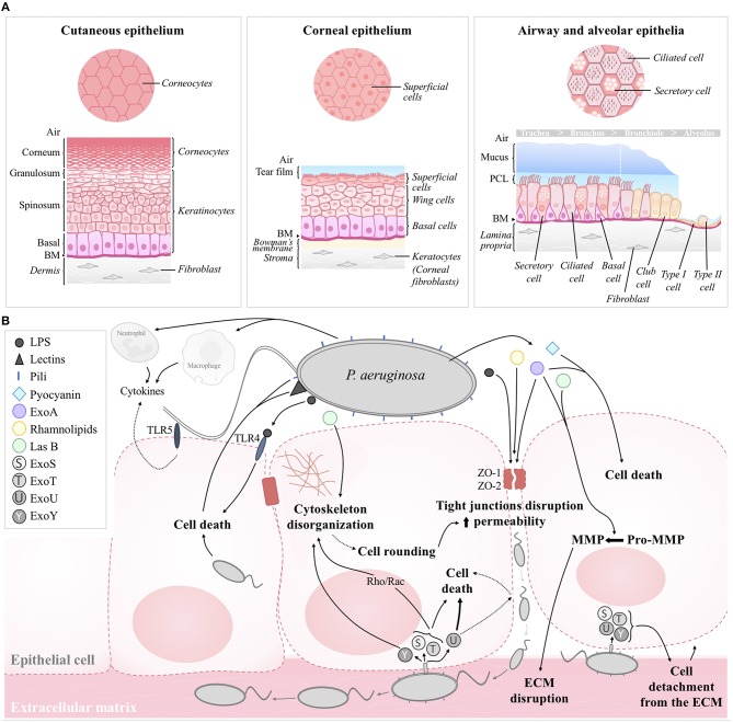
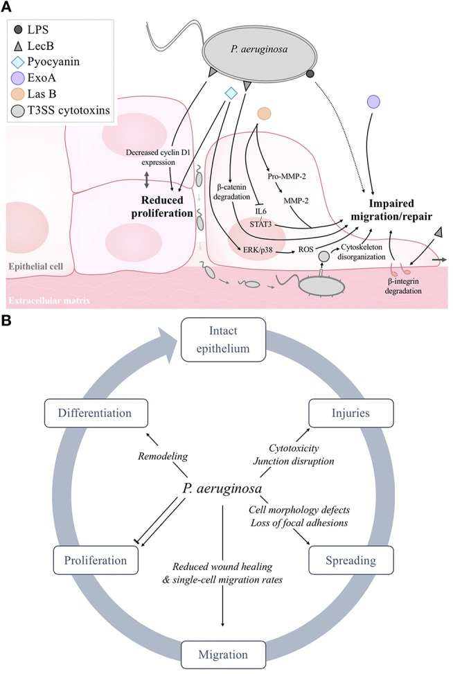
Epithelial tissues protecting organs from the environment are the first-line of defense against pathogens. Therefore, efficient repair mechanisms after injury are crucial to maintain epithelial integrity. However, these healing processes can be insufficient to restore epithelial integrity, notably in infectious conditions. Pseudomonas aeruginosa infections in cutaneous, corneal, and respiratory tract epithelia are of particular concern because they are the leading causes of hospitalizations, disabilities, and deaths worldwide. Pseudomonas aeruginosa has been shown to alter repair processes, leading to chronic wounds and infections. Because of the current increase in the incidence of multi-drug resistant isolates of P. aeruginosa, complementary approaches to decrease the negative impact of these bacteria on epithelia are urgently needed. Here, we review the recent advances in the understanding of the impact of P. aeruginosa infections on the integrity and repair mechanisms of alveolar, airway, cutaneous and corneal epithelia. Potential therapeutic avenues aimed at counteracting this deleterious impact of infection are also discussed.
Keywords: Pseudomonas aeruginosa infections; airway; burn; cornea; epithelial repair; lung; skin; wound repair.
Figures
Similar articles
-
Role of Pseudomonas aeruginosa ExsA in penetration through corneal epithelium in a novel in vivo model.Invest Ophthalmol Vis Sci. 2003 Dec;44(12):5220-7. doi: 10.1167/iovs.03-0229. Invest Ophthalmol Vis Sci. 2003. PMID: 14638720
-
Impact of topical corticosteroid pretreatment on susceptibility of the injured murine cornea to Pseudomonas aeruginosa colonization and infection.Exp Eye Res. 2019 Feb;179:1-7. doi: 10.1016/j.exer.2018.10.010. Epub 2018 Oct 19. Exp Eye Res. 2019. PMID: 30343040 Free PMC article.
-
Fibronectin and alpha5beta1 integrin mediate binding of Pseudomonas aeruginosa to repairing airway epithelium.Eur Respir J. 1999 Jun;13(6):1301-9. Eur Respir J. 1999. PMID: 10445605
-
Pseudomonas aeruginosa adherence to remodelling respiratory epithelium.Eur Respir J. 1996 Oct;9(10):2145-50. doi: 10.1183/09031936.96.09102145. Eur Respir J. 1996. PMID: 8902481 Review.
-
Modulation of lung epithelial functions by Pseudomonas aeruginosa.Trends Microbiol. 2005 Aug;13(8):389-97. doi: 10.1016/j.tim.2005.05.011. Trends Microbiol. 2005. PMID: 15951179 Review.
Cited by
-
Iron Pathways and Iron Chelation Approaches in Viral, Microbial, and Fungal Infections.Pharmaceuticals (Basel). 2020 Sep 25;13(10):275. doi: 10.3390/ph13100275. Pharmaceuticals (Basel). 2020. PMID: 32992923 Free PMC article. Review.
-
Use of Phage Cocktail BFC 1.10 in Combination With Ceftazidime-Avibactam in the Treatment of Multidrug-Resistant Pseudomonas aeruginosa Femur Osteomyelitis-A Case Report.Front Med (Lausanne). 2022 Apr 25;9:851310. doi: 10.3389/fmed.2022.851310. eCollection 2022. Front Med (Lausanne). 2022. PMID: 35547216 Free PMC article.
-
Antibiofilm Activity of PEGylated Branched Polyethylenimine.ACS Omega. 2022 Dec 2;7(49):44825-44835. doi: 10.1021/acsomega.2c04911. eCollection 2022 Dec 13. ACS Omega. 2022. PMID: 36530285 Free PMC article.
-
Bacterial Pyocyanin Inducible Keratin 6A Accelerates Closure of Epithelial Defect under Conditions of Mitochondrial Dysfunction.J Invest Dermatol. 2023 Oct;143(10):2052-2064.e5. doi: 10.1016/j.jid.2023.03.1671. Epub 2023 Apr 10. J Invest Dermatol. 2023. PMID: 37044260 Free PMC article.
-
Effects of elevated levels of intracellular nitric oxide on Pseudomonas aeruginosa biofilm in chronic skin wound and slow-killing infection models.Int Microbiol. 2024 Apr;27(2):349-359. doi: 10.1007/s10123-023-00395-5. Epub 2023 Jul 6. Int Microbiol. 2024. PMID: 37410300
References
-
- Adam D., Bilodeau C., Sognigbé L., Maillé É., Ruffin M., Brochiero E. (2018). CFTR rescue with VX-809 and VX-770 favors the repair of primary airway epithelial cell cultures from patients with class II mutations in the presence of Pseudomonas aeruginosa exoproducts. J. Cyst. Fibros. 17, 705–714. 10.1016/j.jcf.2018.03.010 - DOI - PubMed
Publication types
MeSH terms
Grants and funding
LinkOut - more resources
Full Text Sources