

The transition structure of chromatin fibers at the nanoscale probed by cryogenic electron tomography
- PMID: 31211313
- PMCID: PMC6688845
- DOI: 10.1039/c9nr02042j
The transition structure of chromatin fibers at the nanoscale probed by cryogenic electron tomography
Abstract
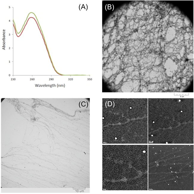
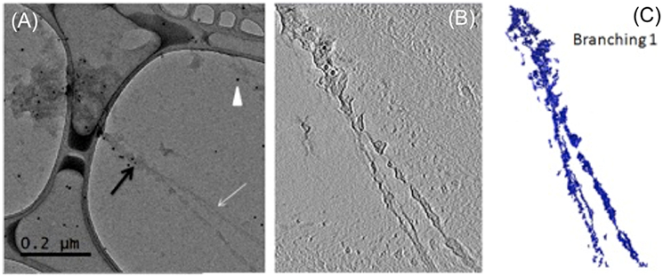
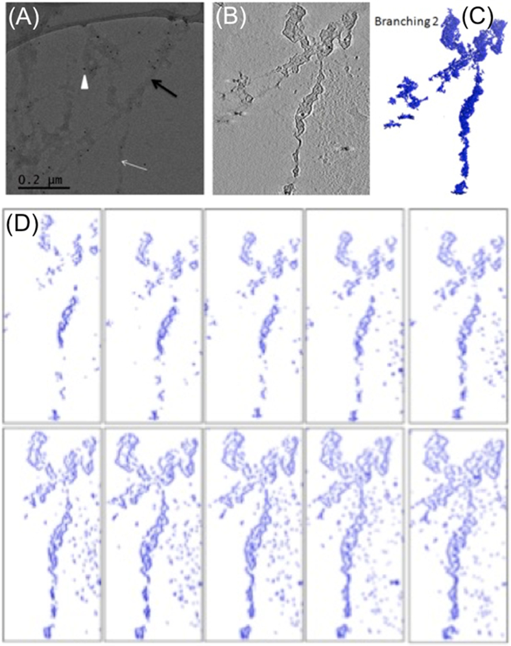
The naked DNA inside the nucleus interacts with proteins and RNAs forming a higher order chromatin structure to spatially and temporally control transcription in eukaryotic cells. The 30 nm chromatin fiber is one of the most important determinants of the regulation of eukaryotic transcription. However, the transition of chromatin from the 30 nm inactive higher order structure to the actively transcribed lower order nucleosomal arrays is unclear, which limits our understanding of eukaryotic transcription. Using a method to extract near-native eukaryotic chromatin, we revealed the chromatin structure at the transitional state from the 30 nm chromatin to multiple nucleosomal arrays by cryogenic electron tomography (cryo-ET). Reproducible electron microscopy images revealed that the transitional structure is a branching structure that the 30 nm chromatin hierarchically branches into lower order nucleosomal arrays, indicating chromatin compaction at different levels to control its accessibility during the interphase. We further observed that some of the chromatin fibers on the branching structure have a helix ribbon structure, while the others randomly twist together. Our finding of the chromatin helix ribbon structure on the extracted native chromatin revealed by cryo-ET indicates a complex higher order chromatin organization beyond the beads-on-a-string structure. The hierarchical branching and helix ribbon structure may provide mechanistic insights into how chromatin organization plays a central role in transcriptional regulation and other DNA-related biological processes during diseases such as cancer.
Conflict of interest statement
Conflicts of interest
There are no conflicts to declare.
Figures
Similar articles
-
Chromatin hierarchical branching visualized at the nanoscale by electron microscopy.Nanoscale Adv. 2020 Nov 13;3(4):1019-1028. doi: 10.1039/d0na00359j. eCollection 2021 Feb 21. Nanoscale Adv. 2020. PMID: 34381959 Free PMC article.
-
Higher-order structure of the 30-nm chromatin fiber revealed by cryo-EM.IUBMB Life. 2016 Nov;68(11):873-878. doi: 10.1002/iub.1568. Epub 2016 Oct 5. IUBMB Life. 2016. PMID: 27704715 Review.
-
Cryo-EM study of the chromatin fiber reveals a double helix twisted by tetranucleosomal units.Science. 2014 Apr 25;344(6182):376-80. doi: 10.1126/science.1251413. Science. 2014. PMID: 24763583
-
Electron microscopy and atomic force microscopy studies of chromatin and metaphase chromosome structure.Micron. 2011 Dec;42(8):733-50. doi: 10.1016/j.micron.2011.05.002. Epub 2011 May 12. Micron. 2011. PMID: 21703860 Review.
-
Structural insights of nucleosome and the 30-nm chromatin fiber.Curr Opin Struct Biol. 2016 Feb;36:106-15. doi: 10.1016/j.sbi.2016.01.013. Epub 2016 Feb 9. Curr Opin Struct Biol. 2016. PMID: 26872330 Review.
Cited by
-
Structure of native chromatin fibres revealed by Cryo-ET in situ.Nat Commun. 2023 Oct 10;14(1):6324. doi: 10.1038/s41467-023-42072-1. Nat Commun. 2023. PMID: 37816746 Free PMC article.
-
Chromatin hierarchical branching visualized at the nanoscale by electron microscopy.Nanoscale Adv. 2020 Nov 13;3(4):1019-1028. doi: 10.1039/d0na00359j. eCollection 2021 Feb 21. Nanoscale Adv. 2020. PMID: 34381959 Free PMC article.
-
Surface structures consisting of chromatin fibers in isolated barley (Hordeum vulgare) chromosomes revealed by helium ion microscopy.Chromosome Res. 2021 Mar;29(1):81-94. doi: 10.1007/s10577-021-09649-2. Epub 2021 Feb 22. Chromosome Res. 2021. PMID: 33615407
-
Cryo-nanoscale chromosome imaging-future prospects.Biophys Rev. 2020 Oct;12(5):1257-1263. doi: 10.1007/s12551-020-00757-7. Epub 2020 Oct 2. Biophys Rev. 2020. PMID: 33006727 Free PMC article. Review.
-
Deciphering the evolution of supramolecular nanofibers in solution and solid-state: a combined microscopic and spectroscopic approach.Chem Sci. 2021 Mar 18;12(16):5874-5882. doi: 10.1039/d0sc07050e. Chem Sci. 2021. PMID: 34168812 Free PMC article.
References
MeSH terms
Substances
Grants and funding
LinkOut - more resources
Full Text Sources