. 2019 Sep 1;160(9):2049-2060.
doi: 10.1210/en.2019-00367.

Dihydrotestosterone Increases Cytotoxic Activity of Macrophages on Prostate Cancer Cells via TRAIL

Affiliations

Dihydrotestosterone Increases Cytotoxic Activity of Macrophages on Prostate Cancer Cells via TRAIL

Geun Taek Lee et al. Endocrinology. .

Abstract

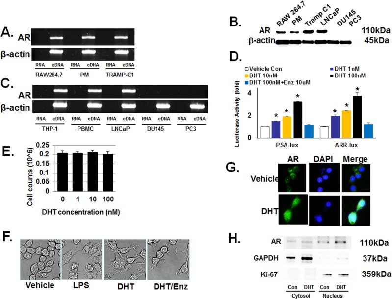
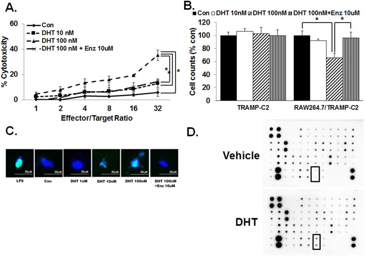
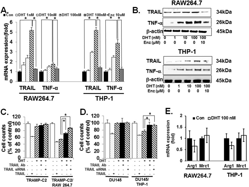
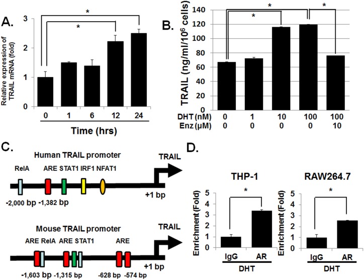
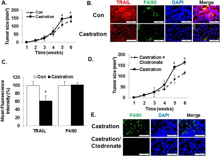
Although androgen deprivation therapy (ADT) and immunotherapy are potential treatment options in men with metastatic prostate cancer (CaP), androgen has conventionally been proposed to be a suppressor of the immune response. However, we herein report that DHT activates macrophages. When the murine macrophage cell line (RAW 264.7), human monocyte cell line (THP-1), and human peripheral blood monocytes were cultured with androgen-resistant CaP cell lines, DHT increased cytotoxicity of macrophages in a concentration-dependent manner. Further studies revealed that DHT induced M1 polarization and increased the expression levels of TNF-related apoptosis-inducing ligand (TRAIL) in macrophages and that this effect was abrogated when TRAIL was neutralized with a blocking antibody or small interfering RNA. Subsequent experiments demonstrated that induction of TRAIL expression was regulated by direct binding of androgen receptor to the TRAIL promoter region. Finally, an in vivo mouse study demonstrated that castration enhanced the growth of an androgen-resistant murine CaP tumor and that this protumorigenic effect of castration was blocked when macrophages were removed with clodronate liposomes. Collectively, these results demonstrate that DHT activates the cytotoxic activity of macrophages and suggest that immunotherapy may not be optimal when combined with ADT in CaP.


Figures

Figure 1.
Figure 1.
Macrophages have a functional conventional AR. Unless specified, cells were treated with the indicated concentrations of dihydrotestosterone (DHT) for 24 h. (A) Through use of RT-PCR, AR mRNA was detected in the murine macrophage cell line RAW264.7 and primary peritoneal macrophages (PM). The androgen-sensitive murine prostate cancer cell line, TRAMP-C1, was used as the positive control cells. Primer sequences are provided in an online repository (25). (B) Immunoblot detected 110-kDa AR protein in RAW 264.7, PM, Tramp C1 (positive control), LNCaP (positive control), DU145 (negative control), and PC3 (negative control). (C) RT-PCR also showed AR mRNA expression in human macrophages. THP-1 human monocyte cell line was differentiated into macrophages by using PMA before isolating mRNA for RT-PCR. LNCaP is a human prostate cancer cell line used as a positive control; DU145 and PC3 are human prostate cancer cell lines used as negative controls. (D) Androgen signaling axis is functional in macrophages. The androgen response reporter vectors, PSA-lux and ARR-lux, were transfected into RAW 264.7 cells and cultured with 0 to 100 nM of DHT and/or enzalutamide (Enz) 10 μM for 24 h. At 1 nM DHT, luciferase activity increased 50% to 100% and a concentration-dependent increase was observed. Enzalutamide at10 μM reversed the induction of luciferase activity. (E) DHT has no significant effect on the proliferative capacity of macrophages. After treatment of RAW264.7 with DHT up to 72 h, no significant change in cell number was detected. The data represent a 72-h study. (F) DHT induced a profound morphological change in macrophages. When RAW264.7 was treated with 100 nM DHT for 24 h, cells developed multiple processes as well as intracellular vacuole-like structures. The morphological change elicited by DHT was nearly identical to that by LPS. Enzalutamide at10 μM inhibited the cellular morphological change by DHT. (G) Nuclear translocation of AR in RAW264.7 after DHT treatment. After treatment with 100 nM DHT for 6 h, AR nuclear translocation was detected by immunofluorescence microscopy. (H) Immunoblot demonstrated increased AR protein in the nuclear but not cytosol fraction. As controls, GAPDH was used for the cytosol fraction and Ki-67 for nuclear fraction. Error bars indicate average ± SEM. *P < 0.05 compared with vehicle-treated control group in all experiments. Control (Con) means only vehicle treatment group. PBMC, human peripheral blood mononuclear cell.
Figure 2.
Figure 2.
DHT increases cytotoxic activity of macrophages and TRAIL expression. (A) When TRAMP-C2 CaP cells and RAW 264.7 cells were cocultured for 3 d with increasing concentration of DHT (0 to 100 nM) and GAPDH release was measured as an indicator of cell viability, there was a concentration-dependent and effector/target ratio-dependent increase in cytotoxic effect. Enzalutamide (Enz) treatment, 10 μM, reversed the increase of cytotoxicity by DHT. Cocultures were carried out for 24 h. Each well contained 5000 target cells and varying number of RAW264.7 cells based on the indicated ratio. (B) Direct cell-cell contact is not required for DHT-induced cytotoxic activity of murine macrophages. When TRAMP-C2 was cocultured with RAW264.7 in the presence of a cell culture insert for 24 h, DHT again decreased the count of TRAMP-C2 in a concentration-dependent manner. Enzalutamide treatment, 10μM, completely reversed the increase of cytotoxic activity in macrophages at 100 nM DHT. (C) Upon DHT treatment, macrophages induced apoptosis in TRAMP-C2 cells. After cocultures RAW264.7 and TRAMP-C2 using the tissue culture inserts, Annexin V-FITC assay was carried out on TRAMP-C2 after removal of the macrophages. The results demonstrated an increase in apoptosis level with increased concentrations of DHT. Enzalutamide, 10 μM, totally abrogated the macrophage-mediated apoptosis induced by 100 nM DHT. Blue indicates DAPI staining; green indicates DNA fragments. (D) Cytokine antibody array including TRAIL was analyzed. After treatment of RAW264.7 cells with 100 nM DHT for 24 h, cell extracts were used to assess the expression of various cytokines. In this array, increased TRAIL protein was detected. Error bars indicate average ± SEM. *P < 0.05 in all experiments. Control means only vehicle treatment group. Con, TRAMP-C2 with vehicle without RAW264.7.
Figure 3.
Figure 3.
DHT induces M1 polarization of macrophages. (A) DHT induced the expression of TNF-α and TRAIL mRNAs in both the murine and human macrophages (RAW264.7 and THP-1, respectively) and enzalutamide (Enz; 10 μM) treatment completely neutralized the increase in TNF-α and TRAIL mRNAs. Cells were cultured with 100 nM DHT for 24 h. Then, RNA was isolated and quantitative PCR was carried out. (B) DHT increased TNF-α and TRAIL protein levels in both RAW264.7 and THP-1 cells. After culturing cells with 100 nM DHT for 24 h, immunoblot was carried out. Again, 10 μM enzalutamide partially blocked the increase of TNF-α and TRAIL proteins. (C) RAW264.7/TRAMP-C2 cocultures using tissue culture inserts were pretreated with the TRAIL neutralizing antibody for 1 h. Subsequently, 100 nM DHT was added and cocultures were incubated for 24 h. After removal of RAW264.7 cells in the top compartment, remaining TRAMP-C2 cells in the lower chamber were counted. As a complementary approach, TRAIL short hairpin RNA was used. After transfecting RAW264.7 cells with short hairpin RNA and incubating for 24 h, they were added to the top compartment of the coculture. Twenty-four h later, the top chamber was removed and the remaining TRAMP-C2 was counted. As control, TRAMP-C2 was cultured without RAW264.7. Where denoted negative (−), appropriate volume of vehicle was used. As a positive control, 100 ng/mL of TRAIL was used. The results demonstrated that both the neutralizing antibody and short hairpin RNA partially restored the decrease in cell count seen in the presence of RAW264.7 and 100 nM DHT. (D) Identical study was carried out using the human system: THP-1 and DU145. Again, TRAIL neutralizing antibody and short hairpin RNA partially reversed the decrease in DU145 cell numbers in the presence of THP-1 and 100 nM DHT. To prevent cell-cell contact, tissue culture inserts were used. (E) After culturing RAW264.7 and THP-1 with 100 nM DHT for 24 h, mRNA was isolated and M2 polarization markers Arg1 and Mrc1 were quantitated with qPCR, and there was no significant effect on M2 polarization of macrophages. Error bars indicate average ± SEM. *P < 0.05 in all experiments. Control means only vehicle treatment group.
Figure 4.
Figure 4.
AR directly activates the transcription of TRAIL mRNA in macrophages. (A) RAW264.7 was treated with 100 nM DHT and cells were harvested after the indicated time and mRNA isolated. TRAIL mRNA was measured by using qPCR. TRAIL mRNA expression values were normalized by subtracting the levels of the vehicle-only group for each time point. The results demonstrated that DHT induced TRAIL mRNA in a time-dependent manner. (B) To measure the effect of DHT on TRAIL expression at the protein level, ELISA was carried out using the conditioned media. RAW264.7 was treated with 1 to 100 nM DHT for 24 h and the conditioned media were collected. The results demonstrated that DHT increased TRAIL protein levels in the conditioned media in a concentration-dependent manner. Enzalutamide (Enz; 10 μM) treatment decreased the DHT-induced TRAIL secretion. (C) There are potential AR binding sites within the human and mouse TRAIL promoter. By using the computer program PROMO, the consensus androgen-responsive element within the TRAIL promoter was assessed. (D) RAW264.7 and THP-1 were treated with 100 nM DHT for 24 h. Then, genomic DNA was isolated ChIP was carried out. The results demonstrated that AR binding to the TRAIL promoter region increased upon treatment with DHT 100 nM. Error bars indicate average ± SEM. *P < 0.05. ARE, androgen response element; Rel-A, transcription factor p65.
Figure 5.
Figure 5.
Macrophages enhance tumor growth of androgen-resistant prostate cancer cells following castration in vivo. (A) To determine the impact of androgens on macrophages in vivo, C57B/6 mice with established androgen-resistant TRAMP-C2 xenograft were castrated and tumor volume was measured weekly. The results demonstrated that castration increased tumor growth. n = 5 per group. (B) At the end of 6 wk, all tumor xenografts were harvested and analyzed. Immunofluorescence microscopy demonstrated a significant increase in the protein levels of TRAIL within the tumor xenografts following castration. However, no detectable effect was seen on the magnitude of macrophage infiltration (F4/80). (C) To quantify the immunofluorescence, mean fluorescence intensity was measured by using ImageJ V1.50i software. TRAIL fluorescence intensity but not that of F4/80 significantly increased with castration. (D) When macrophage was removed with intraperitoneal injections of clodronate liposome every 3 d following castration, the protumorigenic effect of castration in mice with TRAMP-C2 xenograft was reversed. Control mice were injected with PBS liposome. (E) Administration of clodronate-liposome decreased macrophage infiltration in TRAMP-C2 xenografts. Again, the mean fluorescence intensity levels of TRAIL and F4/80 were quantified by using ImageJ V1.50i software. Values are means ± SEM. n = 5 per group. *P < 0.05.

Similar articles

Cited by

References

    1. Wiley SR, Schooley K, Smolak PJ, Din WS, Huang CP, Nicholl JK, Sutherland GR, Smith TD, Rauch C, Smith CA, Goodwin RG Identification and characterization of a new member of the TNF family that induces apoptosis. Immunity. 1995;3(6):673–682. - PubMed
    1. Hesry V, Piquet-Pellorce C, Travert M, Donaghy L, Jégou B, Patard JJ, Guillaudeux T. Sensitivity of prostate cells to TRAIL-induced apoptosis increases with tumor progression: DR5 and caspase 8 are key players. Prostate. 2006;66(9):987–995. - PubMed
    1. Nakajima Y, DelliPizzi AM, Mallouh C, Ferreri NR. TNF-mediated cytotoxicity and resistance in human prostate cancer cell lines. Prostate. 1996;29(5):296–302. - PubMed
    1. Koschny R, Walczak H, Ganten TM. The promise of TRAIL--potential and risks of a novel anticancer therapy. J Mol Med (Berl). 2007;85(9):923–935. - PubMed
    1. Mahalingam D, Szegezdi E, Keane M, de Jong S, Samali A. TRAIL receptor signalling and modulation: are we on the right TRAIL? Cancer Treat Rev. 2009;35(3):280–288. - PubMed

Publication types

Substances