. 2019 Jun 10;17(6):e3000304.
doi: 10.1371/journal.pbio.3000304. eCollection 2019 Jun.

Selective use of primate CD4 receptors by HIV-1

Affiliations

Selective use of primate CD4 receptors by HIV-1

Cody J Warren et al. PLoS Biol. .

Abstract

Individuals chronically infected with HIV-1 harbor complex viral populations within their bloodstreams. Recently, it has come to light that when these people infect others, the new infection is typically established by only one or a small number of virions from within this complex viral swarm. An important goal is to characterize the biological properties of HIV-1 virions that seed and exist early in new human infections because these are potentially the only viruses against which a prophylactic HIV-1 vaccine would need to elicit protection. This includes understanding how the Envelope (Env) protein of these virions interacts with the T-cell receptor CD4, which supports attachment and entry of HIV-1 into target cells. We examined early HIV-1 isolates for their ability to infect cells via the CD4 receptor of 15 different primate species. Primates were the original source of HIV-1 and now serve as valuable animal models for studying HIV-1. We find that most primary isolates of HIV-1 from the blood, including early isolates, are highly selective and enter cells through some primate CD4 receptor orthologs but not others. This phenotype is remarkably consistent, regardless of route of transmission, viral subtype, or time of isolation post infection. We show that the weak CD4 binding affinity of blood-derived HIV-1 isolates is what makes them sensitive to the small sequence differences in CD4 from one primate species to the next. To substantiate this, we engineered an early HIV-1 Env to have high, medium, or low binding affinity to CD4, and we show that it loses the ability to enter cells via the CD4 receptor of many primate species as the binding affinity gets weaker. Based on the phenotype of selective use of primate CD4, we find that weak CD4 binding appears to be a nearly universal property of HIV-1 circulating in the bloodstream. Therefore, weak binding to CD4 must be a selected and important property in the biology of HIV-1 in the body. We identify six primate species that encode CD4 receptors that fully support the entry of early HIV-1 isolates despite their low binding affinity for CD4. These findings will help inform long-standing efforts to model HIV-1 transmission and early disease in primates.


Conflict of interest statement

The authors have declared that no competing interests exist.

Figures

Fig 1
Fig 1. Some primate species encode a CD4 that functions as an entry receptor for an early isolate of HIV-1.
(A) Cryo-EM structure of the Envelope (Env) trimer (blue) from a prototypic early HIV-1 isolate (BG505 [9]) in complex with human CD4 (tan) (PDB:5U1F) [28]. Only the D1 and D2 domains of CD4 are included in this structure, and the D1 domain mediates interaction with Env. Amino acids highlighted in red spheres were previously shown to be evolving under recurrent positive selection and fall at the interaction interface with Env [20,21]. (B) Dog thymocytes (Cf2Th cells) stably expressing human CCR5 and the indicated CD4 receptors (x-axis) were infected with Q23ΔEnv-GFP pseudotyped with BG505 Env, isolated from a newly infected infant [9]. The percent cells infected (GFP-positive) was enumerated by flow cytometry and normalized to the percent infected with human CD4. Error bars represent the mean + SEM from four independent experiments, each with two to three technical replicates. Values above error bars represent fold decrease in viral entry relative to human CD4, only indicated for those samples that passed significance thresholds (one-way ANOVA for repeated measures effect, Dunnett’s test; P < 0.05). Data associated with this figure can be found in the supplemental data file (S1 Data). (Top) Cladogram of species used in this experiment, with the number of amino acid differences in the D1 domain of CD4 (compared to human CD4) noted at the branch tips as well as the amino acid encoded at position 39. CCR5, C-C motif chemokine receptor 5; cryo-EM, cryogenic electron microscopy; GFP, green fluorescent protein; PDB, Protein Data Bank.
Fig 2
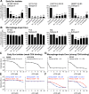
Fig 2. Selective use of primate CD4 receptors is not a result of the transmission bottleneck and instead is seen in many isolates of HIV-1 taken from human blood.
Cells stably expressing human CCR5 and human or primate CD4 (x-axis) were infected with Q23ΔEnv-GFP bearing (A) chronic Envelopes (Envs) [49], (B) early (newly infected; grey bars) and chronic (6 months post infection; black bars) Env pairs derived from the same patient [3,14,50,51], shown for one patient in each panel, or (C) Envs derived from maternal (chronic; black bars)/infant (newly infected; grey bars) transmission pairs [9], with one mother–baby pair shown in each panel. The percent cells infected (GFP-positive) was measured by flow cytometry and normalized to the percent infected with human CD4. Error bars represent the mean + SEM from two independent experiments, each with two to three technical replicates. Data associated with this figure can be found in the supplemental data file (S2 Data). CCR5, C-C motif chemokine receptor 5; GFP, green fluorescent protein.
Fig 3
Fig 3. Macrophage-tropic HIV-1 isolates, which are known to bind human CD4 more tightly, are promiscuous in their use of primate CD4 receptors.
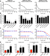
(A, B) Cells stably expressing various primate CD4 receptors (x-axis), along with human CCR5, were infected with Q23ΔEnv-GFP pseudotyped with (A) early HIV-1 Envelopes (Envs) or (B) Envs from common macrophage-tropic HIV-1 isolates. The percent cells infected (GFP-positive) was measured by flow cytometry and normalized to the percent infected with human CD4. Error bars represent the mean + SEM from two independent experiments, each with two to three technical replicates. Values above error bars represent fold decrease in viral entry relative to human CD4, only indicated for those samples that passed significance thresholds (one-way ANOVA for repeated measures effect, Dunnett’s test; P < 0.05). (C and D) Infection of TZM-bl cells with Q23ΔEnv-GFP pseudotyped with early (C) or macrophage-tropic (D) Envs. Each virus was preincubated with increasing concentrations of human soluble CD4 (sCD4) as a competitive inhibitor. TZM-bl cells are HeLa CD4/CCR5 cells that express luciferase in response to HIV-1 tat expression, read in relative light units (RLUs). Error bars represent the mean + SD from one biological replicate (n = 3 to 4 technical replicates), representative of two independent experiments. Error bars are not plotted in instances in which the size of the error bar is less than the symbol size. IC50 values from the soluble CD4 neutralization assays are shown within each panel, for which the mean and SD values reflect the variation of the IC50 calculations from two independent experiments. (E and F) Same as C and D, only neutralization assays were performed with chimpanzee and rhesus macaque sCD4 instead of human sCD4. Data associated with this figure can be found in the supplemental data file (S3 Data). CCR5, C-C motif chemokine receptor 5; GFP, green fluorescent protein.
Fig 4
Fig 4. An early Env engineered to have weak, medium, or tight binding to CD4 becomes progressively more promiscuous in its use of primate CD4 receptors.
(A–C) Amino acid substitutions were introduced into the BG505 Envelope (Env) at position 281 and 375. The headers at the top describing these mutations pertain to panels A, B, and C. In each column, the indicted Env was pseudotyped onto Q23ΔEnv-GFP. (A) Each of the three viruses was preincubated with increasing concentrations of human soluble CD4 (sCD4) and then used to infect TZM-bl cells, which are HeLa CD4/CCR5 cells that express luciferase in response to HIV-1 infection and tat expression, read in relative light units (RLUs). Error bars represent the mean + SD from one biological replicate, representative of two independent experiments. Error bars are not plotted in instances in which the size of the error bar is less than the symbol size. IC50 values are listed on each panel, for which the mean and SD values reflect the variation of the IC50 calculations from two independent experiments. (B) Cells stably expressing the indicated CD4s (x-axis) and human CCR5 were infected with each virus. The percent cells infected (GFP-positive) was measured by flow cytometry and normalized to the percent infected with human CD4. Error bars represent the mean + SEM from two independent experiments, each with three technical replicates. Values above error bars represent fold decrease relative to human CD4 for those samples that passed significance thresholds (one-way ANOVA for repeated measures effect, Dunnett’s test; P < 0.05). (C) Same as panel A but with chimpanzee and rhesus macaque sCD4 instead of human sCD4. (D) Two additional single point mutations were introduced into the BG505 Env, which have also been shown to increase binding affinity for CD4 [77]. These Envs were pseudotyped onto Q23ΔEnv-GFP and used to infect cells stably expressing the indicated CD4 (top of each graph) and human CCR5. The percent cells infected (GFP-positive) was measured by flow cytometry and normalized to the percent infected with human CD4. Error bars represent the mean + SEM from two independent experiments, each with two to three technical replicates. Values above error bars represent fold increase relative to wildtype BG505 Env for those samples that passed significance thresholds (one-way ANOVA for repeated measures effect, Dunnett’s test; P < 0.01). Data associated with this figure can be found in the supplemental data file (S4 Data). CCR5, C-C motif chemokine receptor 5; GFP, green fluorescent protein.
Fig 5
Fig 5. Early isolates of HIV-1 from the blood are deficient in binding and/or fusion with CD4 from some primate species.
(A) 293T cells transiently expressing various Envelope proteins (Envs), and dog thymocytes (Cf2Th cells) stably expressing various primate CD4 receptors and human CCR5, were each transfected with plasmids encoding half of a split Renilla luciferase [79]. These two cell types were then cocultured to induce membrane fusion, which was detected by a luminescent product following the addition of the Renilla luciferase substrate. (B) The percentage of fusion was calculated relative to what was observed with human CD4. Error bars represent the mean + SEM from three to four independent experiments, each with four technical replicates. Values above error bars represent fold decrease relative to human CD4 for those samples that passed significance thresholds (one-way ANOVA for repeated measures effect, Dunnett’s test; P < 0.05). Data associated with this figure can be found in the supplemental data file (S5 Data). CCR5, C-C motif chemokine receptor 5.
Fig 6
Fig 6. Rhesus macaque CD4 alleles are universally defective for HIV-1 entry.
(A) Total RNA was isolated from the blood of 52 rhesus macaque individuals. CD4 was amplified from cDNA, and PCR products were sequenced to yield genotype information for each individual (104 chromosomes). Genotype information was used to phase SNPs using Phase 2.1 [81,82] in DNAsp v5 [83]. The table summarizes the unique CD4 alleles identified. For each allele, the residue encoded at each variable amino acid positions is noted. Unique alleles were cloned and further verified by Sanger sequencing. (B) Dog thymocytes (Cf2Th cells) stably expressing human CCR5 and the indicated rhesus macaque CD4 alleles (x-axis) were infected with HIV-1 (Q23ΔEnv-GFP) bearing a subtype A (BG505) or subtype C (CAP210.2.00.E8) Envelope (Env). The percent cells infected (GFP-positive) was measured by flow cytometry 48 hours post infection. Error bars represent the mean + SEM from two independent experiments, each with three technical replicates. Values above error bars represent fold decrease relative to human CD4 for those samples that passed significance thresholds (one-way ANOVA for repeated measures effect, Dunnett’s test; P < 0.05). (C) 293T cells transiently expressing diverse HIV-1 Envs (see S1 Table) and Cf2Th cells stably expressing the indicated CD4/CCR5 receptors were each transfected with plasmids encoding half of a split Renilla luciferase [79]. These two cell types were then cocultured to induce membrane fusion, which was detected by a luminescent product following the addition of the renilla luciferase substrate. A total of 33 Envs were tested from the following major HIV-1 subtypes: subtype A (n = 12), subtype B (n = 7), subtype C (n = 6), subtype D (n = 6), and subtype A/D (n = 2). The percentage of fusion was calculated relative to what was observed with human CD4/CCR5. Error bars represent the mean + SD, for which n = 2 (two technical replicates from one experiment). Data associated with this figure can be found in the supplemental data file (S6 Data). nt, nucleotide; aa, amino acid; CCR5, C-C motif chemokine receptor 5; GFP, green fluorescent protein.

Similar articles

Cited by

References

    1. Sagar M. HIV-1 transmission biology: selection and characteristics of infecting viruses. J Infect Dis. 2010; 202 Suppl 2: S289–96. - PMC - PubMed
    1. Kearney M, Maldarelli F, Shao W, Margolick JB, Daar ES, Mellors JW, et al. Human Immunodeficiency Virus Type 1 Population Genetics and Adaptation in Newly Infected Individuals. J Virol. 2009; 83: 2715–2727. 10.1128/JVI.01960-08 - DOI - PMC - PubMed
    1. Keele BF, Giorgi EE, Salazar-Gonzalez JF, Decker JM, Pham KT, Salazar MG, et al. Identification and characterization of transmitted and early founder virus envelopes in primary HIV-1 infection. Proc Natl Acad Sci USA. 2008; 105: 7552–7557. 10.1073/pnas.0802203105 - DOI - PMC - PubMed
    1. Abrahams MR, Anderson JA, Giorgi EE, Seoighe C, Mlisana K, Ping LH, et al. Quantitating the multiplicity of infection with human immunodeficiency virus type 1 subtype C reveals a non-poisson distribution of transmitted variants. J Virol. 2009; 83: 3556–3567. 10.1128/JVI.02132-08 - DOI - PMC - PubMed
    1. Derdeyn CA, Decker JM, Bibollet-Ruche F, Mokili JL, Muldoon M, Denham SA, et al. Envelope-constrained neutralization-sensitive HIV-1 after heterosexual transmission. Science. 2004; 303: 2019–2022. 10.1126/science.1093137 - DOI - PubMed

Publication types

MeSH terms

Substances