. 2019 Jun 7;10(1):2510.
doi: 10.1038/s41467-019-10364-0.

CHML promotes liver cancer metastasis by facilitating Rab14 recycle

Affiliations

CHML promotes liver cancer metastasis by facilitating Rab14 recycle

Tian-Wei Chen et al. Nat Commun. .

Abstract

Metastasis-associated recurrence is the major cause of poor prognosis in hepatocellular carcinoma (HCC), however, the underlying mechanisms remain largely elusive. In this study, we report that expression of choroideremia-like (CHML) is increased in HCC, associated with poor survival, early recurrence and more satellite nodules in HCC patients. CHML promotes migration, invasion and metastasis of HCC cells, in a Rab14-dependent manner. Mechanism study reveals that CHML facilitates constant recycling of Rab14 by escorting Rab14 to the membrane. Furthermore, we identify several metastasis regulators as cargoes carried by Rab14-positive vesicles, including Mucin13 and CD44, which may contribute to metastasis-promoting effects of CHML. Altogether, our data establish CHML as a potential promoter of HCC metastasis, and the CHML-Rab14 axis may be a promising therapeutic target for HCC.


Conflict of interest statement

The authors declare no competing interests.

Figures
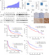
Fig. 1
Fig. 1
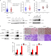
CHML is upregulated in HCC and predicts poor survival. a Real-time PCR analysis of CHML mRNA in 45 paired HCC tumor tissues and normal tissues. b Box plots comparing levels of CHML mRNA in normal human liver tissues and hepatocellular carcinoma tissues in published data sets from oncomine. ***P < 0.001, Student’s t-test. c Western blot analysis of CHML expression in 24 paired normal (N) and HCC tumor (T) tissues. GAPDH is used as internal control. Arrow demonstrates the CHML band, and NS means non-specific band. d Immunohistochemistry staining of CHML in 3 paired normal liver and HCC tumor. Scale bar, 50 μm. e Kaplan–Meier analysis of overall survival of tissue microarray (TMA) data containing 297 patients. f Kaplan–Meier analysis of overall survival data from TCGA liver cancer data containing 364 patients. g, h Kaplan–Meier analysis of recurrence-free survival of the TMA data (g) and TCGA-LIHC data (h). All data are represented as mean ± s.e.m.
Fig. 2
Fig. 2
CHML overexpression promotes HCC cell metastasis and invasion. a Dot plots comparing CHML expression in patients with or without microvascular invasion (MVI) in two published datasets. *P < 0.01, Student’s t-test. b Dot plot analysis of CHML expression in normal, HCC and portal vein tumor thrombus (PVTT) tissues. N normal, T tumor, P PVTT. **P < 0.01 by two-tailed unpaired Student’s t-test. c Western blot analysis of CHML expression in normal, HCC, and PVTT tissues obtained from 4 patients. d Western blot analysis of CHML expression in 5 HCC cell lines. Arrow indicated the CHML band. e Western blot analysis of the overexpression efficiency of CHML in HCC cell line PLC/PRF/5 and YY8103. v vector control. f Boyden chamber and invasion assay are conducted to detect the migratory and invasive ability of control cells and CHML-overexpressed cells. Each representative image is shown. g Quantitative results are respectively illustrated for panel (b). Data are shown as the mean ± s.e.m., **P < 0.01 by two-tailed unpaired Student’s t-test. All data are represented as mean ± s.e.m.
Fig. 3
Fig. 3
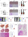
Knockdown of CHML decreased cellular migratory and invasive abilities. a, c Western blot analysis of the knockdown efficiency of CHML in HCC cell line CSQT-2 (a) and LM3 (c). b, d Boyden chamber and invasion assay are conducted to detect the migration and invasion abilities of control cells and CHML-knockdown CSQT-2 cells (b) and LM3 cells (d). Each representative image is shown. Quantitative blot analysis was shown on the right. Data are shown as the mean ± s.e.m., ***P < 0.001 by two-tailed unpaired Student’s t-test. e Left: representative images showing luciferase expression from intrahepatic tumors of both CSQT-2 control and shCHML group. Right: quantification of luciferase expression of intrahepatic tumors. *P < 0.01, Student’s t-test. f Livers of both control and shCHML groups resected from intrahepatic metastasis mouse model. Tissues are photographed, fixed, and stained with hematoxylin and eosin (HE). Scale bar, 60 μm. ***P < 0.001 by two-tailed unpaired Student’s t-test. g Left: representative images showing luciferase expression from lung metastasis of both CSQT-2 control and shCHML group. Right: quantification of luciferase expression of lung metastases. ****P < 0.0001 by two-tailed unpaired Student’s t-test. h HE staining of lungs from both control and shCHML groups resected from tail vein injection metastasis mouse model. Scale bar, 100 μm. ***P < 0.001 by two-tailed unpaired Student’s t-test. All data are represented as mean ± s.e.m.
Fig. 4
Fig. 4
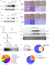
CHML interacts strongly with Rab14. a silver stained gel shows differential bands between control and CHML sample. White arrow indicates differential band, the upper one indicates Flag-CHML. M marker, vec vector control. b Tandem mass spectrum analysis of target band. c, d Interaction between CHML and Rab14 was demonstrated by immunoprecipitation. 293T cells were transfected with Flag-CHML (c) or Flag-Rab14 (d). Immunoprecipitation was performed at 36 h post transfection with Flag antibody. e, f Western blot results showing endogenous CHML interacted with Rab14 in PLC/PRF/5 (e) and YY-8103 (f) cells. Black arrow indicates light chain. g In vitro interaction between CHML and Rab14. Purified 6xHis CHML was incubated with indicated GST fusion proteins, GST pull-down assay was conducted. h Graphic shows the domains in CHML protein. i In vitro interaction assay showing the two GDI domains interacted with Rab14. The truncated domains of CHML were fused with GST, and incubated with Rab14, followed by GST pull-down and WB
Fig. 5
Fig. 5
Function of CHML is Rab14-dependent. a Western blot analysis of the knockout efficiency of Rab14 and overexpression efficiency of CHML in HCC cell line PLC/PRF/5 and YY8103. b Boyden chamber assay was performed to detect the metastatic ability of these indicated cells. Representative images are shown. c Quantitative results are respectively illustrated for panel (b). NS not significant. Data are shown as the mean ± s.e.m., **P < 0.01 by two-tailed unpaired Student’s t-test. All data are represented as mean ± s.e.m.
Fig. 6
Fig. 6
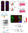
CHML serves to escort Rab14 to membrane. a, b Localization of endogenous Rab14 in PLC/PRF/5 (a) and YY8103 (b) cells was stained and representative images are shown. The distances of signals to nucleus were calculated and statistically presented. ***P < 0.001, Student’s t-test. Scale bar, 20 μm. c, d Rab14-GTP in shCHML CSQT-2 (c) and LM3 (d) cells was pulled down by GST-ΔRCP559-649. Ponceau S indicates GST-ΔRCP559-649. e Schematic showing factors influencing Rab-GTP. f 293T cells were transfected with control vector and Rab14WT, Rab14Q70L, and Rab14S25N plasmid, respectively. After 48 h, immunoprecipitation assay was performed. g 293T cells were transfected with EGFP-Rab14S25N or EGFP-Rab14Q70L plasmid, and after 48 h endogenous CHML was stained. Images were photographed by a laser scanning microscope. Scale bar, 5 μm. White arrowheads indicated merged signal. The percentage of merged signal was counted and presented in bar plot. ***P < 0.001, Student’s t-test. h Schematic showing the function of GDI. i PLC/PRF/5 Membrane fraction was incubated with Flag-CHML purified from 293T cells at 37 °C for 30 min, membrane Rab14 was detected by WB. j CHML immunoprecipitates from 293T cells were incubated with Rab14-depleted membrane, after incubation membrane fraction and supernatant were resolved by Western blot. All data are represented as mean ± s.e.m.
Fig. 7
Fig. 7
Rab14-positive membrane fraction contains metastasis-promotional factors. a, c Western blot analysis of the overexpression efficiency of Rab14WT and its mutants in HCC cell line PLC/PRF/5 and YY8103. V vector control. b, d Metastasis and invasion assays were conducted to detect these indicated cells. e Workflow to identify Rab14-positive vesicle components. f Sucrose-gradient centrifugation was performed to fractionate Rab14 containing endosomes. Rab5A was used as endosome marker. g Immunoprecipitation was performed using anti-Rab14 beads and endosomes. Incubation buffer-pre, endosome-containing buffer before incubating with beads. Incubation buffer-IgG or -Rab14, endosome-containing buffer after incubating with IgG beads or anti-Rab14 beads. h Western blot analysis of eluates from IgG and anti-Rab14 beads. i Venn diagram indicates proteins identified by MS/MS. j Venn diagram analysis of a total of 157 Rab14-positive membrane components. k 157 Rab14-positive membrane components were analyzed with their reported metastatic function in cancer
Fig. 8
Fig. 8
CHML KD influences membrane localization of CD44 and Mucin13. a Representative images showing Mucin13 and CD44 were partially co-localized with EGFP-Rab14 in CSQT-2 cells. Scale bar, 20 μm. White arrowheads indicated merge sites. b, c Western blot analysis of indicated surface protein in YY-8103 (b) and CSQT-2 cells (c). d Representative images showing endogenous distribution of CD44 and Mucin13 both in YY-8103 control and shCHML cells. Scale bar, 20 μm

Similar articles

Cited by

References

    1. Torre LA, et al. Global cancer statistics, 2012. CA Cancer J. Clin. 2015;65:87–108. doi: 10.3322/caac.21262. - DOI - PubMed
    1. Shetty K, He AR. A STORM in a teacup?—The challenges of adjuvant therapy in hepatocellular carcinoma (HCC) Transl. Gastroenterol. Hepatol. 2016;1:2. doi: 10.21037/tgh.2016.03.10. - DOI - PMC - PubMed
    1. Yang JD, Roberts LR. Hepatocellular carcinoma: a global view. Nat. Rev. Gastroenterol. Hepatol. 2010;7:448–458. doi: 10.1038/nrgastro.2010.100. - DOI - PMC - PubMed
    1. Lochan, Reeves, H. L. & Manas, D. M. Surgical management of HCC. In: Practical Management of Chronic Viral Hepatitis (ed Serviddio, G.) (InTech, London, UK, 2013).
    1. Llovet, J. M. et al. Hepatocellular carcinoma. Nat. Rev. Dis. Primers2, 16018 (2016). - PubMed

Publication types

MeSH terms