. 2019 Jul 2;30(1):129-142.e4.
doi: 10.1016/j.cmet.2019.05.006. Epub 2019 May 30.

Acceleration of β Cell Aging Determines Diabetes and Senolysis Improves Disease Outcomes

Affiliations

Acceleration of β Cell Aging Determines Diabetes and Senolysis Improves Disease Outcomes

Cristina Aguayo-Mazzucato et al. Cell Metab. .

Abstract

Type 2 diabetes (T2D) is an age-related disease. Although changes in function and proliferation of aged β cells resemble those preceding the development of diabetes, the contribution of β cell aging and senescence remains unclear. We generated a β cell senescence signature and found that insulin resistance accelerates β cell senescence leading to loss of function and cellular identity and worsening metabolic profile. Senolysis (removal of senescent cells), using either a transgenic INK-ATTAC model or oral ABT263, improved glucose metabolism and β cell function while decreasing expression of markers of aging, senescence, and senescence-associated secretory profile (SASP). Beneficial effects of senolysis were observed in an aging model as well as with insulin resistance induced both pharmacologically (S961) and physiologically (high-fat diet). Human senescent β cells also responded to senolysis, establishing the foundation for translation. These novel findings lay the framework to pursue senolysis of β cells as a preventive and alleviating strategy for T2D.

Keywords: SASP; beta cells; glucose metabolism; insulin resistance; insulin secretion; senescence; senescence signature; senescence-associated secretory profile; senolytic therapies; type 2 diabetes.


Conflict of interest statement

DECLARATION OF INTERESTS

The authors declare no competing interests.

Figures

Figure 1.
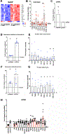
Figure 1.. Generation of a β-cell senescence signature and cellular characteristics.
Islets isolated from 7–8 m old C57Bl/6J male retired breeders were FACS sorted into non-senescent (β-gal negative) and senescent (β-gal positive) subpopulations(A) for RNASeq after gating for enrichment of β-cells (B). Cellular identity of FACS sorted cells was 90% β-cells and 0.5% of immune cells (C). D. As described for senescent cells, β-gal+ β-cell were significantly larger than β-gal-cells. At least 100 cells counted/condition from 3 different fields. For RNA seq data there were 7 sets of paired samples, each set from islets pooled of 30 mice. Senescent β-cells were characterized by a downregulation of hallmark β-cell identity genes (E) and upregulation of “disallowed” genes (F). Senescent β-cells also showed upregulation of specific aging (G) and senescent genes (H).
Figure 2.
Figure 2.. SASP factors are produced and secreted by senescent β-cells.
Senescent β-cells were characterized by an upregulation of SASP factors (A), some of which are significantly increased in conditioned media from β-gal+cells (B). Each point represents a FACS sorting experiment. Mean concentrations of proteins were IL1a 6pg/ml; TNFa 23 pg/ml, IL6 83 pg/ml, CCL5 5 pg/ml. CSCL1 22 pg/ml, CCL3 6 pg/ml, CCL4 4 pg/ml, CXCL10 2 pg/ml. C. Isolated mouse islets were cultured 4d in the presence of CM from β-gal+β-cells had regulation of p16Ink4acompared to those in CM form exposed to CM from β-gal-β-cells, indicating a functional β-cell SASP. CM from 5 FACS sorts with each used on 2–3 separate islet isolations. D. In immortalized β-cell line MIN6 cells senescence was induced by 24 hr exposure to 200 nM doxorubicin; senescence was confirmed by upregulation of p16Ink4a and p21Cis1 mRNA. E. Conditioned media from doxorubicin-treated cells had detectable protein levels of several SASP factors compared to non-senescent MIN6 cells. Each point represents an experiment; *p<0.05. F. MIN6 cell senescence was induced by exposing them to 450 μM H2O2 for 24h; senescence was confirmed by upregulation of p16Ink4a and p21Cis1 mRNA. G. Conditioned media from H2O2-treated cells cells had detectable protein levels of several SASP factors compared to non-senescent MIN6 cells. Each point represents an experiment; *p<0.05. H. MIN6 cells were treated with p16Ink4a siRNA, decreasing its expression by 50%. Several SASP factor mRNAs were significantly changed (red bars) compared to cells treated with Scr siRNA, suggesting their regulation in β-cells is downstream of p16Ink4a. n=4 experiments in triplicate. Means plotted with each point representing a single sample. p<0.05.
Figure 3.
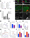
Figure 3.. Insulin resistance accelerates β-cell senescence.
Osmotic minipump administration of the insulin receptor antagonist S961 for 2 wk induced marked hyperglycemia (A) and hyperinsulinemia (B) in 7–m old male C57Bl6/J mice. Islets from treated mice had greater proportion of β-gal+β-cells (C) (12,089–100,000 events/data point) and increased expression of aging (D) and SASP-related (E) genes. A,B,D,E: 7m male C57Bl6/J; C 4m male INK ATTAC. n=5 per group. *p<0.05. F. Image of pancreas showing heterogenous colocalization of insulin and P21CIS1 in S961 treated animals compared to untreated controls. Magnification bar= 50 μm. G. Quantification of cellular P21CIS1 staining intensity presented as mean per β-cells of each islet; at least 30 islets counted per pancreas, n=3 control, 6 S961 animals. Two wk after minipump excision and normalization of blood glucose (H) and insulin (I) levels, some changes induced by S961 were reversed: both the aging (J) and SASP (K) indices decreased with respect to S961 islets, with no change on b cell-related genes (L) (See Suppl Fig 3 for individual values). 7m C57Bl6/J male; n=5 per group. Eight wk of high fat diet (HFD) starting at 8 wk increased body weight (M), fasting glucose (N) and induced glucose intolerance (O). Islets from treated animals showed higher proportion of β-gal+ cells (P)(20,856–40,933 events/data point) and increased aging (Q) and SASP (R) indices. n= 12 C57Bl6/J mice per group. *p<0.05 respect to control diet.
Figure 4.
Figure 4.. Specific deletion of p16Ink4a expressing cells improved metabolic profile, β-cell function and gene expression.
A. Evaluation of deletion protocol with B/B Homodimerizer (10mg/kg) revealed decreased eGFP and Casp8 mRNA in INKATTAC islets. Treatment was two courses of 3 d with 14 d between courses to activate the transgene caspase-8 moiety and lead to cell deletion of p16Ink4a expressing cells in the INKATTAC mice. Each dot represents the islets from an individual animal. B,C. Effects of B/B homodimerizer treatment on deletion of p16Ink4a β-cells were evaluated by quantification of FLAG and insulin co-staining. Pancreas from 5 m old animals (n=6 per group) were stained in parallel and confocal pictures were taken under the same settings such that differences in intensity reflect differences in protein concentration. Magnification bar= 50mm *p<0.05. Old INK-ATTAC mice partially improved glucose metabolism (D) and recovered β-cell function (G) after treatment with B/B homodimerizer. Isolated islets had decreased expression of genes of the aging (E) and SASP (F) indices. 1.3–1.6 y INK-ATTAC female mice. n=7–8 per group. *p<0.05. INKATTAC mice treated with insulin receptor antagonist S961 and B/B homodimerizer had improved metabolic profile (H) as shown by the AUC of their fed glucose levels (I). Improvement of aging (J) and SASP (K) indices was also observed. 9–14 m INK-ATTAC male S961 (20nM/wk) treatment for 2 wk. n=3–4 per group. S961 *p<0.05. (See Suppl Fig 4 for individual values).
Figure 5.
Figure 5.. Beneficial effects of p16Ink4a deletion in HFD INK-ATTAC model.
Changes induced with HFD such as increased body weight (A), fed hyperglycemia (B) and glucose intolerance (C,D) were blunted after B/B homodimerizer. In vivo insulin secretion parameters improved after B/B treatment (E) and although no beneficial changes were seen in aging (F) and SASP (G) indices, islets had enhanced β-cell index (H). (See Suppl Fig 5 for individual values). INK-ATTAC female mice 9m, n=6–7 animals/group at start of the study; at isolation 4–7 animals/group remained. *p<0.05 respect to control; +p<0.05 respect to HFD.
Figure 6.
Figure 6.. Senolytic therapies selectively killed senescent cells and improved glucose metabolism, SASP and β-cell indices.
A. ABT263 specifically killed β-gal+ FACS sorted islet cells in vitro. Islets from male retired breeders C57Bl/6CRL. n= 2–3 experiments with up to 4 replicates/condition; 4 days. When ABT263 was administered in vivo by daily gavage to INK:ATTAC mice treated with S961, circulating blood glucose levels significantly improved (B,C), the percentage of β-gal+cells decreased (D)(31,684–64,940 events per data point) with no change in aging index, SASP and β-cell indices (E, F, G) (See Suppl Fig 6 for individual values). INKATTAC male mice 6–9 m old. n=3–5 animals/group. Administration of ABT263 during 12 wk HFD had no effects on fed glucose levels (H) but decreased the percentage of β-gal+cells (I) as well as the aging and SASP indices (J,K) and unchanged β-cell index (L) compared to those animals receiving HFD without ABT263 treatment (See Suppl Fig 6 for individual values). INK ATTAC 6–9 m old female mice, n= 3–4 each group.
Figure 7.
Figure 7.. Increased load of senescent human β-cells with donor age associated with enrichment of p16INK4A.
A. The percentage of β-gal+cells in human islets increased as the age of the donor increased. Samples from T2D were enriched for senescent cells compared to age-matched non-diabetic donors. B. β-gal+β-cell subpopulation expressed higher levels of P16INK4A mRNA than β-gal- population of the same donor. C. mRNA expression levels of SASP factors from human β-gal+β-cells compared to β-gal-cells. D. In vitro treatment with ABT263 (5 μM) revealed higher cell mortality of β-gal+ human β-cells when compared to β-gal-cells from the same donor. E. Representative pictures of IGF1R stained islets in sections of human pancreas. Sections were stained in parallel and pictures taken in the confocal microscope under the same setting such that differences in intensity reflect differences in protein concentration. F. Quantification of IGF1R intensity in islets from donors of different ages, with and without T2D. G. Representative pictures of nuclear P53BP1 of human pancreas. H. Linear correlation between P53BP1 intensity and BMI in donors without diabetes older than 39 y. I. Increased P53BP1 intensity in islets from donors older than 39 y, excluding those with BMI>33. Each data point is separate donor. Magnification bars = 50μm.

Comment in

  • Targeting β-cell senescence.
    Starling S. Starling S. Nat Rev Endocrinol. 2019 Aug;15(8):438-439. doi: 10.1038/s41574-019-0232-4. Nat Rev Endocrinol. 2019. PMID: 31239521 No abstract available.

Similar articles

Cited by

References

    1. Aguayo-Mazzucato C, van Haaren M, Mruk M, Lee TB Jr., Crawford C, Hollister-Lock J, Sullivan BA, Johnson JW, Ebrahimi A, Dreyfuss JM, et al. (2017). beta Cell Aging Markers Have Heterogeneous Distribution and Are Induced by Insulin Resistance. Cell Metab 25, 898–910 e895. - PMC - PubMed
    1. Baker DJ, Childs BG, Durik M, Wijers ME, Sieben CJ, Zhong J, Saltness RA, Jeganathan KB, Verzosa GC, Pezeshki A, et al. (2016). Naturally occurring p16(Ink4a)-positive cells shorten healthy lifespan. Nature 530, 184–189. - PMC - PubMed
    1. Baker DJ, Wijshake T, Tchkonia T, LeBrasseur NK, Childs BG, van de Sluis B, Kirkland JL, and van Deursen JM (2011). Clearance of p16Ink4a-positive senescent cells delays ageing-associated disorders. Nature 479, 232–236. - PMC - PubMed
    1. Bliss CR, and Sharp GW (1992). Glucose-induced insulin release in islets of young rats: time-dependent potentiation and effects of 2-bromostearate. Am J Physiol 263, E890–896. - PubMed
    1. Bruning JC, Winnay J, Bonner-Weir S, Taylor SI, Accili D, and Kahn CR (1997). Development of a novel polygenic model of NIDDM in mice heterozygous for IR and IRS-1 null alleles. Cell 88, 561–572. - PubMed

Publication types