. 2019 Jul 12;294(28):10942-10953.
doi: 10.1074/jbc.RA119.007929. Epub 2019 May 30.

Phosphatidylinositol 4,5-bisphosphate drives Ca2+-independent membrane penetration by the tandem C2 domain proteins synaptotagmin-1 and Doc2β

Affiliations

Phosphatidylinositol 4,5-bisphosphate drives Ca2+-independent membrane penetration by the tandem C2 domain proteins synaptotagmin-1 and Doc2β

Mazdak M Bradberry et al. J Biol Chem. .

Abstract

Exocytosis mediates the release of neurotransmitters and hormones from neurons and neuroendocrine cells. Tandem C2 domain proteins in the synaptotagmin (syt) and double C2 domain (Doc2) families regulate exocytotic membrane fusion via direct interactions with Ca2+ and phospholipid bilayers. Syt1 is a fast-acting, low-affinity Ca2+ sensor that penetrates membranes upon binding Ca2+ to trigger synchronous vesicle fusion. The closely related Doc2β is a slow-acting, high-affinity Ca2+ sensor that triggers spontaneous and asynchronous vesicle fusion, but whether it also penetrates membranes is unknown. Both syt1 and Doc2β bind the dynamically regulated plasma membrane lipid phosphatidylinositol 4,5-bisphosphate (PIP2), but it is unclear whether PIP2 serves only as a membrane contact or enables specialized membrane-binding modes by these Ca2+ sensors. Furthermore, it has been shown that PIP2 uncaging can trigger rapid, syt1-dependent exocytosis in the absence of Ca2+ influx, suggesting that current models for the action of these Ca2+ sensors are incomplete. Here, using a series of steady-state and time-resolved fluorescence measurements, we show that Doc2β, like syt1, penetrates membranes in a Ca2+-dependent manner. Unexpectedly, we observed that PIP2 can drive membrane penetration by both syt1 and Doc2β in the absence of Ca2+, providing a plausible mechanism for Ca2+-independent, PIP2-dependent exocytosis. Quantitative measurements of penetration depth revealed that, in the presence of Ca2+, PIP2 drives Doc2β, but not syt1, substantially deeper into the membrane, defining a biophysical regulatory mechanism specific to this high-affinity Ca2+ sensor. Our results provide evidence of a novel role for PIP2 in regulating, and under some circumstances triggering, exocytosis.

Keywords: calcium sensor; calcium-binding protein; exocytosis; membrane biophysics; membrane protein; phosphatidylinositol (4,5)-bisphosphate (PIP2); poly-anionic phospholipid; synapse; synaptotagmin; tandem C2 domain protein.


Conflict of interest statement

The authors declare that they have no conflicts of interest with the contents of this article

Figures

Figure 1.
Figure 1.
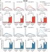
Doc2β penetrates membranes in response to Ca2+. A, schematic diagrams of syt1 and Doc2β. MID, munc13-interacting domain; TMD, transmembrane domain. B, model illustrating the putative membrane penetration activity of Doc2 where the distal tip of Ca2+-binding loop 1 was mutated to cysteine and labeled with the fluorescent dye NBD, shown at right. The shaded stripe in the bilayer leaflet depicts the approximate distribution of the quenching nitroxide on 12-doxyl-PC. Ribbon diagrams show C2A (PDB code 4LCV) and C2B (PDB code 4LDC) of Doc2β from Giladi et al. (52). C, NBD emission spectra from each of the four Ca2+-binding loops of Doc2β C2AB. Graph titles indicate the C2 domain and loop labeled (e.g. C2A*(1)-C2B corresponds to loop 1 of C2A, and C2A*(3)-C2B corresponds to loop 3 of C2A). Labeled C2AB was combined with liposomes (15% PS, 30% PC, 20% PE, and 35% cholesterol) in 500 μm EGTA after which Ca2+ was added (250 μm free [Ca2+]). Ca2+ triggered an intensity increase and blue shift in the emission spectra at all four labeling sites, suggesting burial of the probe into the bilayer. Membrane insertion was confirmed with the use of liposomes containing 15% 12-doxyl-PC, which efficiently quenched the fluorescence at each labeled site. Spectra are representative of data from at least four independent trials.
Figure 2.
Figure 2.
PS and PIP2 synergistically drive Ca2+-independent membrane penetration by syt1 and Doc2β but exert different effects on each protein. A, emission spectra of NBD-labeled Doc2β C2AB before and after the addition of liposomes containing 15 mol % PS and 1 mol % PIP2 in 500 μm EGTA (≤10 nm [Ca]free). Under these conditions, loops 1 and 3 of C2B demonstrate robust increases in emission intensity. Emission from loop 3 is efficiently quenched by 12-doxyl-PC, indicating Ca2+-independent insertion into the bilayer. Spectra are representative of data from at least four independent trials. B, NBD-labeled Doc2β C2AB was combined with the indicated liposomes, and the NBD emission intensity was measured before and after the addition of Ca2+. For each replicate, emission intensity was normalized to the signal from NBD-labeled protein prior to liposome addition. For Doc2β, PS and PIP2 each support Ca2+-dependent penetration activity. However, when combined, PS and PIP2 drove a marked Ca2+-dependent increase in the emission from C2A loop 3. Both PS and PIP2 were required for Ca2+-independent penetration by loops 1 and 3 of C2B (arrows). C and D, same as above but using syt1 C2AB. In contrast to Doc2β C2AB, PS drives penetration of syt1 C2A more efficiently than PIP2 in the presence of Ca2+. The combination of PIP2 and PS did not drive any additional NBD signal increases in C2A but marginally increased NBD signals in C2B. As with Doc2β, both PIP2 and PS were required for robust Ca2+-independent penetration by Syt1 C2B loop 3 (arrow). Error bars, S.E. of four independent trials; *, p < 0.05; **, p < 0.005; ns, p > 0.5; all by Welch's t test.
Figure 3.
Figure 3.
Membrane penetration by full-length syt1 reconstituted into nanodiscs. A, experimental scheme. Full-length syt1 was purified, labeled with NBD on loop 3 of C2A or loop 3 of C2B, and reconstituted into 13-nm-diameter nanodiscs comprising membrane scaffolding protein and POPC (ND-syt1). ND-syt1 was combined with liposomes containing acidic phospholipids in EGTA (500 μm) followed by the addition of Ca2+ to assay Ca2+-independent and Ca2+-dependent membrane penetration activity. B, representative spectra for penetration experiments with ND-syt1. As with syt1 C2AB, Ca2+ and acidic phospholipids caused an increase and blue shift in NBD fluorescence. Likewise, in the presence of both PS and PIP2, Ca2+-independent penetration by C2B, but not C2A, was observed. Spectra are representative of results from four independent trials.
Figure 4.
Figure 4.
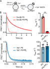
PIP2 slows the disassembly kinetics of C2AB–Ca2+–liposome complexes containing Doc2β but not syt1. A, schematic of disassembly assay. C2AB–Ca2+–liposome complexes were preassembled and then rapidly mixed with EGTA while monitoring FRET between tryptophan residues in C2AB and dansyl-PE acceptors on the liposomes. B, representative traces (left) and rate constants derived from single-exponential fits (right) for disassembly of Doc2β–Ca2+–membrane complexes. The inclusion of 1 mol % PIP2 in liposomes containing 15% PS slowed the observed rates of disassembly by ∼10-fold. C, as above but for syt1 C2AB. In contrast to the case of Doc2β, membrane complex disassembly rates (Kdiss) for syt1 were unchanged with the inclusion of 1 mol % PIP2. Error bars, S.E. of four independent trials; **, p < 0.005; ns, p > 0.5; both by Welch's t test.
Figure 5.
Figure 5.
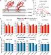
PIP2 markedly deepens membrane penetration by Doc2β but not syt1. A, illustration depicting membrane-bound C2AB and the approximate distributions of nitroxide quenchers. The yellow star represents NBD label, and green spheres represent Ca2+ ions. B, representative emission spectra for nonquenching liposomes along with liposomes containing the indicated doxyl quencher. Relative quenching efficiencies at different probe locations correspond to the average location of the NBD label in the bilayer. Deeper insertion results in stronger quenching by 12-doxyl versus 5-doxyl and HG-doxyl liposomes, whereas shallower insertion results in stronger quenching by HG-doxyl and 5-doxyl liposomes. C, NBD-labeled Doc2β C2AB was combined with liposomes and Ca2+ (250 μm), and quenching efficiencies of doxyl-PC liposomes with and without PIP2 were quantified. Inclusion of PIP2 drives both loops of C2A deeper into the bilayer as evinced by reduced shallow quenching and increased deep quenching. This effect is also apparent for Doc2β C2B. D, same as in C but using NBD-labeled syt1. In contrast to Doc2β, syt1 C2A penetrates deeply in the absence of PIP2 as shown by relatively efficient quenching by 12-doxyl liposomes. In contrast to the case of Doc2β, PIP2 exhibits only a weak tendency to drive additional penetration by syt1. Error bars, S.E. of four independent trials; *, p < 0.05; **, p < 0.005; ns or unmarked, p > 0.5; all by Welch's t test.
Figure 6.
Figure 6.
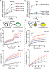
Increasing [PIP2] drives Ca2+-independent penetration by both C2 domains of Doc2β and syt1 and potentiates Ca2+-independent and -dependent vesicle fusion. A, NBD-labeled Doc2β C2AB was combined with liposomes harboring PS and increasing concentrations of PIP2 in the absence of Ca2+, and NBD emission intensity was quantified. Increasing [PIP2] drove substantial intensity increases from NBD labels on all four loops of Doc2β. This effect appeared to reach near-saturation at 5 mol % PIP2. B, as in A but for syt1 C2AB. In addition to robust penetration by C2B loop 3, increasing [PIP2] drove partial penetration by C2A loop 3. C–F, v-SNARE liposomes containing full-length syb2 and full-length syt1 were combined with t-SNARE liposomes containing full-length syntaxin-1A:SNAP-25B (syx:SN25) heterodimer and increasing mol % PIP2. C, scheme of lipid-mixing assay. Fusion of vesicles was monitored by dequenching of NBD. D, results of lipid-mixing assays conducted with increasing mol % PIP2 in the t-SNARE vesicles. Above, full traces; below, Ca2+-free portion of the trace shown on an expanded timescale. PIP2 drove Ca2+-independent and -dependent lipid mixing in a dose-dependent manner. E, scheme of content-mixing assay. Fusion of vesicles was monitored by dequenching of sulforhodamine B. F, results of content-mixing assays conducted with increasing mol % PIP2 in the t-SNARE vesicles. Above, full traces; below, Ca2+-free portion of the trace shown on an expanded timescale. As with lipid-mixing experiments, a dose-dependent effect of PIP2 on Ca2+-independent and -dependent fusion was observed. Error bars, S.E. of four independent trials.
Figure 7.
Figure 7.
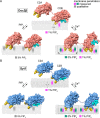
Model of Ca2+-dependent and -independent membrane penetration by Doc2β and syt1 in the presence and absence of PIP2. Calculated membrane penetration depths are illustrated, to scale, for Doc2β and syt1. Models of syt1 and Doc2 were created by rendering the molecular surfaces of the corresponding X-ray or NMR structures (Doc2, as above; syt1, PDB codes 1RSY (C2A) and 1K5W (C2B) from Sutton et al. (53) and Fernandez et al. (54), respectively). The polybasic patch of C2B is rendered cyan in each model. Shaded areas in the bilayer represent the calculated half-widths of the penetration depth measurements for each probe. A, scale drawing of membrane penetration by Doc2β. Prior to binding Ca2+, C2B shallowly penetrates bilayers in the presence of 1% PIP2. After binding Ca2+, all four loops penetrate the bilayer. However, both loops in C2A are relegated to a shallow position unless PIP2 is also present, which enables C2A loop 3 to penetrate 3.7 Å deeper on average into the membrane. In the absence of Ca2+, increases in mol % PIP2 in the target membrane can drive partial penetration of the bilayer by all four loops of Doc2β. B, scale drawing of membrane penetration by syt1. As with Doc2β, 1% PIP2 enables Ca2+-independent penetration by C2B loop 3, and increasing [PIP2] drives penetration by C2A loop 3. Upon binding Ca2+, however, all four loops of syt1 penetrate deeply into the membrane even in the absence of PIP2.

Similar articles

Cited by

References

    1. Südhof T. C., and Rothman J. E. (2009) Membrane fusion: grappling with SNARE and SM proteins. Science 323, 474–477 10.1126/science.1161748 - DOI - PMC - PubMed
    1. Chapman E. R. (2008) How does synaptotagmin trigger neurotransmitter release? Annu. Rev. Biochem. 77, 615–641 10.1146/annurev.biochem.77.062005.101135 - DOI - PubMed
    1. Katz B., and Miledi R. (1967) The timing of calcium action during neuromuscular transmission. J. Physiol. 189, 535–544 10.1113/jphysiol.1967.sp008183 - DOI - PMC - PubMed
    1. Brose N., Petrenko A. G., Südhof T. C., and Jahn R. (1992) Synaptotagmin: a calcium sensor on the synaptic vesicle surface. Science 256, 1021–1025 10.1126/science.1589771 - DOI - PubMed
    1. Bhalla A., Chicka M. C., and Chapman E. R. (2008) Analysis of the synaptotagmin family during reconstituted membrane fusion: uncovering a class of inhibitory isoforms. J. Biol. Chem. 283, 21799–21807 10.1074/jbc.M709628200 - DOI - PMC - PubMed

Publication types

MeSH terms