Review
. 2019 May 24;18(1):101.
doi: 10.1186/s12943-019-1030-2.

Crosstalk between autophagy and epithelial-mesenchymal transition and its application in cancer therapy

Affiliations
Review

Crosstalk between autophagy and epithelial-mesenchymal transition and its application in cancer therapy

Hong-Tao Chen et al. Mol Cancer. .

Abstract

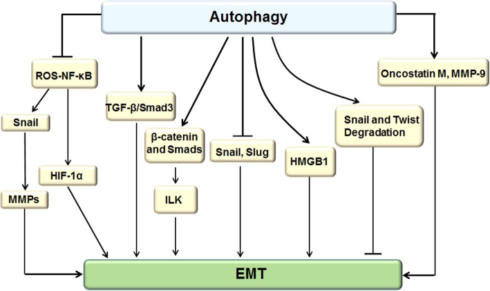
Autophagy is a highly conserved catabolic process that mediates degradation of pernicious or dysfunctional cellular components, such as invasive pathogens, senescent proteins, and organelles. It can promote or suppress tumor development, so it is a "double-edged sword" in tumors that depends on the cell and tissue types and the stages of tumor. The epithelial-mesenchymal transition (EMT) is a complex biological trans-differentiation process that allows epithelial cells to transiently obtain mesenchymal features, including motility and metastatic potential. EMT is considered as an important contributor to the invasion and metastasis of cancers. Thus, clarifying the crosstalk between autophagy and EMT will provide novel targets for cancer therapy. It was reported that EMT-related signal pathways have an impact on autophagy; conversely, autophagy activation can suppress or strengthen EMT by regulating various signaling pathways. On one hand, autophagy activation provides energy and basic nutrients for EMT during metastatic spreading, which assists cells to survive in stressful environmental and intracellular conditions. On the other hand, autophagy, acting as a cancer-suppressive function, is inclined to hinder metastasis by selectively down-regulating critical transcription factors of EMT in the early phases. Therefore, the inhibition of EMT by autophagy inhibitors or activators might be a novel strategy that provides thought and enlightenment for the treatment of cancer. In this article, we discuss in detail the role of autophagy and EMT in the development of cancers, the regulatory mechanisms between autophagy and EMT, the effects of autophagy inhibition or activation on EMT, and the potential applications in anticancer therapy.

Keywords: Anticancer therapy; Autophagy; Cancer metastasis; Epithelial-mesenchymal transition.


Conflict of interest statement

The authors declare that they have no competing interests.

Figures

Fig. 1
Fig. 1
mTOR signaling pathway regulated autophagy and EMT. PI3K activation is induced by interaction with a growth factor receptor, direct binding to Ras, also induced by NF-κB and TGF-β activation. Activation of the PI3K/AKT signaling pathway blocks autophagy by prohibiting mTOR. The PI3K/Akt pathway positively regulates WNT/β-catenin through phosphorylating the serine at residue 552 in β-catenin and the serine at residue 9 in glycogen synthase kinase 3β (GSK3β), which increases intracellular β-catenin levels that combine with E-cadherin to promote EMT. Moreover, The PI3K/Akt pathway activity up-regulates nuclear factors SNAIL and SLUG, contributing to EMT activation. GSK-3β directly induces autophagy by activating LKB1/AMPK and in turn prohibiting the PI3K/AKT/mTOR pathway. It also indirectly triggers autophagy through promoting the hydrolysis of β-catenin protein. LKB1/AMPK activation plays a critical role in stimulating autophagy via decreasing the ratio of p-mTOR/mTOR and p-p70s6k/p70s6k. In addition, LKB1/AMPK hinders EMT by inhibiting Smad2/3 and TGF-β activity. Ras protein mutation not only activates the Ras/Rac1/Mkk7/JNK pathway (with JNK in turn binding to Atg5/Atg7) but also induces the Ras/Raf1/MEK1/2/ERK signaling pathway, which results in autophagy activation and EMT enhancement
Fig. 2
Fig. 2
Beclin-1 signaling pathway regulated autophagy and EMT. The Beclin-1 gene triggers autophagy by forming the PI3K complex and prohibiting EMT through down-regulating ZEB1, WNT1, and NF-κB. Additionally, Beclin-1-induced autophagy accelerated EMT by up-regulating vimentin and Twist expression and decreasing E-cadherin expression
Fig. 3
Fig. 3
P53 signaling pathway regulated autophagy and EMT. Nucleus P53 promotes the up-regulation of autophagy by down-regulating PI3K/AKT/mTOR signaling and enhancing the expression of autophagy-related genes, including Ulk1/2, Atg4, Atg7, and Atg10. Nevertheless, P53 inhibits autophagy in the cytoplasm. The nucleus P53 reduced the expression of ZEB1, ZEB2, and SNAIL by activating the expression of miR-200a and miR-130b, resulting in EMT inhibition. Significantly, mutant P53 triggers EMT and mitochondrial fission that in turn strengthens autophagy
Fig. 4
Fig. 4
JAK/STAT signaling pathway regulated autophagy and EMT. Activation of JAK/STAT protein is stimulated by IL-6, leading to the up-regulation of the expression of MMP-2 and SNAIL and activation of EMT. On the other hand, the extracellular IL-6–mediated JAK/STAT signaling pathway accelerates the cancer process by prohibiting autophagy. Furthermore, autophagy induction hinders EMT through suppressing JAK/STAT signaling
Fig. 5
Fig. 5
The integrin signaling pathway regulated autophagy and EMT. FAK-Src–mediated integrin pathway has been shown to inhibit autophagy and promote EMT. In addition, integrin enhances EMT by stimulating the EGFR-ERK/MAPK signaling pathway. ILK accelerates EMT by activating the WNT/β-catenin pathway. Similarly, ILK can promote EMT development by transferring β-catenin into the nucleus, causing down-regulation of E-cadherin. Additionally, integrin signaling promotes TGF-β1–dependent down-regulation of E-cadherin expression, which is essential for EMT induction. Furthermore, ILK inhibits autophagy by promoting the phosphorylation of AKT and activating mTOR. By contrast, autophagy stimulates β-catenin and Smad signaling to enhance ILK expression, resulting in EMT promotion
Fig. 6
Fig. 6
WNTs signaling pathway regulated autophagy and EMT. The WNTs pathway consists of the classical pathway and nonclassical WNT pathway. The classical pathway (WNT/β-catenin signaling pathway) directly leads to HIF-1α activation, which in turn results in overexpression of SLUG, SNAIL, and TWIST and induction of EMT. The nonclassical WNT pathway mainly contains two WNT proteins, WNT5A and WNT11, which facilitate EMT by inducing p38 (Mapk14) phosphorylation. Dishevelled (Dvl) is a basic and central component of WNT signaling, and it plays an important role in both β-catenin–mediated canonical and β-catenin–independent noncanonical WNT signaling. Dvl expression and stability are negatively controlled by autophagy in the late stages of cancer development, which in turn inhibits the WNT process. On the other hand, autophagy can decrease the stability of TWIST1 protein and hinder EMT
Fig. 7
Fig. 7
NF-κB signaling pathway regulated autophagy and EMT. NF-κB activation increased transcriptional activation of EMT regulator genes expression via binding directly to the sites of EMT transcription factors, including SNAIL1, SLUG, TWIST1, and SIP1 promoter. In addition, TNF-α–mediated stability of SNAIL protein is strengthened by GSK3β activity, which is dependent on NF-κB activation. Furthermore, NF-κB binding to MMPs promotes SNAIL transcription. NF-κB can down-regulate autophagy by inhibiting Beclin-1, an initiator of autophagy. However, autophagy activation suppresses ROS–NF-κB signaling to down-regulate MMPs expression, contributing to EMT inhibition
Fig. 8
Fig. 8
TGF-β signaling pathway regulated autophagy and EMT. Activation of TGF-β/Smad3 in epithelial cells triggers EMT, and the activation of TGF-β/Smad2 signaling pathway also stimulates EMT. Moreover, TGF-β1 induces EMT and cancer metastasis by directly targeting the cytoplasmic domain of E-cadherin (CDH1) and activating WNT/β-catenin signaling. Sometimes, TGF-β cooperates with synergistic factors to induce EMT, such as Ras. Once TGF-β is stimulated, EMT-related transcription factor STAT3 interacts with Ras, which induces SNAIL expression and promotes EMT. Naturally, TGF-β can stimulate the expression of mRNA transcripts of several autophagy-related genes, such as Beclin-1, Atg5, Atg7, and death-associated protein kinase (Dapk), and it induces accumulation of autophagosomes and activation of autophagic flux, which potentiates the induction of the autophagy. It is worthy that autophagy induces TGF-β1 expression and TGF-β1-dependent EMT via triggering cAMP/PKA/CREB signaling, which relies on autophagy-dependent phosphodiesterase 4A (PDE4A) degradation
Fig. 9
Fig. 9
Interplay between cytoskeleton and mitochondria. Cytoskeleton polymerization induced by EMT, which in turn supports mitochondrial fission that are essential for further sustain EMT process by providing energy supplies, and depolymerization of actin cytoskeleton is sufficient for reversing EMT phenotype. Massive activation of autophagy induces mitochondrial fusion and the reconstitution of mitochondrial network, which subsequently reduces the number of available free mitochondria and counteracts EMT. Mitochondrial protein BNIP3 potentially supported mitochondrial fission and turnover through stimulating mitophagy by directly binding to both mitochondria and the autophagosomal protein LC3, but also enhanced cytoskeleton polymerization. The interaction between BNIP3 and cadherin-6 (CDH6) drives EMT, restrains autophagy and promotes mitochondrial fission
Fig. 10
Fig. 10
Regulating EMT by targeting autophagy. The autophagy attenuates EMT by inhibiting the overexpression of SNAIL and SLUG and activation of ROS-NF-κB-HIF-1α pathway. Moreover, ROS-induced NF-κB activation up-regulates SNAIL and MMPs expression to promote EMT. Additionally, autophagy accelerates lysosomal- mediated degradation of SNAIL and TWIST, resulting in EMT inhibition. On the other side, autophagy activation enhances EMT by increasing the expression of HMGB1, metastasis-associated protein oncostatin M and MMP-9, facilitating EMT markers expression in both RNA and protein levels accompany with promotion of TGF-β2/Smad signaling pathway activity, and up-regulating ILK by linking β-catenin and Smad signaling to induce EMT

Similar articles

Cited by

References

    1. Zachari M, Ganley IG. The mammalian ULK1 complex and autophagy initiation. Essays Biochem. 2017;61(6):585–596. doi: 10.1042/EBC20170021. - DOI - PMC - PubMed
    1. Levy JMM, Towers CG, Thorburn A. Targeting autophagy in cancer. Nat Rev Cancer. 2017;17(9):528–542. doi: 10.1038/nrc.2017.53. - DOI - PMC - PubMed
    1. Saitoh M. Involvement of partial EMT in cancer progression. J Biochem. 2018;164(4):257–264. doi: 10.1093/jb/mvy047. - DOI - PubMed
    1. Krebs AM, Mitschke J, Lasierra Losada M, et al. The EMT-activator Zeb1 is a key factor for cell plasticity and promotes metastasis in pancreatic cancer. Nat Cell Biol. 2017;19(5):518–529. doi: 10.1038/ncb3513. - DOI - PubMed
    1. Nieto MA. Epithelial plasticity: a common theme in embryonic and cancer cells. Science. 2013;342(6159):1234850. doi: 10.1126/science.1234850. - DOI - PubMed

Publication types

MeSH terms

Substances

LinkOut - more resources