Review
. 2019 May 22;20(10):2518.
doi: 10.3390/ijms20102518.

Unfolded Protein Response (UPR) in Survival, Dormancy, Immunosuppression, Metastasis, and Treatments of Cancer Cells

Affiliations
Review

Unfolded Protein Response (UPR) in Survival, Dormancy, Immunosuppression, Metastasis, and Treatments of Cancer Cells

Sheng-Kai Hsu et al. Int J Mol Sci. .

Abstract

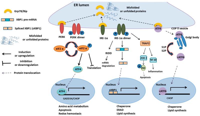
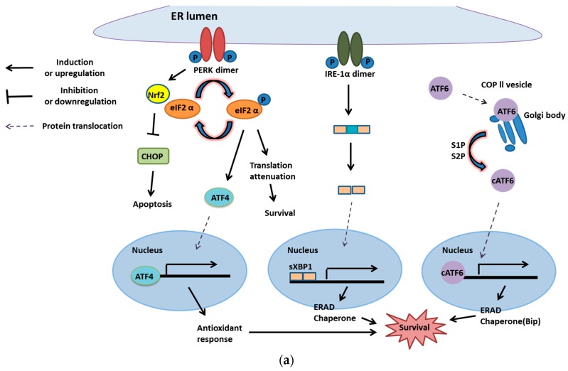
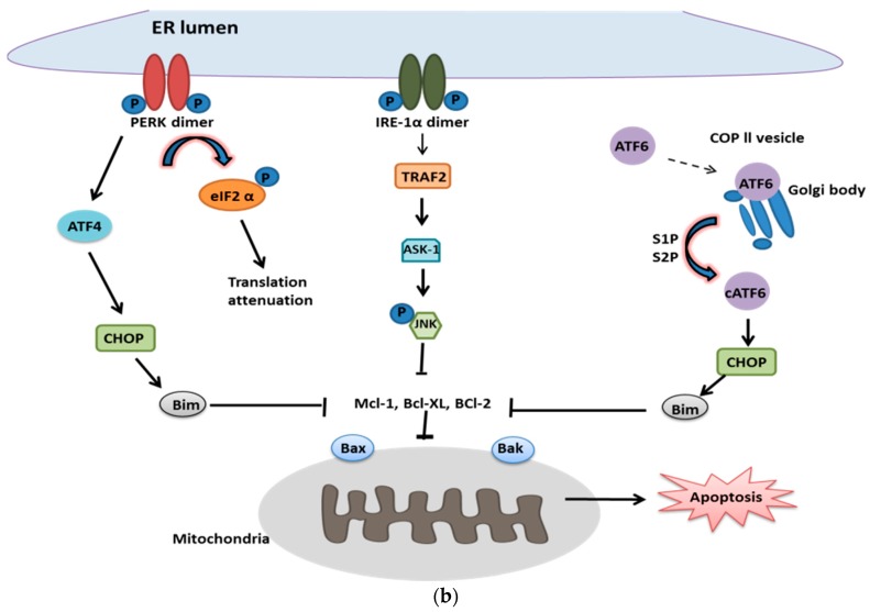
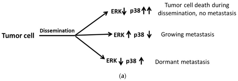
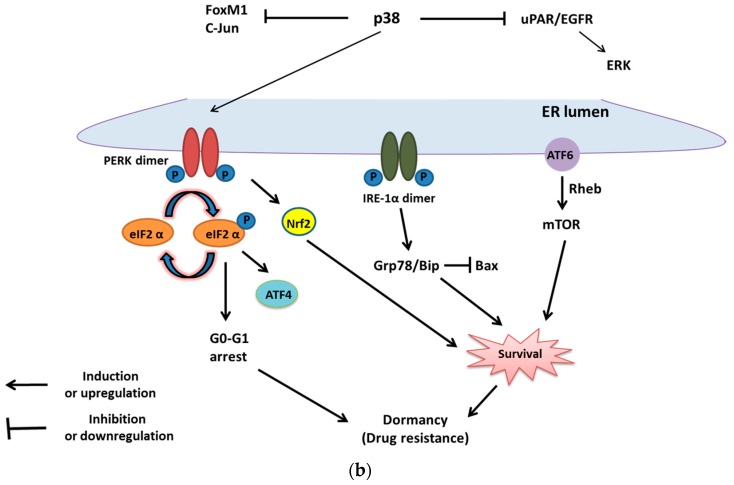
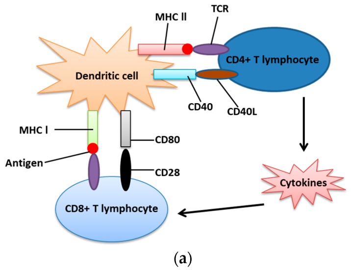
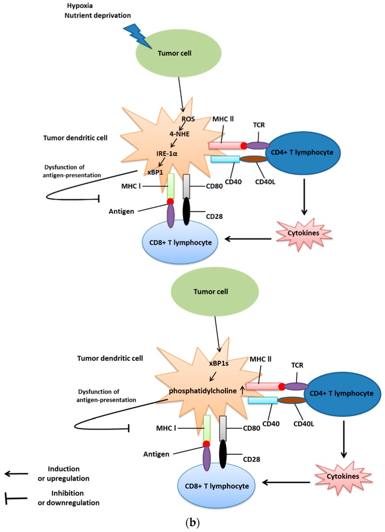
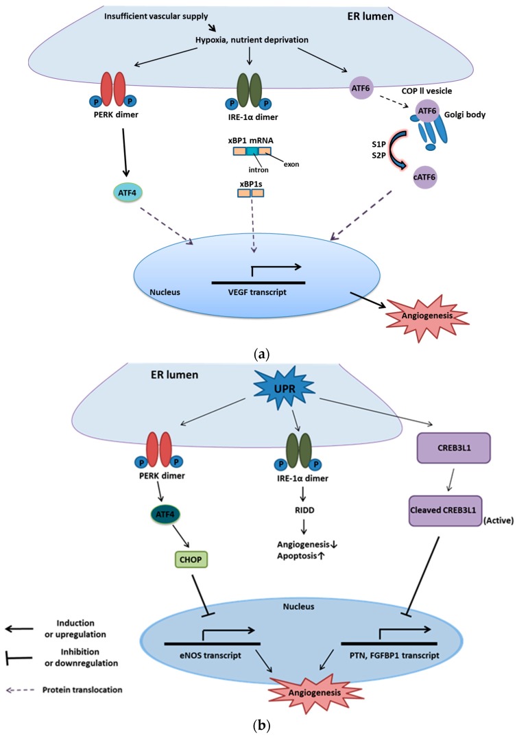
The endoplasmic reticulum (ER) has diverse functions, and especially misfolded protein modification is in the focus of this review paper. With a highly regulatory mechanism, called unfolded protein response (UPR), it protects cells from the accumulation of misfolded proteins. Nevertheless, not only does UPR modify improper proteins, but it also degrades proteins that are unable to recover. Three pathways of UPR, namely PERK, IRE-1, and ATF6, have a significant role in regulating stress-induced physiological responses in cells. The dysregulated UPR may be involved in diseases, such as atherosclerosis, heart diseases, amyotrophic lateral sclerosis (ALS), and cancer. Here, we discuss the relation between UPR and cancer, considering several aspects including survival, dormancy, immunosuppression, angiogenesis, and metastasis of cancer cells. Although several moderate adversities can subject cancer cells to a hostile environment, UPR can ensure their survival. Excessive unfavorable conditions, such as overloading with misfolded proteins and nutrient deprivation, tend to trigger cancer cell death signaling. Regarding dormancy and immunosuppression, cancer cells can survive chemotherapies and acquire drug resistance through dormancy and immunosuppression. Cancer cells can also regulate the downstream of UPR to modulate angiogenesis and promote metastasis. In the end, regulating UPR through different molecular mechanisms may provide promising anticancer treatment options by suppressing cancer proliferation and progression.

Keywords: ATF6; IRE-1; PERK; cancer; endoplasmic reticulum (ER); unfolded protein response (UPR).


Conflict of interest statement

The authors declare no conflict of interest.

Figures

Figure 1
Figure 1
Overview of the UPR process [14]. Upon stimulation of the unfolded protein response, the Grp78/Bip is recruited as a chaperone and leaves its interaction with the three transmembrane proteins of the unfolded protein response (UPR), the IRE-1α, PERK, and ATF6. This allows these proteins to oligomerize and become activated PERK dimerizes and phosphorylates the eIF2α suppressing 5’capped mRNA translation.
Figure 2
Figure 2
Unfolded protein response and cell survival or death. (a) PERK provides cancer cell survival. PERK can activate ATF4, which upregulates the genes with roles in antioxidant response for survival. Moreover, PERK can stimulate Nrf2 to inactivate cell death signaling, CHOP, and phosphorylated elF2α to attenuate translation for survival. Another transmembrane protein of the UPR membrane, IRE-1, and ATF6 also have crucial roles in cancer cell survival. Under the moderate level of ER stress, activated IRE-1 removes the introns of inactivated XBP1 to form spliced XBP1 (XBP1s). XBP1s serves as a transcription factor and binds with the promoter of chaperone and ERAD genes for modifying or degrading misfolded proteins for cell survival. Besides, ATF6 translocates from the ER membrane to the Golgi body. After moving to the Golgi body, ATF6 is cleaved to release the transcription factor (active segment) that induces the expression of chaperones and ERAD [42]; (b) When cells are overloaded with misfolded proteins, three transmembrane proteins of UPR are inclined to trigger cell death signals. Activated PERK phosphorylates elF2 to block protein synthesis. Furthermore, inactive elF2 will induce ATF4, a transcription factor that promotes Noxa and CHOP (both are pro-apoptotic transcription factors). Then, CHOP stimulates Bim, a pro-apoptotic protein of Bcl-2 families, and directly activates Bax and Bak on the membrane of mitochondria to trigger apoptosis. Furthermore, once IRE-1 is phosphorylated by extensive UPR, it will recruit TRAF 2 and activate apoptosis signal-regulating kinase 1 (ASK1) to phosphorylate JNK. Activated JNK can inhibit anti-apoptotic proteins, such as Mcl-1 and Bcl-XL, to trigger cell death signaling. Another pathway, cleaved ATF6, also induces CHOP expression and leads to apoptosis.
Figure 2
Figure 2
Unfolded protein response and cell survival or death. (a) PERK provides cancer cell survival. PERK can activate ATF4, which upregulates the genes with roles in antioxidant response for survival. Moreover, PERK can stimulate Nrf2 to inactivate cell death signaling, CHOP, and phosphorylated elF2α to attenuate translation for survival. Another transmembrane protein of the UPR membrane, IRE-1, and ATF6 also have crucial roles in cancer cell survival. Under the moderate level of ER stress, activated IRE-1 removes the introns of inactivated XBP1 to form spliced XBP1 (XBP1s). XBP1s serves as a transcription factor and binds with the promoter of chaperone and ERAD genes for modifying or degrading misfolded proteins for cell survival. Besides, ATF6 translocates from the ER membrane to the Golgi body. After moving to the Golgi body, ATF6 is cleaved to release the transcription factor (active segment) that induces the expression of chaperones and ERAD [42]; (b) When cells are overloaded with misfolded proteins, three transmembrane proteins of UPR are inclined to trigger cell death signals. Activated PERK phosphorylates elF2 to block protein synthesis. Furthermore, inactive elF2 will induce ATF4, a transcription factor that promotes Noxa and CHOP (both are pro-apoptotic transcription factors). Then, CHOP stimulates Bim, a pro-apoptotic protein of Bcl-2 families, and directly activates Bax and Bak on the membrane of mitochondria to trigger apoptosis. Furthermore, once IRE-1 is phosphorylated by extensive UPR, it will recruit TRAF 2 and activate apoptosis signal-regulating kinase 1 (ASK1) to phosphorylate JNK. Activated JNK can inhibit anti-apoptotic proteins, such as Mcl-1 and Bcl-XL, to trigger cell death signaling. Another pathway, cleaved ATF6, also induces CHOP expression and leads to apoptosis.
Figure 3
Figure 3
The role of dormancy in metastasis and chemoresistance of cancer cells. (a) Dormancy-regulated metastasis affected by the balance between p38 and ERK. When tumor cells disseminate, there are three different outcomes in different microenvironments. Firstly, with upregulation of MAPK p38 combined with decreasing ERK signals, tumor cells degenerate without metastasis during dissemination; Secondly, increasing ERK with decreasing MAPK p38 promotes metastasis; Thirdly, decreasing ERK with increasing p38 brings about metastatic cells dormancy; (b) UPR-induced dormancy is associated with cancer cell survival and chemoresistance. MAPK p38 can suppress FoxM1, c-Jun and the uPAR (Urokinase-type plasminogen activator receptor) transcript, which is crucial for the activation of ERK. Furthermore, it can also trigger the downstream signaling of UPR. Activated PERK can phosphorylate elF2α for the G0–G1 arrest and induce ATF4 for survival. Besides, phosphorylated elF2α can trigger ATF4 for survival. Activated IRE-1α can induce Grp78/Bip for survival as well as block the pro-apoptotic signal, Bax. Furthermore, ATF6α can promote survival through the mTOR signaling pathway. Redrawn from Sosa et al. [48].
Figure 3
Figure 3
The role of dormancy in metastasis and chemoresistance of cancer cells. (a) Dormancy-regulated metastasis affected by the balance between p38 and ERK. When tumor cells disseminate, there are three different outcomes in different microenvironments. Firstly, with upregulation of MAPK p38 combined with decreasing ERK signals, tumor cells degenerate without metastasis during dissemination; Secondly, increasing ERK with decreasing MAPK p38 promotes metastasis; Thirdly, decreasing ERK with increasing p38 brings about metastatic cells dormancy; (b) UPR-induced dormancy is associated with cancer cell survival and chemoresistance. MAPK p38 can suppress FoxM1, c-Jun and the uPAR (Urokinase-type plasminogen activator receptor) transcript, which is crucial for the activation of ERK. Furthermore, it can also trigger the downstream signaling of UPR. Activated PERK can phosphorylate elF2α for the G0–G1 arrest and induce ATF4 for survival. Besides, phosphorylated elF2α can trigger ATF4 for survival. Activated IRE-1α can induce Grp78/Bip for survival as well as block the pro-apoptotic signal, Bax. Furthermore, ATF6α can promote survival through the mTOR signaling pathway. Redrawn from Sosa et al. [48].
Figure 4
Figure 4
Immunocompetence and immunosuppression. (a) Immunocompetence. Dendritic cells (DCs), known as antigen presenting cells, use its CD40 to bind with CD40L (CD40 ligand) on the surface of CD4+ T lymphocyte. Furthermore, DC also uses its MHC-II, combined with antigen, to bind with TCR on CD4+ T lymphocyte. Therefore, CD4+ T lymphocyte is activated by DC to secrete cytokines or to activate B cells. DC can also activate CD8+ T lymphocyte via two signal transductions. One is CD80 and CD28 interaction, and the other is MHC-I and TCR combination; (b) Immunosuppression. When the tumor dendritic cell is under stress, such as hypoxia, nutrient deprivation and accumulation of ROS, it activates 4-NHE to induce IRE1-α and its downstream transcription factor, XBP1. This effect leads to the failure of antigen presentation and, therefore, is not able to activate T lymphocytes. Besides, activated XBP1 (sXBP1) can increase the production of phosphatidylcholine, which inhibits the ability of antigen presentation. Redrawn from Cubillos-Ruiz et al. [32].
Figure 4
Figure 4
Immunocompetence and immunosuppression. (a) Immunocompetence. Dendritic cells (DCs), known as antigen presenting cells, use its CD40 to bind with CD40L (CD40 ligand) on the surface of CD4+ T lymphocyte. Furthermore, DC also uses its MHC-II, combined with antigen, to bind with TCR on CD4+ T lymphocyte. Therefore, CD4+ T lymphocyte is activated by DC to secrete cytokines or to activate B cells. DC can also activate CD8+ T lymphocyte via two signal transductions. One is CD80 and CD28 interaction, and the other is MHC-I and TCR combination; (b) Immunosuppression. When the tumor dendritic cell is under stress, such as hypoxia, nutrient deprivation and accumulation of ROS, it activates 4-NHE to induce IRE1-α and its downstream transcription factor, XBP1. This effect leads to the failure of antigen presentation and, therefore, is not able to activate T lymphocytes. Besides, activated XBP1 (sXBP1) can increase the production of phosphatidylcholine, which inhibits the ability of antigen presentation. Redrawn from Cubillos-Ruiz et al. [32].
Figure 5
Figure 5
UPR and angiogenesis. [75] (a) UPR and pro-angiogenesis. Several low-stress adversities, such as hypoxia and nutrient deprivation, are accessible to activate three branches of UPR (IRE-1, PERK, and ATF6) on the ER membrane. Active IRE-1, PERK, and ATF6 can trigger transcription factors, namely XBP1s, ATF4, and cleaved ATF6, respectively, to upregulate VEGF expression, which leads to endothelial cell survival, proliferation and migration for angiogenesis; (b) UPR and anti-angiogenesis. Severe hypoxia and nutrient deprivation stimulate UPR to activate PERK on the ER membrane. Activated PERK can induce ATF4, a transcription factor, to upregulate the expression of CHOP. CHOP also serves as a transcription factor and is accessible to bind with pro-angiogenic mRNA, such as eNOS, to inhibit angiogenesis. Furthermore, high-intensity ER stresses trigger UPR and stimulate CRE3BL1 to be cleaved by RIP (regulated intermembrane proteolysis) to generate active CRE3BL1. Then, the activated CRE3BL1 translocates to the nucleus and binds with pro-angiogenic mRNA, such as PTN (Pleiotrophin) and FGFBP1 (Fibroblast growth factor-binding protein1), to block angiogenesis.

Similar articles

Cited by

References

    1. Tsai Y.C., Weissman A.M. The Unfolded Protein Response, Degradation from Endoplasmic Reticulum and Cancer. Genes Cancer. 2010;1:764–778. doi: 10.1177/1947601910383011. - DOI - PMC - PubMed
    1. Schwarz D.S., Blower M.D. The endoplasmic reticulum: Structure, function and response to cellular signaling. Cell. Mol. Life Sci. 2016;73:79–94. doi: 10.1007/s00018-015-2052-6. - DOI - PMC - PubMed
    1. Namba T. Regulation of endoplasmic reticulum functions. Aging. 2015;7:901–902. doi: 10.18632/aging.100851. - DOI - PMC - PubMed
    1. Harding H.P., Zhang Y., Ron D. Protein translation and folding are coupled by an endoplasmic-reticulum-resident kinase. Nature. 1999;397:271–274. doi: 10.1038/16729. - DOI - PubMed
    1. Genereux J.C., Qu S., Zhou M., Ryno L.M., Wang S., Shoulders M.D., Kaufman R.J., Lasmezas C.I., Kelly J.W., Wiseman R.L. Unfolded protein response-induced ERdj3 secretion links ER stress to extracellular proteostasis. EMBO J. 2015;34:4–19. doi: 10.15252/embj.201488896. - DOI - PMC - PubMed

LinkOut - more resources